Transcription
TINS-1058; No. of Pages 11OpinionHow the brainstem controls orofacialbehaviors comprised of rhythmicactionsJeffrey D. Moore1,2, David Kleinfeld1,2,3, and Fan Wang41Graduate Program in Neurosciences, UC San Diego, La Jolla, CA 92093, USADepartment of Physics, UC San Diego, La Jolla, CA 92093, USA3Section on Neurobiology, UC San Diego, La Jolla, CA 92093, USA4Department of Neurobiology, Duke University Medical Center, Durham, NC 27710, USA2Mammals perform a multitude of well-coordinated orofacial behaviors such as breathing, sniffing, chewing,licking, swallowing, vocalizing, and in rodents, whisking. The coordination of these actions must occur without fault to prevent fatal blockages of the airway.Deciphering the neuronal circuitry that controls even asingle action requires understanding the integration ofsensory feedback and executive commands. A far greater challenge is to understand the coordination of multiple actions. Here, we focus on brainstem circuits thatdrive rhythmic orofacial actions. We discuss three neuralcomputational mechanisms that may enable circuits fordifferent actions to operate without interfering witheach other. We conclude with proposed experimentalprograms for delineating the neural control principlesthat have evolved to coordinate orofacial behaviors.Neural control of the mammalian face and mouthIt has long been postulated that there is a hierarchicalcontrol structure for motor acts in the nervous system [1,2].Individual motor actions or primitives [3] can be executedsingly or arranged in nested groups to form more complexbehaviors. The nature of the interactions among the neuralcircuits that generate these actions and behaviors has beena topic of long-standing interest to neuroscientists. Interactions between different actions are unavoidable in themammalian face and mouth, which contain sophisticatedmotor plants that serve a variety of basic physiologicalfunctions. These functions include breathing, nutrient ingestion, active sensation, and communication. Effectivebreathing, for example, requires orofacial movements thatmaintain upper airway patency [4], whereas nutrient ingestion requires chewing, licking, lapping, suckling, andswallowing. Sensory exploration also involves licking andchewing for taste, as well as fast breathing, or sniffing, forsmell. In rodents, whisking of the mystacial vibrissae isCorresponding authors: Moore, J.D. (j moore@alum.mit.edu);Wang, F. (fan.wang@duke.edu).Keywords: central pattern generator; vibrissa; orofacial movements; brainstem;pre-Bötzinger complex.0166-2236/ß 2014 Elsevier Ltd. All rights reserved. http://dx.doi.org/10.1016/j.tins.2014.05.001used for touch [5,6]. In humans and some other mammalian species, specialized orofacial movements produce vocalizations or speech. These actions, which are central tomammalian life, must be coordinated with a high degree ofprecision to prevent blockages of the airway and othermaladaptive interactions. For example, the feeding process(eating, drinking, and swallowing) involves spatiotemporally coordinated activities of more than 26 pairs of muscles and five cranial nerves to ensure proper breakdown offood, transfer of food or liquid bolus, and safe swallowing[7]. Consistent with the notion that such precise coordination represents a computationally demanding function ofthe nervous system, defects in orofacial coordination areprominent symptoms of many neurological and neurodegenerative diseases. In Parkinson’s disease for example,impaired coordination of breathing and swallowing contributes to dysphagia (e.g., difficulty in swallowing) andrespiratory impairment [8,9], which form the leading causeof aspiration pneumonia and death in these patients [10].How does the nervous system coordinate the activities ofdifferent orofacial actions such as chewing, swallowing, andbreathing? To answer this question it is first important tonote that many mammalian orofacial behaviors involveperiodic, or rhythmic movement. In fact rhythmicity characterizes some of the most basic, evolutionarily conservedtypes of movements, such as respiration, digestion, andmany forms of locomotion. Considerable insight into thegeneral problem of coordination among different rhythmicmovements is addressed in the pioneering work of von Holst,which surveys the different types of coordinated fin movements in swimming teleost fish [11]. Like swimming, basicrhythmic orofacial movements are thought to depend on thepresence of central pattern generators (CPGs), which couldbe implemented by small networks of neurons in the brainstem. In this review, we evaluate evidence for three possiblemechanisms by which coordination both within and amongorofacial actions can occur: (i) local interactions betweenpotentially co-active circuits (CPGs) ensure their coordination; (ii) a central executive command system arbitrates theexecution and amplitude of different actions; and (iii) peripheral feedback ensures the appropriate timing betweendifferent muscle groups (Figure 1). We believe studies of thebrainstem may teach us general lessons about how nervousTrends in Neurosciences xx (2014) 1–111
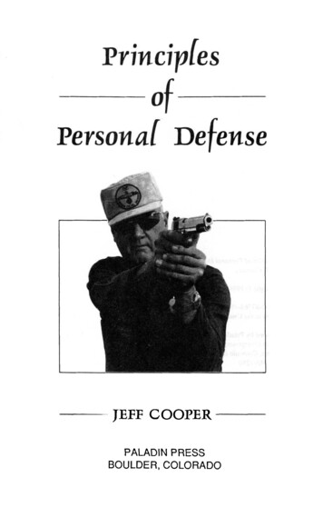
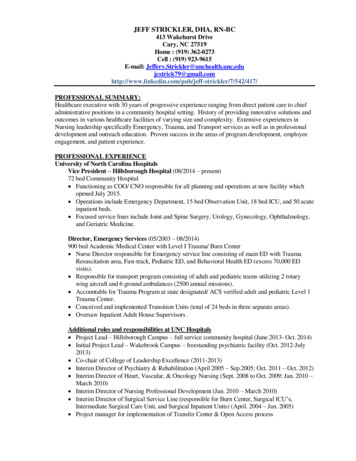
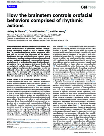
TINS-1058; No. of Pages 11OpinionTrends in Neurosciences xxx xxxx, Vol. xxx, No. xDMododulaulattorsThe ‘Decider’XORA Interac on A’CPGs–FeedbackMotoneuronsM1M2MusclesM1Ac on(s)M2TimeTimeTRENDS in NeurosciencesFigure 1. Schematic of the possible circuit arrangements for execution of differentactions using a shared motor plant. Muscles M1 and M2 can both be used indifferent temporal patterns in two different actions, A and A0 . Possible circuitinteractions include: (1) CPGs interact and coordinate with each other; (2) higherorder centers (D) gate, or select separate CPGs; and (3) peripheral feedback into aCPG alters the phase relation between the muscles. Additionally, neuromodulatorsmay act on either the CPGs themselves or their outputs to affect their frequency oramplitude. CPG, central pattern generator.systems deal with computations that can be performedautonomously but then must interact at times.Coordination of orofacial behaviors with breathingOrofacial behaviors typically involve functions that affectthe upper airway and therefore must be coordinated withbreathing. The nature of this coordination constrains theorganization of the neural circuits that control these behaviors. Rhythmic ingestive behaviors occur at frequenciesthat are faster than the 1–2 Hz frequency of basal respiration in rats. Chewing and mature suckling movementsoccur at 4 Hz [12], and rhythmic licking at 5–7 Hz [13].Rhythmic activities in the trigeminal (V), facial (VII),hypoglossal (XII), and respiratory (cervical) nerve rootletscan be elicited via bath application of NMDA in isolatedbrainstem preparations, suggesting that the brainstemalone is sufficient to generate rhythmic orofacial actions[14,15]. For such preparations, it has further been proposed that the slower breathing rhythm can reset thephase of the faster licking/suckling rhythm [15](Figure 2A). Indeed, in behaving animals it appears thatrhythmic licking and breathing are coordinated despite thedifference in their frequencies [16] (Figure 2B).With regards to rhythmic exploratory behaviors, whisking and sniffing have similar frequencies of 5–10 Hz and2have been reported to occur in a phase-locked, one-to-onemanner in rodents. Specifically, inspiration during sniffingis synchronous with vibrissa protraction, as first describedby Welker in rats [5]. These behaviors involve the use ofcommon muscles in the snout [4,17], and their robust oneto-one coordination suggests that they might depend on acommon rhythm generator. Since Welker’s initial qualitative observations, synchronous sniffing and whisking hasbeen more completely described [18,19] and quantified[20,21] in several subsequent studies in rats. There is alsoevidence that high-frequency sniffing and whisking arephase locked in mice [20]; however, one study reported alack of such coordination in this species [22]. Nonetheless,all of the recent studies of whisking behavior have foundthat whisking, like licking, can also occur during basalrespiration [20–22]. The separable timing of the whiskingand basal breathing motor outputs indicates that theseactions are paced by separate rhythm generators(Figure 2C). During basal respiration, the slow breathingrhythm resets the faster vibrissa protraction rhythm,whereas vibrissa retraction is controlled by the breathingrhythm directly. These results suggest a hierarchical organization in which the breathing rhythm influences thewhisking rhythm but not vice versa [20]. This organizationis consistent with the aforementioned results from isolatedbrainstem preparations that elicit rhythmic hypoglossaloutputs [14,15]. However, it remains to be determinedwhether this hierarchical organization extends to otherorofacial behaviors in behaving animals.Although breathing may exert an influence over someorofacial rhythms, transient events may call for a temporary cessation of breathing that over-rides the importanceof supplying the body with oxygen. For example, noxiousstimuli that may damage the airway can trigger a cessation of breathing and a corresponding pause of the respiratory patterning elements in the medulla [23]. Similarly,swallowing triggers a closure of the epiglottis to preventclogging of the airway, and it appears to modify respiratoryand chewing motor outputs [24,25] (Figure 2D). This hierarchical control between swallowing, breathing, sniffing,chewing, licking, and whisking must be reflected in theinteractions among the neural circuits that generate theseactions. Thus, we now turn our discussion to these putativebrainstem neural circuits.CPGs for breathing, chewing, licking, and swallowing inthe brainstemA CPG is operationally defined as a small network ofneurons, or even a single neuron, whose activity can generate specific movements with correct timing andsequences in the absence of sensory feedback [26,27].Various studies have suggested brainstem central originsfor rhythmic whisking, chewing, and licking. Whisking, forexample, can be generated in the absence of olfactory ortrigeminal sensory input, and also after removal of thecortex [5,18,28,29]. Similarly, chewing [30,31], licking[32,33], and breathing [34] can occur without proprioceptive feedback, and without descending input from thecortex [35]. The major circuits that underlie the generationof rhythmic orofacial actions, including their putativeCPGs, are thought to be located in the pons and medulla
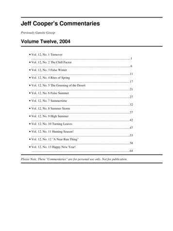
TINS-1058; No. of Pages 11OpinionTrends in Neurosciences xxx xxxx, Vol. xxx, No. x(A)NMDA XIIXIIDrainC5XII0.2 mVC50.2 mV5s(B)Breathing Licking30LicksThermocoupleBreathing Whisking1000.25-0.25Time from inspira on peak (s)1sPhotodiode(C)20Inspira on onset18 000Camera20 1sThermocoupleBreath numberProtrac on onsetSniffBasalDelay (number of cycles)ChewingBreathingBreathing Chewing SwallowingPiezoelectric beltEMGCameras3210Inspira onExpira on3210Jaw openJaw close01s3 Hz0–0.500.51.0Time from inspira on onset (s)1s(D)5 Hzπ2πPhase of swallowing-induceddevia on in cycle (radians)TRENDS in NeurosciencesFigure 2. Coordination between breathing and other rhythmic orofacial actions. (A) An isolated brainstem preparation in which rhythmic bursts of fictive motor activitywere induced via bath application of NMDA (left). Hypoglossal and phrenic motor outputs were monitored electrophysiologically via the XIIth cranial rootlet and the Vthcervical rootlet, respectively (black traces, right). The integrated activity of the XIIth rootlet is shown in green. Phrenic bursts are reported to reset the phase of the fasterhypoglossal rhythm. Adapted from [15,121]. (B) Simultaneous monitoring of licking (green) and breathing (black) in an alert rat (left and middle) shows that the actions arecoordinated (right). The occurrence of a lick is dependent on the phase of the respiratory cycle. Adapted from [16]. (C) Simultaneous monitoring of whisking (red) andbreathing (black) in an alert rat (left and middle) show that the actions are coordinated (right). Protraction and inspiration are upward. Inspiration is synchronous withprotraction on each cycle (top middle) during sniffing but only with a fraction of the cycles during basal respiration (bottom middle), as intervening whisks occur. Rasters ofinspiration onset (black) and protraction onset (red) times relative to inspiration onset for individual breaths are ordered by the duration of the breath (right). At highrespiratory rates, whisking and breathing show a 1:1 temporal relationship, while at lower breathing rates there are additional, intervening whisks between each breath.Adapted from [20]. (D) Simultaneous monitoring of chewing (orange), swallowing (purple), and breathing (black) in an alert rabbit (left and middle) reveal the nature of theircoordination. Although breathing and chewing appear to be asynchronous, swallowing affects both rhythms. The occurrence of a swallowing movement delayssubsequent breathing and chewing cycles. Adapted from [25].3
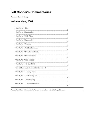
TINS-1058; No. of Pages 11Opinionof the brainstem. These regions contain both the primarysensory input nuclei (Figure 3A) and the final motor outputnuclei (Figure 3B). Detailed descriptions of the main functions of the cranial motor nuclei (V, VII, IX, X, and XII) indriving each of the different orofacial behaviors are provided in Box 1.Locations of CPGs for breathingThe best-characterized brainstem CPG in the mammaliannervous system is the circuitry in the ventral respiratorycolumn that controls breathing [36,37]. The core neuralcircuitry that paces rhythmic breathing is located in thepre-Bötzinger complex (pre-BötC), a small region in themedulla ventral to the nucleus ambiguus. Specific populations of glutamatergic cells in the pre-BötC are both sufficient [38,39] and necessary [40,41] to generate theinspiratory rhythm. The pre-BötC is interconnected withthe parafacial respiratory group (pFRG); a region that hasbeen shown to control active expiration [42,43] (Figure 3C).Sniffing is part of the normal breathing behavior, therefore, it is presumed that pre-BötC also participates in thegeneration of sniffing [20], although the exact circuit mechanism by which the higher frequencies for sniffing aregenerated remains unknown [19]. Similarly, the pre-BötCis likely to be the key CPG for upper airway control duringbreathing, and is also involved in other breathing-relatedrhythms such as gasping and sighing [44–46]. These different respiratory patterns are likely to involve differentneuromodulatory influences [44] (Figure 1).In principle, for rhythmic movements, there could be aseparate central rhythm generator (CRG) that works as aclock, and downstream pattern generators that orchestratethe periodic motor sequences based on input from the clock.Such CPG architectures have been proposed for bothbreathing and locomotion [47–49]. For breathing, it isthought that neurons in the pre-BötC generate the rhythmand neurons in the ventral respiratory group drive theappropriate pools of spinal motoneurons (Figure 3C). However, it has recently been proposed that the pre-BötC itselfcontains both rhythm- and pattern-generating elements(i.e., a separate CRG and CPG) [50]. According to thisproposal, the pre-BötC generates an internal time-keepingreference oscillation that can then be subdivided to generate the fundamental respiratory drive signal. There isanatomical and physiological evidence to suggest thatthe respiratory drive signal is then ‘broadcast’ to multipleCPG elements further downstream [51,52].Putative locations of the CPGs for ingestive andexploratory orofacial behaviorsAs a starting point to identify the specific neuronal components of orofacial CPGs, there have been many efforts tosurvey ‘pre-motor’ interneurons that project to motoneurons in different cranial motor nuclei. Early studies involved injecting classic retrograde neural tracers intocranial motor nuclei to label neurons projecting directlyto those nuclei [53,54]. Later, replication-competent pseudorabies or rabies viruses were injected into muscles ofinterest, and as the viruses spread retrogradely acrosssynapses, they labeled both pre-motoneurons and neuronsoligosynaptically connected with motoneurons [55,56].4Trends in Neurosciences xxx xxxx, Vol. xxx, No. xMost recently, the use of glycoprotein-deleted deficientrabies viruses (DG-rabies) in combination with geneticcomplementation has enabled the selective identificationof vibrissa, jaw, and tongue pre-motoneurons [57,58]. Incontrast to earlier techniques, this use of DG-rabies allowsfor trans-synaptic retrograde labeling of only pre-motoneurons via intramuscular injection. These tracing studieshave identified locations of various orofacial pre-motoneurons in the brainstem (Figure 3C). Details of the anatomicallocations of key groups of putative pre-motoneurons aresummarized in Box 2.The locations of pre-motoneurons arising from thesetracing studies have been used to guide functional observational and manipulation studies to identify orofacialCPGs. Using fictive rhythmic chewing preparations inguinea pigs, it was suspected that the minimal patterngenerating circuitry for mastication included the reticularformation between the rostral extent of the V nucleus andthe caudal extend of the VII nucleus [59,60]. This work ledto the hypothesis that chewing involves a CRG in the oraldivision of the medial gigantocellular reticular formation(Gi/GcO) that provides input to a more caudal CPG regionin the parvocellular reticular formation (PCRt) to coordinate the timing between jaw opening and jaw closing [61].Other experiments demonstrate that neurons in the dorsalprincipal trigeminal nucleus (dPrV) burst rhythmicallyduring fictive chewing in anesthetized and paralyzed rabbits [62] and raised the possibility that the chewing CPG isin the dPrV [63]. In contrast to both these possibilities, amore recent study by Travers and colleagues demonstratedthat inactivation of the PCRt and the intermediate reticular formation (IRt) between the VII and XII nucleidiminishes chewing activity and food intake in alert rats,whereas injections into Gi/GcO had no effect [64]. Thisstudy suggests the alternative possibility that the chewingCPG may be located more caudally in the medulla, andthat the role of Gi/GcO may be to relay cortical commandsto this medullary CPG rather than to generate the chewingrhythm itself (Figure 3D). Nonetheless, differentiatingbetween these hypotheses will require manipulations thatdemonstrate sufficiency and necessity of these variousregions in alert, behaving animals.Like chewing, rhythmic licking involves centrally generated, coordinated actions of the jaw opener, tongue protruder, and tongue retractor muscles [13,65]. Interneuronsthat are presynaptic to XII motoneurons are concentratedin the IRt. This region is dorsomedial to the pre-BötC andventrolateral to the XII motor nucleus [52,66]. Extracellular recording found that the spiking activity of units in thisregion is phase-locked to rhythmic licking [67], and infusion of an inhibitory agonist into the IRt between the VIIand XII nuclei blocks licking [68]. Furthermore, injection ofa m-opioid agonist in the same region alters the frequencyof licking [69]. Thus the CPG for licking, and possibly theCRG as well, is thought to be located in the IRt (Figure 3D).This region overlaps with the IRt region necessary forchewing, consistent with the fact that both behaviorsrequire coordinated jaw and tongue movements.In addition to its role in the control of ingestive orofacialmovements, the IRt has been implicated in exploratorymovement. A recent study provides experimental evidence
TINS-1058; No. of Pages 11OpinionTrends in Neurosciences xxx xxxx, Vol. xxx, No. x(A)Brainstem sensory nuclei involved in orofacial eminalmesencephalic on(Propriocep on)Solitarynucleus(Taste)Ventral(B)Frontal planeHorizonta
1 s 1 s 10 20 30-0.25 0 0.25 XII C5 20 1 s 1 s –0.5 0 0 0.5 1.0 Basal Sniff 18 000 Breath number Delay (number of cycles) T