Transcription
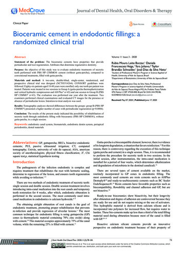
Journal of Dental Health, Oral Disorders & TherapyClinical PaperOpen AccessBioceramic cement in endodontic fillings: arandomized clinical trialAbstractVolume 11 Issue 3 - 2020Statement of the problem: The bioceramic cements have properties that provideperiradicular and root regeneration. Attributes that determine regenerative dentistry.Rúbia Moura Leite Boczar,1 DanielaFrancescato Veiga,1 Yara Juliano,2 TaylorBrandão Schnaider,1 José Dias da Silva Neto1Purpose: the objective of this study was to evaluate endodontic treatments of necroticteeth performed with PBS HP CIMMO cement (without gutta-percha), compared toconventional treatments, filled with gutta-percha.Materials and method: A two-arm, double-blind, single-center, randomized, andprospective clinical trial was designed (NCT03514264). CONSORT guidelines werefollowed. Eighty-six patients aged 18-60 years were enrolled; only one tooth per patient wastreated. Patients were treated in two sessions in Group A (gutta-percha thermoplasticizationand vertical hydraulic compresssion and AH Plus n 43) and one session in Group B (PBSHP CIMMO n 43). The evaluation was performed one year after the treatment. Twoexaminers performed clinical examinations and evaluated CT images for the presence orabsence of periradicular lesion. Intention-to-treat analysis was used.Student of Professional Master’s Program in Science Applied ofHealth, University of Vale do Sapucaí, Brazil2Department of Biostatistics, University of Vale do Sapucaí,Brazil1Correspondence: Jose Dias da Silva Neto, ProfessionalMaster’s Program in science Applied of Health, Universidadedo Vale do Sapucaí, Pouso Alegre-MG, Av. Prefeito Tuany Toledo470, Fátima I, CEP 37550-000, Brazil, Tel 5535-344-99248,5535984321520, EmailReceived: May 07, 2020 Published: June 17, 2020Results: Tomographic analyzes showed differences between the groups: group B (PBS HPCIMMO ) presented a higher number of cases with periradicular regeneration (p 0.0004).Conclusion: The results of the present study indicated the possibility of rehabilitation ofnecrotic teeth through endodontic filling with bioceramic (PBS HP CIMMO ), withoutgutta-percha, in a single session.Keywords: endodontic canal system, biomaterials, endodontic dentin system, periapicalperiodontitis, dental materialsAbbreviations: GP, guttapercha; BECs, bioactive endodonticcements; PUI, passive ultrasound irrigation; CT, computedtomography; Univás, university of vale do sapucaí; ASA, americansociety of anesthesiologists; physical fitness classification; X2, Chisquare test;p, statistical hypothesis testingIntroductionThe pathogenesis of the infection endodontic is complex andrequires treatment that rehabilitates the root with hermetic sealing,determine to regression of the lesion, and ensures tooth regenerationwhile avoiding re-infection.1–5There are two methods of endodontic treatment of necrotic teeth:single session and double session. Double session treatment involvesintroducing intra-canal medication into the root canals and temporarytooth restoration for 4 weeks, after which, endodontic obturation isperformed in the second session. The most commonly used intracanal medication in endodontics is calcium hydroxide.6–9The obtaining airtight obturation of root canals is the goal ofendodontic treatment, promoting proper rehabilitation that preventsre-infection and provide regression of alveolar lesions.1 The mostcommon technique for endodontic filling is using guttapercha (GP)cones (a thermoplastic material containing 70% zinc oxide) alongwith cement.10 This material occupies approximately 75% of the canalvolume, while the remaining 25% is filled with cement.11Submit Manuscript http://medcraveonline.comJ Dent Health Oral Disord Ther. 2020;11(3):78‒84.Gutta-percha is a biocompatible element; however, there is evidenceof its longterm degradation, a situation that favors reinfection.12 For thisreason, there is controversy regarding the execution of this technique(gutta percha and cement) in a single session. Thus, it is recommendedto perform the procedure for necrotic teeth in two sessions. In theinitial session, after instrumentation, the intra-canal medication isinstalled for a period of four weeks, which determines alkalinizationand degradation of microbiota in the dentinal canaliculi.13There are several types of cement available on the market,routinely incorporated to GP cones in endodontic filling. Themost commonly used cements are resin cements such as AHPlusDentsply 10 and ready-to-usebioceramic cements such as BC SealerEndoSequence .14 Resin cements have favorable properties such asbiocompatibility, flowability and channel adhesion and GP, but arenot bioactive.10Ready-to-use bioceramics show bioactivity, but their longevityafter obturation and degree of adhesion are controversial because theyare ready for use and do not require mixing or the use of activators.This hydrophilic material is derived from calcium silicate and isincorporated into polymers for increased flow, but requires water toharden. These bio-cements make up less than a third of the total fillingmaterial used during obturation because most of the canal is filledwith GP.10–15Bioactive calcium silicate cements provide an innovativeperspective on endodontic treatment because of their property of78 2020 Boczar et al. This is an open access article distributed under the terms of the Creative Commons Attribution License, whichpermits unrestricted use, distribution, and build upon your work non-commercially.
Bioceramic cement in endodontic fillings: a randomized clinical trialCopyright: 2020 Boczar et al.79volumetric expansion and their ability to trigger the nucleationof hydroxyapatite deposits to reduce marginal porosities and sealdiscrepancies between the dentin and the obturation material.16,17The primary outcome of the study was to assess the presence orabsence of periradicular injury by cone beam computed tomographyafter one year of treatment.These bioactive endodontic cements (BECs) are consideredsynthetic dentin because they facilitate root and periradicularregeneration and establish definitive alkalinization of the root,rendering the environment inhospitable to microbial proliferation.18The regeneration is established on two root surfaces: external(periodontal ligament and alveolar bone) and internal (cement anddentinal wall).18–21The secondary outcome was the clinical examination: presence offistula, pain on apical palpation and percussion.Recent studies have developed a bioactive cement with theaddition of adjunctive materials, which are important to create astrong material that can resist masticatory forces. This cement is calledPBS HP CIMMO . Composed of mineral oxides in the form of finehydrophilic particles. It has excellent marginal sealing capacity andalkaline pH that prevent the migration of microorganisms and fluidsinto the root canal. Composition: SiO2, K2O, Al2O3, Na2O, Fe2O3,SO3, BaSO4, CaO, MgO. A national patent application has been filedin the National Intellectual Property Institute (registration number:BR 102017 022748). Experimental studies have been performed todefine the regenerative capacity of PBS HP CIMMO cement infurcation lesions.22,23Clinical studies with PBS HP CIMMO cement have been carriedout to evaluate the regeneration of teeth subjected to parendodonticsurgery.24,25 The results of these studies have demonstrated that theclinical use of PBS HP CIMMO cement is feasible because itfacilitates regeneration of bone and periodontal ligament structuresin the long-term and is more resistant to masticatory forces due to thepresence of additives.24,25Therefore, the objective of this study was to evaluate endodontictreatments of necrotic teeth performed with PBS HP CIMMO cement (without gutta-percha), compared to conventional treatments,filled with gutta-percha.20,21,24–25Materials and methodThis study was a randomized, non-inferiority trial, two-arm,double-blind, single-center trial that was conducted from November2017 to June 2019 at the Endodontics Clinic of the BrazilianAssociation of Dentistry at Vale Sapucaí University (Univás), PousoAlegre, Minas Gerais, Brazil.The project was approved by the Ethics Committee of the Univás,mind nº, CAAE-77495717.7.0000.5102. The study was enrolled atClinicalTrials.gov (NCT 03514264). The financing obtained to carryout the study was by the Company MJS Industry and Commerce ofMaterials for Health ltda cnpj: 24.752.361/0001-52.It was calculated the sample size using the Lee Sample softwareby comparing two proportions with a dichotomous response. Asa parameter, it was cosidered the failure index for the endodonticobturation technique with cement and gutta-percha at 20%.26–28The researchs by Silva Neto et al.20–22,25 were used as a parameter todefine the percentage of failure for the (PBS HP CIMMO PousoAlegre, Minas Gerais, Brazil) group at 10%. The test was consideredmonocaudal with a test power of 90% and a level of significance of5%, was allocated 43 patients per group.The Endodontics Team of the Univás Graduate Clinic examinedthe patients through anamnesis, clinical examination and radiographic.Inclusion criteria were as follows; age: 18-60 years, gender: male orfemale. The preoperative diagnosis was made through clinical andradiographic exams that showed pulp necrosis with the presenceof periapical lesions, one tooth per patient. Single root or multipleroots and all with periapical lesion. The preoperative images wereradiographic due to the high cost of tomography. It was decided toperform CT scans only after one year. The exclusion criteria were asfollows; patients who did not agree to participate in the study and/or who did not sign the informed consent form, pregnant women,patients with periodontal disease and patients with ASA III, IV andV status (American Society of Anesthesiologists; physical fitnessclassification). Patients were allocated into 2 groups, A or B, at a1:1 ratio according to a random computer generated sequence byRandomization.com (https://www.randomization.com).Opaque envelopes, sequentially sealed and numbered, containingthe group name, as well as the material to be used for each patient,were used to ensure allocation confidentiality. Each envelope wasdelivered to the professional who performed the intervention, atthe initial moment of care. In all, 4 professional dental surgeonsspecializing in endodontics performed the interventions.The professionals who treated group A subjects (gutta-percha)instructed their patients, after the first session, to return 30 dayslater for their second obturation session. Group B patients (PBSHP CIMMO Pouso Alegre, Minas Gerais, Brasil) underwent theintervention in a single session.The 86 patients were instructed to return one year after endodonticobturation, for clinical examinations and cone-beam computedtomography. Clinical examinations and tomography analyzes, wereperformed, independently by 2 endodontist examiners with more than20 years of clinical experience.In order to ensure participants’ adherence to the research, freepreventive dental care was offered to all patients who returned for thecollection of results.The recommended techniques for each group are as follows:Group A - Endodontic obturation with (AHPlus Dentsply Pirassununga, São Paulo, Brazil) Cement and Gutta-percha(Dentsply Pirassununga, São Paulo, Brazil).First session: Anesthesia was administered with (Articaine, DFL Industry and Commerce- Taquara, Rio de Janeiro, Brazil), openingand absolute isolation. Then, instrumentation, intra-canal medicationinstallation and temporary restoration with (Fgm Maxxion R A2 Cruzeiro, São Paulo, Brazil), were performed.The endodontic instruments used for the instrumentation were:manual instruments (K 10 Dentsply Pirassununga, São Paulo,Brazil) and mechanized instruments (Prodesign Logic Easy BeloCitation: Boczar RML,Veiga DF, Juliano Y, et al. Bioceramic cement in endodontic fillings: a randomized clinical trial. J Dent Health Oral Disord Ther.2020;11(3):78‒84. DOI: 10.15406/jdhodt.2020.11.00523
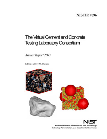
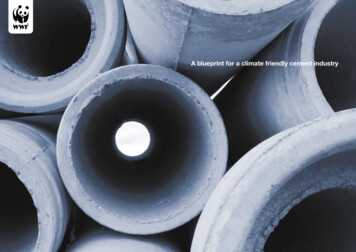
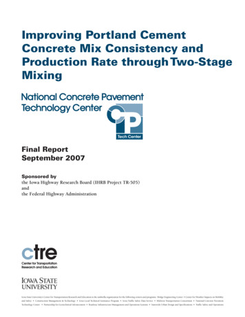
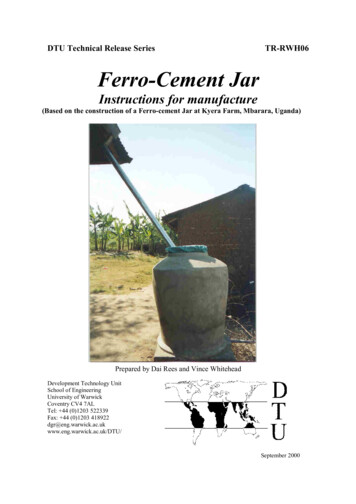
Copyright: 2020 Boczar et al.Bioceramic cement in endodontic fillings: a randomized clinical trialHorizonte, Minas Gerais, Brazil), according to the needs of the toothto be treated (2501, 1505, 2504, 2505), coupled to the endodonticmotor (Easy SI Belo Horizonte, Minas Gerais, Brazil).The crown-down instrumentation technique was used, dentistrywas performed with Novapex appliance and the final foraminaldilation was (25). The auxiliary chemical solution for irrigation was5.5% sodium hypochlorite. Passive ultrasound irrigation (PUI) (DabiAtlante ProfiNeo Ribeirão Preto, São Paulo, Brazil) and ultrasonictip (ESI 015 - Trinks Pirituba, São Paulo, Brazil) with 5.5% sodiumhypochlorite and EDTA T (Formula & Action São Paulo, São Paulo,Brazil) were performed at the end of the instrumentation. Intracanalmedication (Callen PMCC SSWhite ) was then placed into the canalsby syringe (ML Endodontic SSWhite Rio de Janeiro, Rio de Janeiro,Brazil) and the tooth was provisionally restored with restorative glassionomer (Fgm Maxxion R A2 Cruzeiro, São Paulo, Brazil).The systemic medication administered followed the determinationof each case. When there was systemic involvement and acuteinflammatory process, amoxicillin potassium clavulanate 875mg, 1tablet every 12 hours for 7 days, was prescribed. In case of allergyto amoxicillin, 300 mg of clindamycin, 1 capsule every 8 hours for7 days, was prescribed. The antiinflammatory drug of choice wasdexamethasone 4mg, 1 tablet every 12 hours for 3 days or 400 mgnimesulide, 1 tablet every 12 hours for 3 days for patients in whomdexametazone was contraindicated. The second session occurred 4weeks after the first.Second session (endodontic obturation)After 30 days,6,7,9,13 patients in group A returned to the obturationsession. The same anesthesia and absolute isolation procedureswere performed. After temporary removal of the restoration,intracanal medication was withdrawn by irrigation with 5.5% sodiumhypochlorite and use of the final instrumentation file. The canals weredried with absorbent paper cones and the main gutta-percha coneswere coupled to the channels with final calibration (25). Cone-proofradiography was performed to certify calibration. After handlingthecement (AHPlus Dentsply ), the gutta-percha cones were insertedinto the channels associated with the cement. The filling endodontictechnique performed was gutta-percha thermoplasticization andvertical hydraulic compression using a device (Touch ‘n Heat 5004 SybronEndo Orange, CA 92867, EUA). After cleaning the pulpchamber, the teeth were restored with composite resin and the patientswere instructed to return to proservation.Group B - Endodontic obturation with PBS CIMMO HP Cement.Single sessionThe pacients of Group B received the same treatment as groupA: anesthesia, coronary opening, absolute isolation, endodonticinstrumentation technique, auxiliary chemicals (5.5% sodiumhypochlorite and EDTA T Formula & Action ) and PUI. The canalswere dried with absorbent paper cones and filled with PBS HPCIMMO cement as a single shutter element.The endodontic fillings with PBS CIMMO HP were performedaccording to the following protocol: one cement portion and two dropsof distilled water were dispensed onto a glass plate. Agglutination ofthe cement in the water was performed using a flexible spatula number8024 (Duflex Rio de Janeiro, Rio de Janeiro, Brazil); it was followedmanufacturer’s recommendations, up to to achieve consistency (flow).The Macspaden (Flex Pack Easy Belo Horizonte, Minas Gerais,Brazil) instruments, coupled to the micro engine of the dental officeintra system, were used to install the intra canal cement. Schilder(double niti Easy Belo Horizonte, Minas Gerais, Brazil) condenserswere used to condense the intra canal cement. These instruments arecalibrated according to the anatomy of each instrumented channel:green (0.35 and 0.70), black (0.40 and 0.80), yellow (0.50 and 1.00)and blue (0,60 and 1.20).Similar to group A, the teeth were restored with composite resin.Systemic medication was only used in acute cases with systemicinvolvement and followed the same protocol as group A.After one year, both group A and group B patients returned to theendodontic clinic for clinical examinations, which were performedby two endodontists with more than twenty years of experience,independently and blinded to the group to which the patientparticipated. The patients were referred for tomographic exams.The analysis of the tomographic exams was performed by the twoendodontists independently and blind to the group belonging. The grayscale was used to characterize the CT image, since the preoperativeimages were radiographic due to the high cost of the tomography.Statistical analysisFor categorical variables (gender, presence or absence of fistula,presence or absence of periapical lesion, analyzed by tomography),the chi-square test was applied. Analyzes were performed using theBioestat ver program. 5.3 (Mamirauá Institute, Pará and Amazonas,Brazil) and the significance level was set at p 0.05 or 5%.Results43 patients were allocated to each group. There were 9 losses,3 in group A and 6 in group B. These patients did not return to theexamination after one year (Figure 1).There was no significant difference between groups in terms ofgender, age, location [intraoral (upper or lower arch)] or number ofroots of treated teeth.Two evaluators independently performed clinical examinationsand found no clinical evidence determined in the secondary outcomeof the present study. They evaluated the cone beam computedtomography and disagreed with the CT findings in two cases treatedin group B, but agreed with 100% confirmation of the positive resultsafter a consensus meeting.Table 1 shows the comparison between the groups regardingtomography. There was significance (p 0.004) for group B.Figure 2 shows a failure related to the presence of periapical lesion(primary study outcome).In group A and Figure 3 shows success in group B (absence ofperiapical lesion, also related to the primary outcome). The intentionto-treat analysis method was used (patients who did not show up forevaluation after one year of treatment were considered unsuccessfulfor the statistical analysis: 3 for group A and 6 for group B.Citation: Boczar RML,Veiga DF, Juliano Y, et al. Bioceramic cement in endodontic fillings: a randomized clinical trial. J Dent Health Oral Disord Ther.2020;11(3):78‒84. DOI: 10.15406/jdhodt.2020.11.00523
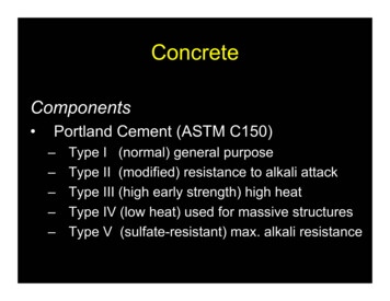
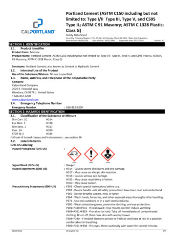
Bioceramic cement in endodontic fillings: a randomized clinical trialCopyright: 2020 Boczar et al.81Figure 1 Consort 2010 flow diagram.Figure 2 Tooth 2.5 of patient 27 (group A).2(A)Periapical radiography before treatment.2 (B) Periapical radiography immediately after treatment; (arrowsindicate periapical lesion).2 (C)Cone beam tomography one year later, with presence of periapical lesion.Citation: Boczar RML,Veiga DF, Juliano Y, et al. Bioceramic cement in endodontic fillings: a randomized clinical trial. J Dent Health Oral Disord Ther.2020;11(3):78‒84. DOI: 10.15406/jdhodt.2020.11.00523
Copyright: 2020 Boczar et al.Bioceramic cement in endodontic fillings: a randomized clinical trial82Figure 3 tooth 4.7 of patient 35 (group B). 2 (A) Periapical radiography before treatment. 2 (B) Periapical radiography immediately after treatment; (arrowsindicate periapical lesion). 2 (C) Cone beam tomography one year later, with presence of periapical lesion.Table 1 Comparison of groups A and B: comparison between the groups oncomputed tomographyn (%)FailureSuccessTotalGroup A835 (43)43Group B043 (43)43Total878 (86)86Group A x Group B(Chi-square test)X2 0.00; p 0.004DiscussionStandard endodontic obturation consists of combining endodonticcement with gutta-percha (GP) cones. GP is an inert material andrepresents 75% of the volume of the obturation, while the remainder25% of the obturation consists of cement that connects the GP
May 07, 2020 · necrotic teeth through endodontic filling with bioceramic (PBS HP CIMMO ), without gutta-percha, in a single session. Keywords: endodontic canal system, biomaterials, endodontic dentin system, periapical periodontitis, dental materials Journal of Dental Health