Transcription
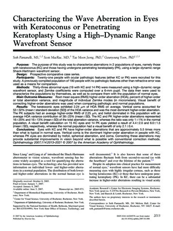
Characterizing the Wave Aberration in Eyeswith Keratoconus or PenetratingKeratoplasty Using a High–Dynamic RangeWavefront SensorSeth Pantanelli, MS,1,2 Scott MacRae, MD,3 Tae Moon Jeong, PhD,2 Geunyoung Yoon, PhD1,2,3Purpose: The purpose of this study was to characterize aberrations in 2 populations of eyes, namely thosewith keratoconus (KC) and those having undergone penetrating keratoplasty (PK), using a large– dynamic rangeShack-Hartmann wavefront sensor.Design: Prospective comparative case series.Participants: Twenty-one people with ocular pathologic features (either KC or PK) were recruited for thisstudy. A previously compiled population of 190 people with no pathologic features other than refractive error wasused as a means for comparison.Methods: Thirty-three abnormal eyes (19 with KC and 14 PK) were measured using a high– dynamic rangewavefront sensor, and Zernike coefficients were computed over a 6-mm pupil. The data then were used tocharacterize the populations by themselves, as well as to compare them with the population of normal eyes.Main Outcome Measures: Root mean square (RMS) higher-order aberration (HOA), percent of higher-orderor total aberration variance, and magnitude of individual Zernike modes (in micrometers). Visual benefit ofcorrecting higher-order aberrations was used when comparing pathologic and normal populations.Results: The keratoconic eyes exhibited 2.24 m of HOA RMS on average. Vertical coma accounted for53 32% (mean standard deviation [SD]) of the HOA variance and was the most dominant higher-order aberration.The PK subjects had an average higher-order RMS of 2.25 m, and trefoil dominated in this population with anaverage HOA variance contribution of 38 23% (mean SD). The KC and PK higher-order aberrations represented16 20% and 16 13% (mean SD) of the total aberration variance, whereas the ratio was only 1 1% in the normalpopulation. A visual benefit calculation on 15 KC eyes and 14 PK eyes yielded a result of 4.4 2.0 and 6.0 1.5(mean SD), respectively, whereas the normal population had a visual benefit of only 2.1 0.4.Conclusions: Eyes with KC and PK have higher-order aberrations that are approximately 5.5 times morethan what is typical in normal eyes. Vertical coma is the dominant higher-order aberration in people with KC,whereas PK eyes are dominated by trefoil, spherical aberration, and coma. Correcting these aberrations mayprovide substantial improvements in vision beyond what is possible with conventional correction methods.Ophthalmology 2007;114:2013–2021 2007 by the American Academy of Ophthalmology.Since Liang1 and Liang et al2 introduced the Shack-Hartmannaberrometer to vision science, wavefront sensing has become widely accepted as a tool for quantifying the aberrations in human eyes. The technology also has provided newinformation on individual lower- and higher-order aberrations in a normal population. The distribution of both lowerand higher-order aberrations in the normal human eye isOriginally received: October 8, 2005.Final revision: January 7, 2007.Accepted: January 8, 2007.Available online: June 5, 2007.Manuscript no. 2005-973.1Department of Biomedical Engineering, University of Rochester, Rochester, New York.2Center for Visual Science, University of Rochester, Rochester, New York.3Department of Ophthalmology, University of Rochester, Rochester, New York.Supported by the National Institutes of Health, Bethesda, Maryland (grantno. R01-EY014999); New York State Office of Science, Technology, and 2007 by the American Academy of OphthalmologyPublished by Elsevier Inc.well documented.3 It is also known that some of theseaberrations fluctuate both from second-to-second (as withthe heartbeat)4 and over the lifetime of the patient.5,6Despite its adoption into clinical practice for measurementof normal eyes,3 wavefront sensors have had more difficultymeasuring eyes with highly irregular corneas, such as thosehaving keratoconus (KC) or those that have undergone penetrating keratoplasty (PK). In KC, there can be a substantialincrease in higher-order aberrations resulting in visual acuityAcademic Research’s Center for Electronic Imaging Systems, Rochester,New York (grant no. 5-23576); Research to Prevent Blindness, New York,New York; and Bausch & Lomb, Rochester, New York.The authors have no proprietary interests in the research or instrumentsdescribed herein.Correspondence to Seth Pantanelli, MS, 261 Meliora Hall, Center forVisual Science, University of Rochester, Rochester, NY 14627. E-mail:seth pantanelli@urmc.rochester.edu.ISSN 0161-6420/07/ –see front matterdoi:10.1016/j.ophtha.2007.01.0082013
Ophthalmology Volume 114, Number 11, November 2007loss that is not correctable with conventional spectacles or softcontact lenses. Penetrating keratoplasty may induce a largeamount of astigmatism and higher-order aberrations similar inscale to keratoconic patients. The lack of dynamic range inwavefront sensors needed to measure these aberrations accurately makes it difficult to correct them using customizedablation. This in turn leaves some eyes that have undergonecorneal transplantation with a clear graft but suboptimal vision.In 2002, Maeda et al7 evaluated eyes with mild andmoderate KC (n 35) using a KR-9000 aberrometer (Topcon, Tokyo, Japan). The study was one of the first to look ata population of people with KC and to identify dominanthigher-order aberrations (i.e., coma). Similarly, in 2003,Shah et al8 also published measurements and comparisonsof KC and PK eyes with those of normals. In both of theseanalyses, however, physical limitations of the wavefrontsensor used prevented measurement of the wavefront aberration in eyes with various stages of KC over a large pupilsize ( 6 mm). The same limitation on dynamic range alsoprevented measuring the PK eyes over a large pupil in thestudy of Shah et al. This constraint limits the value of themeasurement, because correction with customized contactlenses or laser refractive surgery may require characterization of the aberration outside an optical zone size of 6.00mm or more, depending on their scotopic pupil sizes.9The limit in dynamic range mentioned above occursbecause of constraints, namely a tradeoff between the dynamic range and measurement sensitivity, in the design of aconventional Shack-Hartmann wavefront sensor. Increaseddynamic range of a Shack-Hartmann system can be obtained by using a shorter focal length lenslet array, but thiscauses a decrease in measurement sensitivity.10 Many methods, which are described elsewhere,11–16 have been proposed for overcoming the shortcomings that limit a wavefront sensors ability to measure KC and PK eyes. Thesemethods, however, usually come at a cost. We previouslyreported a newly developed Shack-Hartmann wavefrontsensor that has a large dynamic range and no sacrifice ofmeasurement sensitivity (Pantanelli S, et al. Large dynamicrange Shack-Hartmann wavefront sensor for highly aberrated eyes. Abstract presented at: Association for Researchin Vision and Ophthalmology Annual Meeting, April 2003,Fort Lauderdale, Florida).17 The goal of this study was touse this system to investigate dominant higher-order aberrations in eyes with KC and those that have undergone PKover a pupil size of 6.00 mm or more. In addition, thewavefront aberration and theoretical visual benefit of correcting higher-order aberrations in these populations wasstudied and compared with a population of normal eyes.from a conventional Shack-Hartmann wavefront sensor; no significant difference in measuring higher-order aberration was found.17Wavefront aberrations in the pre-LASIK normal populationwere obtained from a previously existing database provided byBausch & Lomb, Inc. (Rochester, NY). These aberrations weremeasured using a Bausch & Lomb Zywave IIz Shack-Hartmann–based wavefront sensor. Subjects in this control population had anaverage refractive error of 3.69 1.62 diopters (D; mean standarddeviation [SD]) sphere and 0.55 0.46 D cylinder. No subject hadmore than 7.74 D and 3.58 D of sphere or cylinder, respectively.The Zernike aberrations, up to the fifth order, were calculated for eachsubject based on the central 6.0 mm of the pupil.Informed written consent was obtained from the 21 participantswith abnormal eyes. All of the subjects underwent dilation with 1.0%tropicamide. Aberrations in the abnormal groups were measuredusing a large– dynamic range Shack-Hartmann wavefront sensor described previously.17 A minimum of 10 measurements were obtainedfor each eye, and each measurement contained 145 spots over a6.00-mm pupil. To ensure adequate dynamic range of the wavefrontsensor, it was verified that each spot fell within its respective subaperture on the charge-coupled device. The Zernike coefficients for upto tenth-order aberrations were determined for each picture over thecentral 6.0-mm portion of the pupil. The 5 best measurements werechosen based on the quality of the wavefront sensing spot pattern.These were then averaged to produce a single set of Zernike coefficients, reported in the Optical Society of America standard format,18for each abnormal eye. Finally, to compensate for the enantiomorphism that is present3,18 when comparing the aberrations in left andright eyes, the sign of Zernike modes that are not symmetric about thevertical axis was reversed in all data sets acquired from left eyes (i.e.,Z2 2, Z31, Z33, etc.). A Student t test ( , 0.05) was used to determinewhich Zernike modes in the KC and PK populations were statisticallydifferent from normal.The magnitude of individual Zernike aberrations and the rootmean square (RMS) of the Zernike coefficients were used tocompare the normal, KC, and PK populations. However, it isimportant to remember that RMS is not an ideal predictor of imagequality,19 nor is it mathematically appropriate to use when assessing the proportion an individual aberration has on the total magnitude of aberration (Chen L, Porter J, Singer B, et al. Predictingsubjective image quality from the eye’s wave aberration. Abstractpresented at: Association for Research in Vision and Ophthalmology Annual Meeting, April 2003, Fort Lauderdale, Florida). Instead, the total variance (sum of the square of all Zernike coefficients second order and above) and higher-order variance (sum ofthe square of all Zernike coefficients third order and above) areused when calculating the contribution an individual aberration hason the total measured aberration. Therefore, the contribution anindividual Zernike mode with radial index n and azimuthal frequency m has on the total aberration (PTOT) and higher-orderaberration (PHOA) is defined byPTOT 2(Z mn)n i兺n 2Patients and MethodsThe protocol for this study received institutional review boardapproval from the University of Rochester School of Medicine andDentistry. The populations used included 14 PK eyes from 11patients, 19 eyes with KC from 10 patients, and 378 pre-LASIKnormal eyes from 190 patients. None of the normal eyes had anyocular pathologic features, aside from refractive errors. The measurement accuracy of the large– dynamic range wavefront sensorwas verified previously by direct comparison with results obtained20142(Z mn)andPHOA 2(Z mn)n i兺n 3,2(Z mn)where i is the highest radial index recorded for the population (i 5 for normals and i 10 for abnormals).From the measured wave aberration, the RMS, point spreadfunction, and modulation transfer function (MTF) can be calculated. All of these can be used as image quality metrics, although
Pantanelli et al 䡠 Characterizing the Wave Aberration in KC and PKone is usually better than the other, depending on the application.For instance, retinal plane-based metrics such as the point spreadfunction and MTF provide a better prediction of retinal imagequality compared with a metric based on the pupil plane, such asRMS.20,21 To quantify the benefit that these 3 populations mightexperience from perfect correction of their higher-order aberrations, a metric called visual benefit was used. This metric is basedon another function mentioned above, the volume MTF (vMTF),which quantifies the optical quality of a system over the broadrange of spatial frequencies that it encounters. To calculate visualbenefit for each eye, the volume under the MTF was calculated forspatial frequencies between 0 and 60 cycles/degree under whitelight conditions. First, this was carried out assuming optimizedcorrection of second-order aberrations only, where sphere, cylinder, and axis values were chosen that maximized the vMTF.Second, the vMTF was calculated assuming correction of allsecond-order and higher-order aberrations up to the tenth order forabnormal eyes. Higher-order aberrations up to only the fifth orderwere used for normal eyes because the aberration data higher thanthe fifth order were not available. However, as mentioned below,it is insignificant in normal eyes because of the small contributionof higher-order aberrations above the fifth order to degradation ofimage quality.22 The ratio of the second- and higher-order corrected vMTF (vMTFcustomized) over the second-order correctedvMTF (vMTFsecond order) provides the estimate of visual benefit forcorrecting the higher-order aberrations. Therefore, a visual benefitof 1 indicates no benefit of correcting the higher-order aberration.This calculation is summarized byvisual benefit vMTFcustomizedvMTF2nd Order.ResultsKeratoconic PopulationFigure 1 plots both the average second-order and higher-orderaberrations of this abnormal population. In all of the eyes exceptfor 2, the defocus was positive (5.15 4.20 m or 3.96 3.23 Dsphere, mean SD) for a 6-mm pupil, which corresponded to theneed for myopic correction. The astigmatism terms for the population when taking into account the sign were Z2 2 0.59 1.09 m (mean SD) and Z22 0.14 1.82 m (mean SD), equivalent to 2.06 1.15 D cylinder (mean SD) for a 6-mm pupil.Higher-order aberrations were significantly larger than those typically found in the normal population; the keratoconic eyes had anaverage higher-order RMS of 2.24 1.22 m (mean SD) over a6.00-mm pupil, which was 5.5 times more than the 0.41 0.16 m(mean SD) found in the normal eyes (P 0). The most dominanthigher-order aberrations in the keratoconic group were verticalcoma, trefoil, and spherical aberration, in that respective order.Most notably, the vertical coma was negative in all of the eyesexcept for 1 of the 19 and accounted for 53 32% (mean SD) ofthe higher-order variance. This average vertical coma was morethan 1000% more than that found in normal eyes (P 0). Whenboth horizontal and vertical coma terms were considered, theyaccounted for 62 30% (mean SD) of the higher-order variance.Trefoil and spherical aberration accounted for 18 27% and6 13% (mean SD) of the higher-order variance, respectively.Secondary astigmatism also was statistically different from normal(magnitude 0.18 0.16 m [mean SD]; P 0.005) in thispopulation. Finally, 16 20% of the total variance in keratoconiceyes was due to higher-order aberrations, whereas only 12 12%(mean SD) of this variance came from astigmatism.Penetrating Keratoplasty PopulationFourteen eyes that had undergone PK were measured using thelarge– dynamic range Shack-Hartmann wavefront sensor. The datawere processed using the method described above for the keratoconic eyes and are presented in Figure 2. The PK subjects hadsecond-order astigmatism values of Z2 2 1.69 2.61 m andZ22 0.54 2.65 m, or 4.24 1.48 D of cylinder (mean SD).All except for 2 of the eyes had positive defocus (3.95 3.12 mor 3.04 2.40 D sphere [mean SD]) and were myopic. The corneal transplant eyes also had 5.5 times more higher-order RMS(2.25 0.75 m, mean SD) than normal eyes (0.41 m) over aFigure 1. Graphs demonstrating the average Zernike coefficient values for a population of keratoconic eyes. The most dominant aberration, accountingfor approximately 53% of the total higher-order variance, was vertical coma. Defocus was consistently positive. Zernike modes that were statisticallydifferent from normal (P 0.05) are designated by an asterisk (*).2015
Ophthalmology Volume 114, Number 11, November 2007Figure 2. Graphs demonstrating the average Zernike coefficient values for a population of penetrating keratoplasty (PK) eyes. The most dominanthigher-order aberrations were trefoil, coma, and spherical aberration. Two penetrating keratoplasty eyes were not included in this plot because a lack ofleft–right designation prevented compensation for enantiomorphism. Zernike modes that were statistically different from normal (P 0.05) are designatedby an asterisk (*).6.0-mm pupil (P 0). The most dominant higher-order aberrations were trefoil, coma, and spherical aberration, which accountedfor 38 23%, 20 17%, and 12 14% (mean SD) of the higherorder variance, respectively. These eyes had a higher-order varianceto-total variance ratio of 16 13%; however, this time the percentage was smaller than the contribution of cylinder to the totalvariance: 41 21% (mean SD).Comparison with Normal EyesFigure 3 plots the magnitude of the higher-order Zernike coefficients for keratoconic (n 19), PK (n 14), and normal eyes(n 378) over a 6.0-mm pupil. The plot reinforces what has beenmentioned previously: that abnormal eyes have larger amounts ofhigher-order aberrations when compared with normal eyes. In fact,the average magnitude of every aberration up to the fifth order inabnormal eyes was between 2 and 7 times the magnitude found innormal eyes. Dominant higher-order aberrations in each population are labeled; keratoconic eyes have the greatest magnitude ofcoma of the 3 populations and PK eyes have the greatestmagnitude of trefoil and spherical aberration. Perhaps the clearest objective indication that higher-order aberrations are moresevere in abnormal eyes than in normal eyes is the ratio of thehigher-order aberration variance over the total aberration variance. In KC and PK eyes, the higher-order aberration contributed 16 20% and 16 13% (mean SD), respectively, to theamount of total aberration in the eyes. In the normal population,the higher-order aberration was only 1 1% (mean SD) of theFigure 3. Graph demonstrating comparison of abnormal and normal eyes. Abnormal eyes have greater amounts of higher-order aberrations whencompared with normal eyes.2016
Pantanelli et al 䡠 Characterizing the Wave Aberration in KC and PKFigure 4. Graph demonstrating the root mean square (RMS) wavefront error versus Zernike order for keratoconic and penetrating keratoplasty (PK)populations. The magnitude of the RMS wavefront error from the third to the tenth Zernike order is plotted for each abnormal eye population. Incomparison with Liang’s work on normal eyes,22 the abnormal populations have between 2 and 6 times greater magnitude RMS for each of the Zernikeorders.total aberration. Moreover, the comparison is underappreciatedbecause the KC and PK populations also had 70% and 48%,respectively, more second-order variance than the normalpopulation.To determine which aberrations play a significant role in imagequality degradation, the magnitude of the RMS versus Zernikeorder was plotted. Figure 4 plots the higher-order RMS for eachZernike order in the normal, KC, and PK populations over a6.0-mm pupil.The differences between the abnormal and normal populationsalso are evident when looking qualitatively at the wavefront maps.In Figure 5, 2 representative higher-order wavefront maps fromindividual eyes of each of the 3 populations are shown. The samecolor scale was used for all 6 of the maps so that they could becompared directly. One can note qualitatively what has alreadybeen demonstrated objectively: coma dominates the KC population and trefoil dominates in the PK population. Most notably, thesheer magnitude of the phase advances and phase lags seen in theKC and PK populations dwarf the higher-order aberrations ofrepresentative eyes from the normal population.re
Characterizing the Wave Aberration in Eyes with Keratoconus or Penetrating Keratoplasty Using a High–Dynamic Range Wavefront Sensor Seth Pantanelli, MS,1,2 Scott MacRae, MD,3 Tae Moon Jeong, PhD,2 Geunyoung Yoon, PhD1,2,3 Purpose: The purpose of this study was to characterize aberrations in 2 populations of eyes, namely those with keratoconus (KC) and those having