Transcription
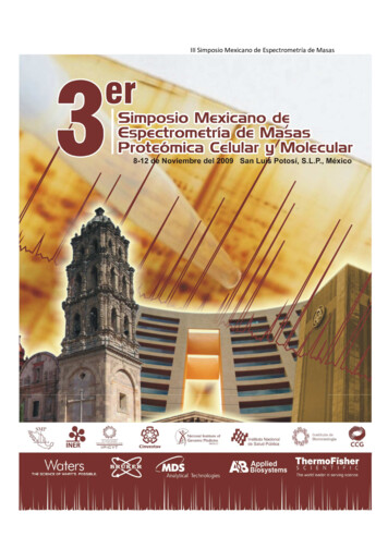
III Simposio Mexicano de Espectrometría de Masas1
III Simposio Mexicano de Espectrometría de MasasIII Simposio Mexicano deEspectrometría de MasasProteómica Celular y MolecularCiudad de San Luis Potosí8 al 12 de Noviembre 20092
III Simposio Mexicano de Espectrometría de Masas3
III Simposio Mexicano de Espectrometría de Masas4
III Simposio Mexicano de Espectrometría de Masas5
III Simposio Mexicano de Espectrometría de Masas6
III Simposio Mexicano de Espectrometría de rg7
III Simposio Mexicano de Espectrometría de MasasPrograma y ResúmenesIII Simposio MexicanoMexicano deEspectrometría de MasasProteómica Celular y MolecularCiudad de San Luis Potosí8 al 12 de Noviembre 2009Sociedad Mexicana de Proteómica ACSan Luis Potosí, S.L.P., Méxicowww.smp.org.mx8
III Simposio Mexicano de Espectrometría de MasasINFORMACIÓN GENERALRegistroSe llevará a cabo en el Salón Duque del Hotel WestinConferenciasLas conferencias Magistrales y Orales se llevarán a cabo en el SalónReal San Luis “A” y los stands y posters en el Salón Real San Luis “B”.Se anexa mapa del HotelComidasLas comidas (desayuno, comida) se incluyen a los participantes quese hospeden en el Hotel Westin.El comité organizador ofrecerá un Coctel de bienvenida en el SalónReal San Luis y Patio central el día 8 de Noviembre después de laConferencia Inaugural.También ofrecerá una Cena de Clausura el 12 de Octubre a las 20:00horas en el Hotel WestinServicios de CaféDurante los días del Simposio, habrá café y refrescos disponiblesdurante los recesos programados.Entrega de ConstanciasLas constancias para los expositores (ponentes) serán entregadas porel coordinador de cada sesión. Las constancias para los asistentes seentregarán el jueves 12 de octubre9
III Simposio Mexicano de Espectrometría de MasasÍNDICEPág.Mensaje de la Mesa Directiva2Comité Organizador3Programa del Simposio4Resúmenes de Conferencias Plenarias11Resúmenes de Sesiones Orales47Resúmenes de Presentaciones en Cartel62Índice de autores1101
III Simposio Mexicano de Espectrometría de MasasMensaje de la Mesa DirectivaEstimados participantes,La presente mesa directiva de la Sociedad Mexicana de Proteómica desea darlesla más cordial bienvenida al 3er Simposio de Espectrometría de Masas,Proteómica Molecular y Celular. En conjunto hemos preparado un programadonde participarán destacados científicos internacionales y nacionales con el finde escuchar diferentes temas de interés en nuestro campo de estudio. Esindudable que los avances de la tecnología proteómica han sido espectaculares ysus aplicaciones constituyen una promesa para estudiar el proteoma de humanos,microorganismos, plantas y animales. Sus aplicaciones a la medicina, entre otrasmuchas funciones, son por ahora una excelente alternativa en: a) la identificaciónde nuevos biomarcadores, b) descubrir mecanismos moleculares involucrados enla patogenia de enfermedades aun poco entendidas, y c) en el descubrimiento denuevos fármacos. Por todo esto, deseamos hacer de este Congreso un encuentroplacentero basado en un programa científico de muy buena calidad y ampliasoportunidades de interactuar entre grupos de trabajo de la academia e iniciativaprivada en México y otros países. En esta ocasión, además, hemos dedicado unespacio especial a los trabajos de los estudiantes e investigadores que se inicianen el área de la proteómica, conscientes de que ellos son el futuro de estadisciplina en nuestro país. Finalmente, deseamos mostrar nuestro agradecimientoa las compañías que con su apoyo han contribuido al éxito de este Congreso.AtentamenteMesa Directiva (2008-2010).Sociedad Mexicana de Proteómica2
III Simposio Mexicano de Espectrometría de MasasSOCIEDAD MEXICANA DE PROTÉOMICA A.C.Presidente: Luis M. Terán, INERVice-Presidente: Humberto Lanz Mendoza, INSPTesorero: Gerardo Corzo, IBT-UNAMSecretaria: Ana Paulina Barba, IPICyTVocales:José Luis Gallegos Pérez, InmegenBronwyn Jane Barkla, IBTLuis González de la Vara, Cinvestav-IrapuatoRoberto Arreguín Espinosa, Instituto de Química, UNAMCOMITÉ ORGANIZADOR LOCALAna Paulina Barba de la Rosa, IPICyTFabiola León Galván, IPICYTAlicia Chagolla, CINVESTAV-IrapuatoIng. Gabriela Lizbeth Melendez Govea, IPICYTL.C.C. María Teresa Gallegos Cepeda, IPICyTL.D.G. Sofía González Cabrera, IPICYT3
III Simposio Mexicano de Espectrometría de MasasPROGRAMAIII Simposio Mexicano de Espectrometría de MasasRegistroDomingo 8 y lunes 9 de noviembre: Salón Duque a partir de las 9:00 hDomingo 8 de Noviembre8:30 - 9:50RegistroCurso Pre-Simposio, Salón Real San Luis “A”9:00 – 10:30Programa Educacional IPrincipios básicos de cromatografía y su acoplamiento aEspectrometría de masas.Francisco Rojo, Facultad de Química, UNAM10:30 – 11:30Conceptos básicos en espectrometría de masas, CLmultidimensional y otras metodologías analíticas utilizadas enel análisis proteómicoJosé Luis Gallegos Pérez, INMEGEN11:30 – 12:00Receso12:00 – 13:00Programa Educacional IIMétodos de análisis de proteínas por espectrometría demasas (PMF y métodos MSMS)Alicia Chagolla, CINVESTAV-Irapuato13:00 – 14:00Programa Educacional IIIBioinformáticaErnesto Pérez Rueda, IBT-UNAM4
III Simposio Mexicano de Espectrometría de Masas13:30 – 15:30LUNCH16:50 – 17:00Inauguración17:00 – 18:00Conferencia InauguralPresentador: Dr. Luis M. TeránDr. Hans Peter MockEvaluation of plant genetic resources using proteomicapproaches18:00 – 20:00Inauguración de la Sección de PostersCoctel de BienvenidaLunes 9 de noviembre9:00 – 9:50Lectura Plenaria IIDr. Richard CaprioliMolecular Profiling/Imaging of compounds in tissues bymass spectrometry: Assessing spatial and temporalchanges.9:50 – 10:40Lectura Plenaria IIIDr. Cesar BatistaProteomics analysis of amphibian skin secretion usinghigh resolution mass spectrometry.10:40 – 11:00Receso5
III Simposio Mexicano de Espectrometría de Masas11:00 – 12:15Simposio 1: Proteómica ClínicaPresentador: Dr. Luis M. Terán1. Dr. Richard CaprioliBiomarker discovery using MALDI mass spectrometry: Astudy of human melanoma2. Dr. Genaro PimientaProteomic identification of novel biomarkers for HER2positive breast cancer3. Dr. Genaro PimientaProteomic investigation of inactive and active JNK212:15 – 13:30Simposio 2: Proteómica BiomédicaPresentador: Dr. Humberto Lanz Mendoza1. Dr. Juan Pablo Reyes GrajedaProteomic analysis of oxidative stress in a diet-inducedhypercholesterolemic model2. Dra. Rosa M. del AngelProteómica del Dengue3. Dr. Sergio EncarnaciónProteomics for gynecological cancer study13:30 – 15:30Comida15:30 – 16:50Sesiones Orales I17:00 – 19:00Sesión de Posters19:00Cena6
III Simposio Mexicano de Espectrometría de MasasMartes 10 de noviembre9:00 – 9:50Lectura Plenaria IVDr. Jack Throck WatsonInterleafing chemistry and mass spectrometry tocharacterize the structure of folding intermediates ofcysteinyl proteins9:50 – 10:40Lectura Plenaria VDr. Sixue ChenProteomics of plant redox regulatory networks10:40 – 11:00Receso11:00 – 12:15Simposio 3: Proteómica de enfermedades infecciosasPresentador: Dr. Gerardo Corzo1. Dra. Victoria PandoProteomics technology for virus research2. Dr. Manuel RodríguezFunctional characterization of ubiquitylated proteincomplexes using Tandem ubiquitin binding entities(TUBEs): Toward the ubiquitin interactome3. Dra. Cecilia Silva SánchezFlujo de trabajo para el descubrimiento de biomarcadores enproteómica7
III Simposio Mexicano de Espectrometría de Masas12:15 – 13:30Simposio 4: Proteómica de PlantasPresentador: Dra. Bronwyn Barkla1. Dra. Bronwyn BarklaQuantitative proteomics of tonoplast from the ssociations and functions of glycolytic enzymes in plantsalt tolerance2. Dra. Silvia ValdésAnálisis proteómico de la respuesta al estrés hídrico encélulas de Bouteloua gracilis3. Dr. Luis González de la VaraSeparation of calcium-dependent protein kinases frombeetroot plasma membranes according to theirhydropathy, and their identification by massspectrometry13:30 – 15:30Comida15:30 – 16:50Sesiones Orales I17:00 – 19:00Sesión de Posters19:00CenaMiércoles 11 de noviembre9:00 – 9:50Lectura Plenaria VIDr. Hans Peter MockCharacterization of plant co-chaperones with homology tothe human protein HOP (heat shock-organizing protein)8
III Simposio Mexicano de Espectrometría de Masas9:50 – 10:40Lectura Plenaria VIIDra. Maria C. Prieto ConawayAdvantages of high mass accuracy, resolving power anddynamic range of the MALDI LTQ Orbitrap for tissueimaging experiments.10:40 – 11:00Receso11:00 – 12:10Simposio 5Presentador: Dr. Luis González de la Vara1. Dr. Hans VissersA data independent approach towards the qualitativeand quantitative profiling of complex protein mixtures2. María C. Prieto ConawayA novel pressure ion trap for high throughput proteinidentification and highly sensitive analysis of drugs andmetabolites12:10 - 13:30Sesión de Negocios13:30 – 15:30Comida16:50 – 19:00VISITA GUIADA AL CENTRO DE SAN LUIS19:00CenaJueves 12 de noviembre9:00 – 9:50Lectura Plenaria VIIIDr. Luis M. TeránProteómica de secreciones nasales de niños coninfluenza A9
III Simposio Mexicano de Espectrometría de Masas9:50 – 10:40Lectura Plenaria IXDr. Robert Ventzki3D-Gel electrophoresis, a new approach to proteinanalysis10:40 – 11:00Receso11:00 – 12:15Simposio 6: Proteómica de microorganismosPresentador: Dr. Sergio Encarnación1. Dra. Yolanda López VidalProteínas únicas y comunes entre la micobacterias notuberculosas de origen clínico y ambiental.2. Dr. Sergio EncarnaciónProteome, phosphoproteome,Rhiobium etli.andsecretomein3. Dra. Clara Inés Espitia PinzónInteracciones patógeno-hospedero en tuberculosis. Unabordaje proteómico.12:15 – 13:30Simposio 7: Nuevas tecnologíasPresentador: Dr. José Luis Gallegos Pérez1. Dr. Michael L. EasterlingTop-Down Proteomics, a multiple-platform primer2. Dr. Jack Throck WatsonRecent Developments in MS/MS Technology13:30 – 15:30Comida15:30 – 16:50Sesiones Orales17:00 – 18:00Clausura18:00 – 19:00Cena Clausura10
III Simposio Mexicano de Espectrometría de MasasRESÚMENESCONFERENCIAS PLENARIASSalón Real San Luis “A”11
III Simposio Mexicano de Espectrometría de MasasL01EVALUATION OF PLANT GENETIC RESOURCES USING PROTEOMICAPPROACHESKATJA W ITZEL1, STEPHANIE KASPAR1, DIANA W EIER1, W INNIE W ESCHKE1, UDOSEIFFERT2, ANDREA MATROS1 & HANS-PETER MOCK11Leibniz Institute of Plant Genetics and Crop Plant Research (IPK), Gatersleben,Germany. 2 Fraunhofer-Institute IFF, Magdeburg, GermanyThe major focus of the IPK Gatersleben is the collection, preservation andevaluation of plant genetic resources with an emphasis on cereals. The widegenetic diversity represented in plant gene-banks is central for further breedingefforts towards a larger number of traits, such as enhanced stress tolerance orimproved nutritional quality of crop plants. Within recent years, methods for thecomprehensive analysis of transcripts, proteins and metabolites of cereals havebeen introduced and are constantly improved to gain novel information withinfundamental or applied research. The presentation will focus on two examples forthe application of proteomics performed on barley. In a first approach, the geneticdiversity of barley for contrasting salt tolerance was evaluated using mappingpopulations. Differences in the proteome were followed in contrasting parental linesand selected off-springs to identify potential candidates related to salt tolerance.Furthermore, the heterologous yeast system was used to identify barley geneproducts complementing a salinity sensitive mutant strain. Candidate genes arecurrently verified in transgenic approaches in barley and by mutant studies usingArabidopsis. In a second study changes in the proteome of developing barleygrains were followed using a label-free LC-MS approach, which yielded a highlycomplex multidimensional data-set. For the elucidation of statistically significantand objective kinetic patterns during grain developmental processes multivariatestatistics was applied.12
III Simposio Mexicano de Espectrometría de MasasL02MOLECULAR PROFILING/IMAGING OF COMPOUNDS IN TISSUES BY MASSSPECTROMETRY: ASSESSING SPATIAL AND TEMPORAL PROCESSESRichard CaprioliVanderbilt University School of Medicine, Nashville, TN USAThe spatial and temporal aspects of molecular processes in cells andtissues play an enormous part in the biology that defines living systems. Profilingand Imaging MALDI MS provides an effective means to measure and assess thosedimensions on a molecular basis, including peptides, proteins, lipids, metabolite aswell as others. The technology is extraordinarily high throughput with highmolecular specificity and is an excellent discovery tool. It provides the capability ofmapping the location of specific molecules directly from fresh frozen and formalinfixed tissue sections. For example, utilization of this technology provides spatialinformation across a tissue section for a target protein expression or for a signatureof multiple proteins and can be used to correlate changes in expression levels withspecific disease states or drug response. Molecular patterns can be directlycorrelated to known histological regions within the tissue, a technique termedhistology directed molecular profiling.In the case of frozen tissues specimens, cryostat sections ( 10 µm thick)are thaw-mounted on flat metallic target plates, and matrix automatically deposited.This can be done in a histology-directed manner to bring into play the expertiseand experience of pathologists to obtain molecular profiles from discrete areas oftissue. Formalin fixed tissues can also be analyzed by first exposing them to anantigen retrieval step. In the imaging mode, high density laser ablation of anordered array of spots over the entire tissue gives rise to a 2-dimensional iondensity map (or image) with 30-80 µm lateral resolution in which location andrelative abundance of a given analyte is displayed. From the analysis of a singlesection, images at virtually any molecular weight may be obtained.This presentation will discuss several applications of this technology,including examples of discovery in mouse developmental models and the profilingof human tumors, characterizing protein differences between tumor grades, and forthe creation of 3-D protein images. In addition to MALDI ToF MS and MS/MSinstrumentation, the capabilities of ion mobility MS and FTICR MS for profiling andimaging of tissues will be discussed. The technology has been applied to drugtargeting and metabolic studies and the measurement of concomitant proteinchanges in specific tissues after systemic drug administration. Finally, we explorethe correlation of lipid and protein changes in several systems in both health anddisease.13
III Simposio Mexicano de Espectrometría de MasasL03PROTEOMICS ANALYSIS OF AMPHIBIAN SKIN SECRETIONS USING HIGHRESOLUTION MASS SPECTROMETRYCesar V. F. BatistaUnidad de Proteómica, Instituto de Biotecnología, Universidad Nacional Autónomade México, Cuernavaca, México. E. mail: fbatista@ibt.unam.mxOver 20 years have past since peptides with antimicrobial activity were firstidentified in skin secretions of the African clawed frog Xenopus laevis. Since thattime, several hundred such antimicrobial peptides have been isolated from the skinof anurans (frogs and toads) belonging to several families ranging from theprimitive Leiopelmatidae (tailed frogs) to the highly derived Ranidae (true frogs).However, the number of species investigated and the number of bioactivecompounds are still small. Mexico is one of the 18-mega diverse countries of theworld. With over 200,000 different species, Mexico is home of 10–12% of theworld's biodiversity. Mexico ranks first in biodiversity in reptiles with 707 knownspecies, second in mammals with 438 species, and fourth in amphibians with 290endemic species. Nevertheless, almost 400 different amphibian species can befound in this country. Only in Morelos state 38 species were classified in differentfamilies, which is estimated to contain over four thousand unknown bioactivecomponents in the skin secretion of these amphibians. Frog skin secretion hasbeen found to be a rich source of agents with antimicrobial (bacteria, fungi,parasite and virus), anticancer, insulin releaser and smooth muscle contractorproperties, among others. The total secretion contains approximately one hundredcomponents constituting a very complex molecular mixture that require efficientmethods and technologies for large-scale analysis. Mass spectrometry hasbecome a powerful tool to analyze complex mixture of peptides and proteins duringthe last decade, basically due to developing of high resolution and versatile massanalyzers. In this work we present a large-scale analysis of the skin secretionsfrom Mexican amphibians using a high resolution Orbitrap-FT mass spectrometerto determine the number of components and the chemical structure of novelbioactive peptides. Hundreds of components were partially characterized andmany full sequences were obtained from the species Hyla eximia, Pachymedusadacnicolor, Lithobates spectabilis, Lithobates forreri and Lithobates spustulosus.Antibacterial activities were also accessed using resistant bacteria from clinicalisolates. Three peptides isolated from L. spectabilis showed a potent activityagainst multi-resistant Pseudomonas aeruginosa to all commercially availableantibiotics in Mexico.Acknowledge: This work was partially financed by grants from DGAPA IN-221508to CVFB.14
III Simposio Mexicano de Espectrometría de MasasL04PROTEOMIC INVESTIGATION OF INACTIVE AND ACTIVE JNK21,2Pimienta G, Ficarro SB, Gutierrez GJ, Bhoumik A, Peters EC, Ronai Z,2Pascual J.1Current Address: Department of Molecular Biophysics and Biochemistry, YaleUniversity New Haven, CT. USA2Previous Address: Burnham Institute for Medical Research, La Jolla, CA. USAThe c-Jun N-terminal kinases (JNKs) are ubiquitous proteins that phosphorylatetheir substrates, such as transcription factors, in response to physical stress,cytokines or UV radiation. This leads to changes in gene expression, ensuingeither cell cycle progression or apoptosis. Active phospho JNK1 is the main in vivokinase component of the JNK cascade, whereas JNK2 is presumed not toparticipate as a kinase during JNK signalling. However, there is evidence that JNKisoforms interact functionally in vivo. Also, a recent chemical genetics investigationhas confirmed that JNK transient activation leads to cellular proliferation, whereasa sustained one is pro-apoptotic. Here we investigate the phosphorylation patternof JNK2, with protein biochemistry tools and tandem mass spectrometry. Wechoose to focus on JNK2 because of its reported constitutive activity in gliomacells. Our results indicate that purified JNK2 from transfected nonstressed 293Tcells is a mixture of the mono-sites pThr183 and pTyr185 of its activation loop andof pThr386 along its unique C-terminal region. Upon UV stimulation, itsphosphorylation stoichiometry is upregulated on the activation loop, generating amixture of mono-pTyr185 and the expected dual-pThr183/pTyr185 species, withthe pThr386 specie present but unaltered respect to the basal conditions.15
III Simposio Mexicano de Espectrometría de MasasL05BIOMARKER DISCOVERY USING MALDI MASS SPECTROMETRY: A STUDYOF HUMAN MELANOMARichard Caprioli and William HardestyVanderbilt University School of MedicineThe aim of this work is to determine a specific protein signature from stageIII melanoma tumors that distinguish and classify tumor and control lymph nodeand that correlate to aggressive tumors through patient survival data. Patients thatpresent with stage III disease, metastasis to the regional lymph nodes, are at amuch higher risk for recurrence and disease progression. Currently, these patientsexhibit a range in survival and treatment response. Diagnostic and prognosticclinical approaches to distinguish this stage of disease for are predominantlymacroscopic and molecular classification has focused mainly focused on geneticmutations.In this study, proteomic data from 69 stage III melanoma and 17 diseasefree lymph nodes acquired by histology-directed MALDI IMS is used to classifytumor from control lymph node and to molecularly sub-classify stage III disease bytumor aggressiveness. The histology directed MALDI technology allows apathologist to examine a serial, histological stained tissue section and guide andconfidently target the cellular regions of interest. MALDI matrix is then applieddirectly onto the tissue at the regions selected by the pathologist. Results ofmultiple classification models between control lymph node and stage III melanomashow a return of an average recognition capability of 97% and a cross-validationresult of 92.5%. Using a Cox-Proportional Hazard model, 9 proteins were foundsignificantly (p 0.05) associated with survival and 3 proteins associated withdisease recurrence. This set was combined to generate a multiplex molecularsignature to group patients into poor (lower 50%) and favorable (upper 50%)survival and recurrence sets.16
III Simposio Mexicano de Espectrometría de MasasL06PROTEOMIC IDENTIFICATION OF NOVEL BIOMARKERS FOR HER2- POSITIVEBREAST CANCERф12331Pimienta Genaro , Yien Gu , Xu Sun , Jianjun Hu , Kim Min-Sik , Chaerkady1445Raghothama , Gucek Marjan , Cole Robert N , Sukumar Saraswati , Pandey1,6Akhilesh2¶and Chen Hexin1McKusick-Nathans Institute of Genetic Medicine and Department of Biological Chemistry,Johns Hopkins School of Medicine, Baltimore, Maryland, USA2Department of Biology, University of South Carolina, Columbia, South Carolina, USA3Department of Computer Science and Engineering, University of South Carolina,Columbia, South Carolina, USA4The Johns Hopkins School of Medicine, Mass Spectrometry and Proteomics Facility,Baltimore, Maryland, USA5Sidney Kimmel Comprehensive Cancer Center, Johns Hopkins School of Medicine,Baltimore, Maryland, USA6Departments of Pathology and Oncology, Johns Hopkins School of Medicine, Baltimore,Maryland, USAфCurrent address:Department of Molecular Biophysics and Biochemistry, Yale University, New Haven,Connecticut, USAThe receptor tyrosine kinase HER2 is an oncogene commonly amplified in breastcancer and in many cases indicative of poor prognosis in patients. Accordingly, itsover-expression in mammary epithelial cell lines is determinant of a tumorigenicphenotype. Various quantitative proteomic studies have been reported that explorethe role of HER2 in conferring a cancerous proteomic phenotype. Most of theseinvestigations have entailed comparing differences in protein profile by combiningtwo-dimensional gel electrophoresis and tandem mass spectrometry. In this studywe have used SILAC-based proteomics to determine the proteomic profile ofHER2-expressing mammary epithelial cells isolated from a transgenic mouse.From a short-list of 23 proteins with a relevant annotated function and a distinctiveSILAC ratio, we highlight that the up-regulation of Plastin 2, Thymosin beta 4 andTumor protein D52 supports the notion of a tumorigenic phenotype that we observeby cell characterization. In addition, we show by in-silico microarray data analysisthat the down-regulation of Gelsolin 1 and the up-regulation of Retinol-bindingprotein 1 can be used to predict the probability of metastasis-free survival in breastcancer patients. These two proteins may thus be considered novel cancerbiomarkers.17
III Simposio Mexicano de Espectrometría de MasasL07PROTEOMIC ANALYSIS OF OXIDATIVE STRESS IN A DIET-INDUCEDHYPERCHOLESTEROLEMIC MODELJ. P. Reyes-Grajeda1, K. Calderon-Gonzalez1, J. L. Gallegos-Perez1, A.Jiménez-Corona2, A. Moreno2, S. Damián-Zamacona3, & Jaime Mas-Oliva31. Unidad de Proteómica Médica. Instituto Nacional de Medicina Genómica,INMEGEN, 2. Instituto de Química, Universidad Nacional Autónoma de México(UNAM), 3. Instituto de Fisiología Celular, Universidad Nacional Autónoma deMéxico (UNAM). E-mail: jreyes@inmegen.gob.mxIn Mexico the general prevalence of hypercholesterolemia in the open population isin the order of 26.5 % with direct evidence of its involvement in the process ofatherosclerosis.This disease presenting a multifactorial etiology involves the interaction betweengenetic and environmental factors that in turn modulate the various functions ofdifferent cell types and inflammatory molecules within the vessel wall. Since thespecific participation of the different cell types in the formation of theatherosclerotic plaque, directly correlated to the presence of both normal andabnormal proteins is still under active investigation, the need for efficient detectionsystems in plasma are becoming critical.Here, we report a proteomic analysis of differentially expressed proteins in theplasma of normal and hypercholesterolemic rabbits (New Zealand), fed either witha control or a high-cholesterol diet for a period of sixteen weeks.The proteomic analysis of plasma proteins carried out with both groups ofexperimental animals shows the fact that several proteins reach statisticalsignificance for differential expression based on a DIGE gel analyses (using athreshold level of two-fold and -two-fold) obtained from both samples after beingfed with the high-cholesterol diet.The MS data analysis from this last group clearly shows an increase in a series ofproteins associated with the presence of oxidative stress, process that has beenassociated with the development of atherosclerosis.Therefore, the principal aim of this line of investigation has been the search ofhomologous proteins related to human disease such as the ones involved withabnormalities in lipid metabolism and cardiovascular calcification, both processesdirectly related to the development and progression of atherosclerosis.18
III Simposio Mexicano de Espectrometría de MasasL08PROTEÓMICA DEL DENGUEJorge Reyes-del Valle, Martha Yocupicio-Monroy, Sofía Alcaraz-Flores,Raúl Agis-Juárez, Salvador Chávez-Salinas, Rosa M. del AngelLa fiebre del dengue es la infección viral transmitida por mosquitos más ampliamentedistribuida en el mundo. Se estima que anualmente esta enfermedad afecta a 100millones de personas, siendo 2.500 millones las que viven en áreas de riesgo detransmisión de la enfermedad. La infección por dengue puede manifestarse en tres formasclínicas de gravedad creciente, la fiebre clásica del dengue, que se caracteriza por fiebrealta, cefalea, artralgia, mialgia, linfadenopatía y leucopenia; el dengue hemorrágico quese distingue de la fiebre clásica por la fuga plasmática y la trombocitopenia simultanea y elsíndrome de choque por dengue que se manifiesta por hemo-concentración y en loscasos más graves, por fallo circulatorio, estado de choque (síndrome de choque pordengue) y la muerte en cierta proporción de casos. El agente causal de la fiebre deldengue y sus complicaciones es el virus del dengue (DEN), miembro de la familiaFlaviviridae y del género flavivirus. Se reconocen cuatro serotipos del virus (DEN-1, DEN2, DEN-3 y DEN-4) y dentro de cada serotipo varios genotipos. Morfológicamente, DEN esuna partícula esférica de aproximadamente 50 nm de diámetro, la cual contiene unacápside de 30 nm rodeada por una envoltura lipídica. La superficie del virus contiene dosproteínas, la proteína E (de la envoltura) y la proteína M (de membrana). La glicoproteínaE contiene la mayoría de los determinantes antigénicos del virus y es indispensable en laentrada viral. El genoma de DEN consiste en una cadena de RNA de polaridad positiva deaproximadamente 11kb, con una estructura Cap tipo I, m7GpppAmpN2,, en su extremo 5’,y carece de la cola de poli (A) en su extremo 3’. En lugar de poli (A), DEN tiene unaestructura de tallo y burbuja muy estable y rica en nucleótidos CU. En el extremo 5’ delgenoma viral, la región no traducida (RNT) está constituida por aproximadamente 100bases (de 89 nt para DEN-2 y de 101 nt para DEN-4), mientras que en el extremo 3’ laRNT comprende aproximadamente 400 nucleótidos (384 para DEN-4 a 466 para DEN-1).Como para todos los virus de cadena positiva, el RNA genómico de DEN es infectivo. Latraducción del genoma viral da origen a una poliproteína, que es procesada co- y posttraduccionalmente, para dar origen a 10 proteínas virales maduras (C, prM y E, y lasproteínas no estructurales NS1, NS2A, NS2B, NS3, NS4A, NS4B y NS5). Dado queDEN, como muchos otros virus, codifica para un número limitado de proteínas tiene queutilizar diversas proteínas y organelos celulares para entrar, traducirse, replicarse y formarnuevas partículas virales. Estas moléculas celulares determinan en gran medida que elvirus sea infectivo, el tropismo, la susceptibilidad y la virulencia. Con la finalidad deidentificar algunas de las proteínas celulares que participan en la entrada de DENaislamos proteínas con afinidad a la proteína de la envoltura E mediante cromatografía deafinidad. El análisis por MALDI-ToF de las proteínas de membranas aisladas nos permitióidentificar a las proteínas de choque térmico HSP90 y HSP70. Por otro lado, separamoslas proteínas con afinidad a la RNT3’ de DEN-4 con la idea de conocer algunas de lasproteínas celulares que participan en la replicación viral. El análisis por MALDI-ToF nospermitió identificar a las proteínas: la proteína de unión al tracto de polipirimidinas, laproteína disulfuro isomerasa y a calreticulina. La función de estas proteínas en el cicloreplicativo viral ha sido confirmada mediante su silenciamiento y/o su sobreexpresión.19
III Simposio Mexicano de Espectrometría de MasasL09PROTEOMICS FOR GYNECOLOGICAL CANCER STUDYSergio Encarnación, Juan Carlos Higareda, Alberto Checa Rojas, MagdalenaHernández y Alberto Carlos Ramírez TorresLaboratorio de proteomica. Centro de Ciencias Genómicas. Cuernavaca, MéxicoEmail: encarnac@ccg.unam.mxDuring the carcinogenic process, several genetic and epigenetic changes occur inthe cell, all of which derive in altered exp
Espectrometría de masas. Francisco Rojo, Facultad de Química, UNAM 10:30 - 11:30 Conceptos básicos en espectrometría de masas, CL multidimensional y otras metodologías analíticas utilizadas en el análisis proteómico José Luis Gallegos Pérez, INMEGEN 11:30 - 12:00 Receso 12:00 - 13:00 Programa Educacional II