Transcription
EXPERIMENTAL AND THERAPEUTIC MEDICINE 11: 1184-1188, 20161184Pathophysiology of valvular heart disease (Review)YI ZENG1, RONGRONG SUN1, XIANCHI LI2, MIN LIU3, SHUANG CHEN2 and PEIYING ZHANG21Xuzhou Clinical Medical College of Nanjing University of Chinese Medicine; 2Department of Cardiology,Xuzhou Central Hospital, The Affiliated Xuzhou Hospital of Medical College of Southeast University;3Department of Cardiology, Xuzhou Clinical School of Xuzhou Medical College, Xuzhou, Jiangsu 221009, P.R. ChinaReceived November 9, 2015; Accepted February 5, 2016DOI: 10.3892/etm.2016.3048Abstract. Valvular heart disease (VHD) is caused by eitherdamage or defect in one of the four heart valves, aortic,mitral, tricuspid or pulmonary. Defects in these valves can becongenital or acquired. Age, gender, tobacco use, hypercholesterolemia, hypertension, and type II diabetes contribute tothe risk of disease. VHD is an escalating health issue with aprevalence of 2.5% in the United States alone. Consideringthe likely increase of the aging population worldwide,the incidence of acquired VHD is expected to increase.Technological advances are instrumental in identifyingcongenital heart defects in infants, thereby adding to thegrowing VHD population. Almost one‑third of elderly individuals have echocardiographic or radiological evidence ofcalcific aortic valve (CAV) sclerosis, an early and subclinicalform of CAV disease (CAVD). Of individuals ages 60, 2% suffer from disease progression to its most severe form,calcific aortic stenosis. Surgical intervention is thereforerequired in these patients as no effective pharmacotherapiesexist. Valvular calcium load and valve biomineralization areorchestrated by the concerted action of diverse cell‑dependent mechanisms. Signaling pathways important in skeletalmorphogenesis are also involved in the regulation of cardiacvalve morphogenesis, CAVD and the pathobiology of cardiovascular calcification. CAVD usually occurs without anyobvious symptoms in early stages over a long period of timeand symptoms are identified at advanced stages of the disease,leading to a high rate of mortality. Aortic valve replacementis the only primary treatment of choice. Biomarkers such asasymmetric dimethylarginine, fetuin‑A, calcium phosphateproduct, natriuretic peptides and osteopontin have been usefulin improving outcomes among various disease states. ThisCorrespondence to: Dr Peiying Zhang, Department of Cardiology,Xuzhou Central Hospital, The Affiliated Xuzhou Hospital ofMedical College of Southeast University, 199 South Jiefang Road,Xuzhou, Jiangsu 221009, P.R. ChinaE‑mail: zpying58@126.comKey words: valvular heart disease, calcific aortic valve disease,aortic valve stenosis, congenital heart disease, endothelial cells,biomineralization, calcificationreview, highlights the current understanding of the biologyof VHD, with particular reference to molecular and cellularaspects of its regulation. Current clinical questions and thedevelopment of new strategies to treat various forms of VHDmedically were e of cardiac valve and pathogenesis of CAVDHistopathology of CAVDInflammation and activation of CAVD pathogenesisGenetics of CAVDTreatment choices and biomarkers1. IntroductionValvular heart disease (VHD) is a major health problemafflicting the elderly in particular, with a prevalence of 2.5%in the United States. VHD occurs due to congenital defects orbecause of acquired pathology (1). Calcific aortic valve disease(CAVD) is initiated as aortic valve sclerosis (AVSc), which isa mild thickening of the valve, to aortic valve stenosis (AVS),which results in severe impairment of the valve motion. CAVDis increasingly present in the aging population, reachingepidemic proportions, with approximately one third of individuals aged 65 years, showing sub‑clinical evidence ofCAVD, in the form of aortic sclerosis (2). As a large proportionof the worldwide population is becoming aged, the prevalence of acquired forms of VHD is expected to rise (3). Age,gender, tobacco use, hypercholesterolemia, rheumatic heartdisease and hypertension constitute significant risk factors ofacquired CAVD. Congenital CAVD primarily results from thedisturbed expression of genes that are involved in normal heartvalve development. Congenital valve abnormalities comprisealmost 50% of the cases of congenital heart defects (CHD) (4).Advances in the identification of these defects and in the associated care for infants suffering from CHDs is on the rise, thusincreasing the net incidence and burden of congenital valvediseases (4). Type II diabetes is considered an important riskfactor for native CAVD (5). The pathogenesis of congenitaland acquired CAVD is likely due to the interplay of geneticand environmental influences, even though the precise mechanisms are not known.
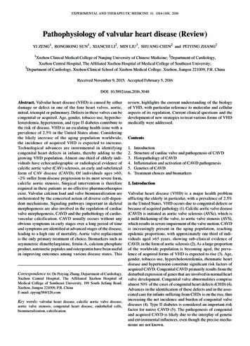
ZENG et al: PATHOPHYSIOLOGY OF VALVULAR HEART DISEASE1185Figure 1. Pathogenic pathways involved in calcific aortic valve disease. Mechanical stress or injury on the aortic valve along with other atherosclerotic riskfactors causes valvular endothelial dysfunction. This leads to lipid deposition in the subendothelium where they are oxidized and factors such as oxidizedlow‑density lipoprotein (oxLDL) are formed. Inflammatory cells such as monocytes, infiltrate the valve tissue and form foam cells by phagocytosis of the lipids.Inflammatory cytokines are released which promote remodeling of the extracellular matrix. Fibroblasts transdifferentiate into valvular myofibroblasts with anosteoblast‑like phenotype, and cause calcification. Biomarkers are associated with different stages of CAVD and can be useful in following the pathogenesis ofthe disease. TNFα, tumor necrosis factor α; TGF‑β, transforming growth factor‑β; IL‑1β, interleukin‑1β; MMP, matrix metalloproteases; ECM, extracellularmatrix; ADMA, asymmetric dimethylaminoarginine; tPA, tissue plasminogen activator; CRP, C‑reactive protein; CaxP, calcium phosphate product.Although the incidence of VHD is high, therapeuticapproaches for this disease are limited. The only availableprimary clinical approach for valve repair or replacement issurgery as the primary treatment (6,7). In fact, aortic valvereplacement is the second most frequent cardiac surgeryfollowing coronary artery bypass grafting (8). CAVD advancesto calcific aortic stenosis (CAS), which is the most severe formof the disease. It is extremely debilitating affecting as manyas 2% of individuals 60 years of age, requiring surgery topreclude death, once the symptoms become evident (9).CAVD is mainly diagnosed by clinical examination, echocardiography and cardiac catheterization. There are also manypotential biomarkers that provide clinically useful informationregarding the extent, severity, progression and prognosis ofCAVD (8).valves and any derangements in these morphological unitscan have detrimental effects on the complicated structuresof valves that open and close approximately 100,000 timesdaily in order to maintain proper directionality of blood flowthrough the heart chambers (11). The protective endotheliumover the surface of the valve leaflets is formed by the VECs,which communicate with VICs in the underlying layer andregulate their response to alterations in the blood flow (10).Genetic or acquired/environmental causes that disrupt thenormal organization and composition of the ECM and communication between VECs and VICs alter valve mechanics andinterfere with the valve leaflet function, culminating in heartfailure (11).2. Structure of cardiac valve and pathogenesis of CAVDThe histopathologic heterogeneity of CAVD indicates theinvolvement of diverse cell‑dependent mechanisms thatregulate calcium load on the valve leaflets (12), as well asthe participation of different cell types, including interstitialcells, endothelial cells and cardiac chondrocytes, in valvebiomineralization (13) (Fig. 1). Histopathologic studies (12)demonstrated the presence of calcified nodules composedof amorphous calcium phosphate, without any organizationinto specific histological structures. In these affected valves,similar to atherosclerosis, there are signs of inflammation andbone morphogenetic protein‑2 expression (12). Additionally,woven and lamellar bone with osteoblast matrix productionand vascularization has been identified in calcifying nativeaortic valves (14). During the pathogenesis of CAVD, one ofThe atrioventricular valves (mitral and tricuspid) and thesemilunar valves (aortic and pulmonic) are two types ofmature heart valves. These valves consist of an outer layerof valve endothelial cells (VECs) surrounding three layersof extracellular matrix each with specialized function andinterspersed with valve interstitial cells (VICs) (10). Changesin the functionality and localization of matrix componentspotentially lead to VHD, since the proper organization ofextracellular matrix (ECM) is essential in maintaining overallvalve morphology and normal valve function. The three layersof ECM, consisting of collagens, proteoglycans and elastin,collectively contribute to the biomechanical support for the3. Histopathology of CAVD
1186EXPERIMENTAL AND THERAPEUTIC MEDICINE 11: 1184-1188, 2016Figure 2. Crosstalk between the Notch and Wnt signaling pathways in calcific aortic valve disease. Notch 1 signaling, which represses BMP2 and Runx2 expression and impedes β‑catenin stabilization and signaling, is useful in the prevention of valvular calcification. In calcific aortic valve disease, elevated levels ofWnt3a activate the coreceptor complex formed by Frizzled and Lrp5/6, leading to the stabilization of β‑catenin and promoting the osteogenic transition of valveinterstitial cells and valvular calcification. BMP, bone morphogenetic protein; NICD, Notch intracellular domain; Runx2, runt‑related transcription factor 2.the earliest events following endothelial cell dysfunction isthe accumulation of lipids and subendothelial matrix at theventricular surface of the valve with downward displacementof the subjacent elastic lamina while plaque‑like subendothelial deposits occur on the aortic surface of the valve. It hasbeen suggested that VECs via the endothelial‑mesenchymaltransition (EMT), can contribute to calcifying vascular celltypes, in response to stimuli that promote arterioscleroticcalcification (15). Athough the contribution of acquired immunity to the progression of CAVD remains to be determined,recent data indicate that an adaptive immune response is likelyactivated in CAVD as clonally expanded effector‑memoryT‑cell populations are observed in the valve and in the circulation of patients with severe CAS (16) (Fig. 1).The earliest amorphous calcium phosphate depositionoccurs in a stippled pattern on the fibrosal interface with thefibro‑fatty expansion of the valve spongiosa (17) and thesecalcium deposits form readily via epitaxial mineral deposition on a number of nidi, including cholesterol crystals (18),collagen and fragmented elastin fibers (19). Coexpression ofcollagen and alkaline phosphatase, which in the elastin‑richenvironment can trigger mineralization, has been demonstrated in CAVD by immunogold electron microscopy (20).Biomineralization also occurs in the absence of alkalinephosphatase, as matrix vesicles contain molecules such asannexin A5, annexin A6, and phosphatidylserine, whichreadily bind calcium and nucleate mineral deposition (21)while the absence of inhibitors of mineralization such aspyrophosphate, phosphoosteopontin, and fetuin can furtherpromote the deposition of calcium (22). However, followingthe initiation of mineral deposition, circulating osteoprogenitors (COPs) derived from myeloid cell lineage arrive to the siteand play an important role in the subsequent stages of diseaseresponse.T These COP cells most likely originate from thebone marrow. The presence of type I collagen ( ) CD45 ( )COP cells in valves has been detected at the fibroproliferativeand neovascularization phases of disease, whereas CD45( ) cells are observed in ossifying and non‑ossifying valvesegments (23). Circulating myeloid calcifying cells, which arepositive for alkaline phosphatase and osteocalcin are elevatedin type II diabetes patients, and may contribute to the increasedincidence of CAVD in these patients (24).4. Inflammation and activation of CAVD pathogenesisInflammation is known to play a significant role in manytypes of macrovascular calcification, including CAVD (25).A number of the inflammation‑associated factors, includingtumor necrosis factor, interleukin 1‑β, advanced glycosylation‑end products, and oxidized low‑density lipoprotein(oxLDL) cholesterol, activate vascular biomineralizationand vascular osteogenic signaling processes (26) (Fig. 1).Aggravated fibrocalcific responses have been observed inCAVD in association with increased levels of oxLDL (27). Byemploying histological studies on human samples and mousemodels, it has been demonstrated that reactive oxygen species,specifically hydrogen peroxide, has a pro‑osteogenic andpathogenic role in CAVD and that a number of the enzymaticmechanisms that counteract oxidative stress are downregulated in valves during the pathogenesis of CAVD (28).Specifically, hydrogen peroxide has been shown to activateosteogenic Cbfa1/ runt‑related transcription factor 2 (Runx2)and Msx2/Wnt signaling pathways to promote mineralizationand these pathways are activated in calcifying human aorticvalves (28).5. Genetics of CAVDAdvances in genomic technologies have led to the identification of several genes that contribute to the normal developmentand function of the four heart valves and to the identificationof many genetic abnormalities in some of these genes in
ZENG et al: PATHOPHYSIOLOGY OF VALVULAR HEART DISEASEcongenital form of CAVD (29). The most common congenitalvalve anomalies are bicuspid aortic valve (BAV) and mitralvalve prolapse (MVP). BAV is estimated to have a prevalenceof 1‑2% (30). Normal aortic valve develops until there are threecusps, whereas in BAV disease there is a fusion of two of theleaflets during development, leading to significant morbidityprimarily through valve calcification. NOTCH1, a memberof the Notch signaling pathway, was one of the first mutatedgenes identified in BAV. The Jagged/Notch signaling pathway,which plays an important role in bone formation is also centralto valve morphogenesis and CAVD. Jagged1 signals fromendothelial cells support the Notch1‑mediated EMT necessaryfor cardiac valve morphogenesis during the development ofheart (31). Heterozygous loss‑of‑function NOTCH1 mutationssegregate with the disease in families with autosomal dominant BAV (32). Ex vivo, Notch can suppress Runx2 signalingand mineralization (Fig. 2) in VICs (33).MVP affects 2‑3% of the population and is manifested bysystolic displacement of a thickened mitral valve leaflet intothe left atrium. This condition is normally observed in adultsand often associated with the fibromyxomatous degenerationof the leaflets, valve regurgitation, congestive heart failure,arrhythmias, and infective endocarditis (34). Involvement oftransforming growth factor‑β (TGF‑β), which is needed forremodeling and maintenance of mitral valve, in MVP pathogenesis has been suggested (35). TGF‑β, bone morphogeneticproteins, and Wnt, which act via signaling through ALK‑ andLDL receptor‑related protein‑receptor complexes to promotebone formation, mineralization, and skeletal homeostasis(Fig. 2), also play an important role in the earliest stages of aorticvalve morphogenesis (36). A recent genome‑wide associationstudy that included CAVD and mitral annular calcificationpatients identified a single‑nucleotide polymorphism (SNP) inthe lipoprotein (a) locus only in CAVD patients (37).6. Treatment choices and biomarkersAs mentioned previously, surgical valve replacement is thewidely accepted treatment of choice for AVS, with eithermechanical or biological prostheses. Although there are othertreatment options, including balloon aortic valvuloplasty orpercutaneous valve replacement, they have many limitations.For example, aortic valvuloplasty showed non‑trivial complication rates, but moderately high rates of aortic insufficiencywith much higher rates of recurrence (38). Percutaneousaortic valve replacement is currently being examined inseveral studies and appears to be suitable for select groups ofpatients (39).Numerous biomarkers have been suggested for followingthe pathogenesis of aortic valve disease, but not all biomarkersare clinically useful candidates. Presently, asymmetric dimethylarginine, fetuin‑A, calcium phosphate product, natriureticpeptides and osteopontin are the most promising candidates.A circulating level of asymmetric dimethylarginine, which isinvolved in endothelial cell dysfunction, was found to correlate with the extent of AVS (40). Fetuin‑A, an inhibitor of softtissue calcification, also seems to be a good candidate as itsserum levels show strong inverse correlation with the extentof valve degeneration and calcification (41). Osteopontin isdirectly associated with the ectopic calcification process,1187which occurs during the latter stages of CAVD making thisprotein a specific biomarker for CAVD (42).In conclusion, CAVD and other types of VHDs are reachingepidemic status in their prevalence in many developed anddeveloping countries. CAVD ranges from AVSc, i.e., mildthickening of the valve, to AVS, which is severe impairmentof the valve motion. Risk factors for acquired valve diseasesinclude age, gender, tobacco use, hypercholesterolemia, hypertension, and type II diabetes mellitus. Diverse cell‑dependentmechanisms and signaling pathways orchestrate valvebiomineralization with the participation of different cell typesincluding interstitial cells, endothelial cells, cardiac chondrocytes, and COPs. In addition, several genetic mutations thatcause congenital valve diseases have been identified alongwith specific SNPs associated with CAVD. Despite the manyadvances, there is still a lack of pharmacological treatmentsfor the valve diseases and the most widely accepted approachis surgery. Recent advances in the identification of molecularmechanisms involved in the development and pathogenesis ofvalvular disease are making a significant impact in our understanding of the heart valve disease.References1. Go AS, Mozaffarian D, Roger VL, Benjamin EJ, Berry JD,Borden WB, Bravata DM, Dai S, Ford ES, Fox CS, et al; AmericanHeart Association Statistics Committee and Stroke StatisticsSubcommittee: Heart disease and stroke statistics ‑ 2013 update:a report from the American Heart Association. Circulation 127:e6‑e245, 2013.2. Rajamannan NM, Evans FJ, Aikawa E, Grande‑Allen KJ,Demer LL, Heistad DD, Simmons CA, Masters KS, Mathieu P,O'Brien KD, et al: Calcific aortic valve disease: not simply adegenerative process: a review and agenda for research from theNational Heart and Lung and Blood Institute Aortic StenosisWorking Group. Executive summary: Calcific aortic valvedisease‑2011 update. Circulation 124: 1783‑1791, 2011.3. d'Arcy JL, Prendergast BD, Chambers JB, Ray SG andBridgewater B: Valvular heart disease: The next cardiacepidemic. Heart 97: 91‑93, 2011.4. Le Gloan L, Mercier LA, Dore A, Marcotte F, Ibrahim R,Mongeon FP, Asgar A, Miro J, Poirier N and Khairy P: Recentadvances in adult congenital heart disease. Circ J 75: 2287‑2295,2011.5. Katz R, Wong ND, Kronmal R, Takasu J, Shavelle DM,Probstfield JL, Bertoni AG, Budoff MJ and O'Brien KD:Features of the metabolic syndrome and diabetes mellitus aspredictors of aortic valve calcification in the Multi‑Ethnic Studyof Atherosclerosis. Circulation 113: 2113‑2119, 2006.6. Itagaki S, Adams DH and Anyanwu AC: Triggers for surgicalreferral in degenerative mitral valve regurgitation. Circ J 77:28‑34, 2013.7. Maeda K, Kuratani T, Mizote I, Shimamura K, Takeda Y,Torikai K, Nakatani S, Nanto S and Sawa Y: Early experiencesof transcatheter aortic valve replacement in Japan. Circ J 77:359‑362, 2013.8. Beckmann E, Grau JB, Sainger R, Poggio P and Ferrari G:Insights into the use of biomarkers in calcific aortic valve disease.J Heart Valve Dis 19: 441‑452, 2010.9. Nightingale AK and Horowitz JD: Aortic sclerosis: Not aninnocent murmur but a marker of increased cardiovascular risk.Heart 91: 1389‑1393, 2005.10. Tao G, Kotick JD and Lincoln J: Heart valve development, maintenance, and disease: The role of endothelial cells. Curr Top DevBiol 100: 203‑232, 2012.11. Hinton RB Jr, Lincoln J, Deutsch GH, Osinska H, Manning PB,Benson DW and Yutzey KE: Extracellular matrix remodelingand organization in developing and diseased aortic valves. CircRes 98: 1431‑1438, 2006.12. Mohler ER III, Gannon F, Reynolds C, Zimmerman R,Keane MG and Kaplan FS: Bone formation and inflammation incardiac valves. Circulation 103: 1522‑1528, 2001.
1188EXPERIMENTAL AND THERAPEUTIC MEDICINE 11: 1184-1188, 201613. Boström KI, Jumabay M, Matveyenko A, Nicholas SB and Yao Y:Activation of vascular bone morphogenetic protein signaling indiabetes mellitus. Circ Res 108: 446‑457, 2011.14. Srivatsa SS, Harrity PJ, Maercklein PB, Kleppe L, Veinot J,Edwards WD, Johnson CM and Fitzpatrick LA: Increasedcellular expression of matrix proteins that regulate mineralization is associated with calcification of native human andporcine xenograft bioprosthetic heart valves. J Clin Invest 99:996‑1009, 1997.15. Balachandran K, Alford PW, Wylie‑Sears J, Goss JA, Grosberg A,Bischoff J, Aikawa E, Levine RA and Parker KK: Cyclic straininduces dual‑mode endothelial‑mesenchymal transformation ofthe cardiac valve. Proc Natl Acad Sci USA 108: 19943‑19948,2011.16. Winchester R, Wiesendanger M, O'Brien W, Zhang HZ,Maurer MS, Gillam LD, Schwartz A, Marboe C and StewartAS: Circulating activated and effector memory T cells areassociated with calcification and clonal expansions in bicuspidand tricuspid valves of calcific aortic stenosis. J Immunol 187:1006‑1014, 2011.17. Otto CM, Kuusisto J, Reichenbach DD, Gown AM andO'Brien KD: Characterization of the early lesion of ‘degenerative’valvular aortic stenosis. Histological and immunohistochemicalstudies. Circulation 90: 844‑853, 1994.18. Laird DF, Mucalo MR and Yokogawa Y: Growth of calciumhydroxyapatite (Ca‑HAp) on cholesterol and cholestanol crystalsfrom a simulated body fluid: A possible insight into the pathological calcifications associated with atherosclerosis. J ColloidInterface Sci 295: 348‑363, 2006.19. Schoen FJ and Levy RJ: Founder's Award, 25th Annual Meetingof the Society for Biomaterials, perspectives. Providence, RI,April 28‑May 2, 1999. Tissue heart valves: Current challengesand future research perspectives. J Biomed Mater Res 47:439‑465, 1999.20. Rajamannan NM, Subramaniam M, Rickard D, Stock SR,Donovan J, Springett M, Orszulak T, Fullerton DA, Tajik AJ,Bonow RO, et al: Human aortic valve calcification is associatedwith an osteoblast phenotype. Circulation 107: 2181‑2184,2003.21. Kapustin AN, Davies JD, Reynolds JL, McNair R, Jones GT,Sidibe A, Schurgers LJ, Skepper JN, Proudfoot D, Mayr M, et al:Calcium regulates key components of vascular smooth musclecell‑derived matrix vesicles to enhance mineralization. CircRes 109: e1‑e12, 2011.22. Jahnen‑Dechent W, Heiss A, Schäfer C and Ketteler M: Fetuin‑Aregulation of calcified matrix metabolism. Circ Res 108:1494‑1509, 2011.23. Egan KP, Kim JH, Mohler ER III and Pignolo RJ: Role forcirculating osteogenic precursor cells in aortic valvular disease.Arterioscler Thromb Vasc Biol 31: 2965‑2971, 2011.24. Fadini GP, Albiero M, Menegazzo L, Boscaro E, Vigili deKreutzenberg S, Agostini C, Cabrelle A, Binotto G, Rattazzi M,Bertacco E, et al: Widespread increase in myeloid calcifying cellscontributes to ectopic vascular calcification in type 2 diabetes.Circ Res 108: 1112‑1121, 2011.25. Wallby L, Janerot‑Sjöberg B, Steffensen T and Broqvist M:T lymphocyte infiltration in non‑rheumatic aortic stenosis: Acomparative descriptive study between tricuspid and bicuspidaortic valves. Heart 88: 348‑351, 2002.26. Towler DA: Molecular and cellular aspects of calcific aorticvalve disease. Circ Res 113: 198‑208, 2013.27. Stewart CR, Stuart LM, Wilkinson K, van Gils JM, Deng J,Halle A, Rayner KJ, Boyer L, Zhong R, Frazier WA, et al: CD36ligands promote sterile inflammation through assembly of aToll‑like receptor 4 and 6 heterodimer. Nat Immunol 11: 155‑161,2010.28. Miller JD, Chu Y, Brooks RM, Richenbacher WE, Peña‑Silva Rand Heistad DD: Dysregulation of antioxidant mechanismscontributes to increased oxidative stress in calcific aortic valvularstenosis in humans. J Am Coll Cardiol 52: 843‑850, 2008.29. Lincoln J and Garg V: Etiology of valvular heart disease-geneticand developmental origins. Circ J 78: 1801-1807, 2014.30. McBride KL and Garg V: Heredity of bicuspid aortic valve: Isfamily screening indicated? Heart 97: 1193‑1195, 2011.31. Hofmann JJ, Briot A, Enciso J, Zovein AC, Ren S, Zhang ZW,Radtke F, Simons M, Wang Y and Iruela‑Arispe ML: Endothelialdeletion of murine Jag1 leads to valve calcification andcongenital heart defects associated with Alagille syndrome.Development 139: 4449‑4460, 2012.32. Foffa I, Ait Alì L, Panesi P, Mariani M, Festa P, Botto N, Vecoli Cand Andreassi MG: Sequencing of NOTCH1, GATA5, TGFBR1and TGFBR2 genes in familial cases of bicuspid aortic valve.BMC Med Genet 14: 44, 2013.33. Nus M, MacGrogan D, Martínez‑Poveda B, Benito Y,Casanova JC, Fernández‑Avilés F, Bermejo J and de la Pompa JL:Diet‑induced aortic valve disease in mice haploinsufficient forthe Notch pathway effector RBPJK/CSL. Arterioscler ThrombVasc Biol 31: 1580‑1588, 2011.34. Freed LA, Levy D, Levine RA, Larson MG, Evans JC, Fuller DL,Lehman B and Benjamin EJ: Prevalence and clinical outcome ofmitral‑valve prolapse. N Engl J Med 341: 1‑7, 1999.35. Lincoln J and Garg V: Etiology of valvular heart disease‑geneticand developmental origins. Circ J 78: 1801‑1807, 2014.36. Armstrong EJ and Bischoff J: Heart valve development:Endothelial cell signaling and differentiation. Circ Res 95:459‑470, 2004.37. Thanassoulis G, Campbell CY, Owens DS, Smith JG, Smith AV,Peloso GM, Kerr KF, Pechlivanis S, Budoff MJ, Harris TB, et al;CHARGE Extracoronary Calcium Working Group: Geneticassociations with valvular calcification and aortic stenosis.N Engl J Med 368: 503‑512, 2013.38. Balmer C, Beghetti M, Fasnacht M, Friedli B and Arbenz U:Balloon aortic valvoplasty in paediatric patients: Progressiveaortic regurgitation is common. Heart 90: 77‑81, 2004.39. Webb JG, Chandavimol M, Thompson CR, Ricci DR, Carere RG,Munt BI, Buller CE, Pasupati S and Lichtenstein S: Percutaneousaortic valve implantation retrograde from the femoral artery.Circulation 113: 842‑850, 2006.40. Vallance P, Leone A, Calver A, Collier J and Moncada S:Accumulation of an endogenous inhibitor of nitric oxide synthesisin chronic renal failure. Lancet 339: 572‑575, 1992.41. Koos R, Brandenburg V, Mahnken AH, Mühlenbruch G,Stanzel S, Günther RW, Floege J, Jahnen‑Dechent W, Kelm Mand Kühl HP: Association of fetuin‑A levels with the progressionof aortic valve calcification in non‑dialyzed patients. EurHeart J 30: 2054‑2061, 2009.42. Yu PJ, Skolnick A, Ferrari G, Heretis K, Mignatti P, Pintucci G,Rosenzweig B, Diaz‑Cartelle J, Kronzon I, Perk G, et al:Correlation between plasma osteopontin levels and aortic valvecalcification: Potential insights into the pathogenesis of aorticvalve calcification and stenosis. J Thorac Cardiovasc Surg 138:196‑199, 2009.
ZENG et al: PATHOPHYSIOLOGY OF VALVULAR HEART DISEASE 1185 Although the incidence of VHD is high, therapeutic approaches for this disease are limited. The only available primary clinical approach for valve repair or replacement is surgery as the primary treatment (6,7). In fact, aortic valve replacement is the second most frequent cardiac surgery