Transcription
(2022) 8:7Patil et al. 00133-2Open AccessREVIEWMediastinal irradiation and valvular heartdiseaseShivaraj Patil1, Srinath‑Reddi Pingle2, Khalid Shalaby1 and Agnes S. Kim1,3*AbstractAnticancer therapy has the potential to cause unwanted cardiovascular side effects. Utilization of radiation therapyto treat tumors near the heart can result in radiation-induced valvular heart disease among other cardiovascularpathologies. The aim of this review is to describe the epidemiology, pathophysiology, risk prediction, non-invasiveimaging modalities and management of radiation-induced valvular heart disease with a focus on pre-operative riskassessment and contemporary treatment options.Highlights Mediastinal radiation therapy improves cancer survival but increases the risk of developing radiation-induced valvu‑lar heart disease, often in combination with other radiation-induced cardiovascular pathologies, ultimately leading toincreased cardiovascular morbidity and mortality. Radiation-induced valvular heart disease presents after a prolonged, albeit variable, latent period and poses uniqueclinical challenges, underscoring the need for timely monitoring by a cardio-oncologist. There is an elevated risk of morbidity and mortality after valvular operations in patients with prior mediastinal radia‑tion therapy beyond what is estimated by current preoperative risk stratification models. Careful patient selection and management by a multidisciplinary heart team is paramount in optimizing outcomesof radiation-induced valvular heart disease. In challenging cases associated with high operative risk, transcathetertherapies may be considered.Keywords: Mediastinal irradiation, Valvular heart disease, Cancer, Radiation-induced heart diseaseIntroductionRadiation therapy (RT) plays an integral role in thetreatment of various thoracic malignancies. Tumorsnear the heart, such as left-sided breast cancer, lymphoma, lung cancer and esophageal cancer, can exposethe nearby cardiac tissue to irradiation, which may beunavoidable when delivering radiation. Historically,*Correspondence: akim@uchc.edu3Department of Medicine, Calhoun Cardiology Center, Universityof Connecticut School of Medicine, 263 Farmington Avenue, Farmington,CT 06030, USAFull list of author information is available at the end of the articlethe myocardium was believed to be resistant to theeffects of RT due to the post-mitotic state of myocytes.However, in the middle of the twentieth century, evidence began to emerge of cardiac toxicity secondaryto RT [1]. Substantial doses of mediastinal irradiationcan potentially result in injury to any component ofthe heart. The spectrum of radiation-induced heartdisease is wide-ranging from early manifestations suchas acute pericarditis to delayed effects: vasculopathy, valvular heart disease (VHD), pericardial disease,conduction system dysfunction and pulmonary venoocclusive disease [2]. The Author(s) 2022. Open Access This article is licensed under a Creative Commons Attribution 4.0 International License, whichpermits use, sharing, adaptation, distribution and reproduction in any medium or format, as long as you give appropriate credit to theoriginal author(s) and the source, provide a link to the Creative Commons licence, and indicate if changes were made. The images orother third party material in this article are included in the article’s Creative Commons licence, unless indicated otherwise in a credit lineto the material. If material is not included in the article’s Creative Commons licence and your intended use is not permitted by statutoryregulation or exceeds the permitted use, you will need to obtain permission directly from the copyright holder. To view a copy of thislicence, visit http:// creat iveco mmons. org/ licen ses/ by/4. 0/. The Creative Commons Public Domain Dedication waiver (http:// creat iveco mmons. org/ publi cdoma in/ zero/1. 0/) applies to the data made available in this article, unless otherwise stated in a credit line to the data.
Patil et al. Cardio-Oncology(2022) 8:7The overall success of RT, used alone or in combination with other modalities, has resulted in large cohorts ofcancer survivors. Patients treated for Hodgkin lymphoma(HL) and breast cancer are subject to late complicationsfrom chest irradiation. Among these patients, valvulardysfunction is a frequently encountered non-coronarypathology resulting in significant morbidity and mortality [3–6]. Patients with prior RT who experience cardiacmanifestations often require complex high-risk surgicalinterventions, posing a clinical challenge [7–11]. Treatment for VHD continues to evolve with advances in transcatheter valve therapy complementing traditional surgicalapproaches, resulting in expansion of available treatmentmodalities and the potential to transform patient care.Literature search methodsWe conducted a PubMed search using the combinationsof the words “Mediastinal Radiation”, “Chest Radiation”,“Thoracic Radiation”, “Valvular Heart Disease”,” Valvular Dysfunction”, “Aortic Stenosis”, “Aortic regurgitation”,“Mitral Stenosis”, “Mitral regurgitation”, “Tricuspid Stenosis”, “Tricuspid Regurgitation”, “Pulmonary Stenosis”,and “Pulmonary Regurgitation”. All identified manuscripts, including reviews and case series, were considered for inclusion in this review. We also reviewed thebibliographies of all identified manuscripts to find additional relevant publications. We limited the scope of ourwork to studies on humans, published in English since1950. Authors reviewed all identified manuscripts for relevance to this review, and articles deemed relevant wereincluded.EpidemiologyThe reported prevalence of radiation-induced VHD ishighly variable in part due to the differences in studydesign, baseline characteristics of various cancer survivorcohorts, heterogeneous cardiotoxic exposures, and lackof established methodologies used to characterize valvedysfunction. In a cohort of five-year childhood cancersurvivors who had received mediastinal RT (MRT), theobserved prevalence of one or more valvular abnormalities at the time of first echocardiographic evaluation was43.1% at a median age of 22 years, after a median followup of nearly 15 years [12]. Studies among survivors of HLwith a history of MRT have shown the prevalence of valvular abnormalities as detected by echocardiography torange from 2.8–61% [13, 14].A cross-sectional study of 82 HL survivors by Bijlet al. compared valvular dysfunction among patientswho received MRT with those who did not and foundthat left-sided valvular lesions, aortic regurgitation(38.2%) followed by mitral regurgitation (36.7%), werethe most common abnormalities [14]. Left-sided valvularPage 2 of 11pathologies are generally more common in the generalpopulation, likely due to the higher-pressure system onthe left side of the heart as compared with the right sideand may explain why the mitral and aortic valves aremore commonly affected than pulmonary and tricuspidvalves [15, 16]. Tricuspid regurgitation (20.4%) was themost common right-sided valvular lesion. Aortic stenosis(AS) was the most common obstructive valvular dysfunction followed by mitral stenosis. In contrast, a study byCella et al. reported a higher occurrence of mitral (25.0%)and tricuspid (14.3%) compared with aortic (5.4%) valvedisease among HL survivors. This discrepancy is possiblydue to the relatively shorter follow-up period at the timeof echocardiographic assessment in the latter study [17].For all types of VHD, the prevalence and severity werehighest in the group of patients who had received RTmore than 20 years prior to evaluation, a finding alsosupported by the study of nearly 300 asymptomatic survivors by Heidenreich et al., highlighting the latency inthe development of valvular lesions [18]. In breast cancersurvivors, the prevalence of VHD has been reported tobe between 0.4–4.2% among patients exposed to RT. Therisk of developing VHD is increased by approximatelyfifty percent in left-sided, compared with right-sided,breast cancer, highlighting the implication of mean heartdose in the development of radiation-induced toxicity [5,19, 20]. In a large, population-based cohort comprising70,230 surgically treated breast cancer patients receiving adjuvant RT, death due to VHD was more frequent(standardized mortality ratio 1.28, 95% CI 1.08–1.52)compared with the general population [21].Risk factors and pathophysiologyThe strongest risk factor for developing radiationinduced VHD is the total radiation dose delivered tothe mediastinum, more specifically the total dose delivered to the heart valves. In a nested cohort study of 1852patients treated for HL between 1965 and 1995, 5% hadVHD (N 89) with 74% of lesions being severe or lifethreatening (N 66) [22]. A dose–response relationshipof progressively nonlinear increases in clinically significant VHD was noted with relative risks of 1.4, 3.1, 5.4,and 11.8 for affected heart valves receiving 30 Gy, 31to 35 Gy, 35 to 40 Gy, and 40 Gy, respectively. Withadvancements in RT in the current era, the cumulativerisk of developing VHD is estimated at 1.4% at 30 yearsamong patients receiving MRT for HL with a standardradiation dose of 20 or 30 Gy [22]. The volume of heartirradiated is also a major risk factor for the development of VHD. In one analysis of patients treated between2002 and 2008 with chemotherapy and involved-fieldRT, radiation to cardiac sub-volumes correlated with the
Patil et al. Cardio-Oncology(2022) 8:7Page 3 of 11occurrence of VHD. The volume of left atrium exceeding25 Gy and the volume of left ventricle exceeding 30 Gypredicted mitral and aortic valve dysfunction [17].A meta-analysis of 40,781 breast cancer survivorswho had received modern RT showed that the risk ofdeveloping of VHD was 1.97 compared to those whodid not undergo RT (95% CI: 1.07–3.67, p 0.03). Themean heart dose reported was 6.3 Gy for the wholeheart, which is higher than contemporary radiationdoses [23, 24]. There is a higher whole heart radiationdose exposure in individuals receiving RT for leftsided breast cancer compared to right-sided breastcancer, primarily affecting the left ventricle, left anterior descending artery and right ventricle due to theiranatomic proximity [25]. Techniques like prone positioning, respiratory gating, and deep inspiration breathhold displace the breast away from the heart and therefore reduce radiation burden to the heart (Table 1).Table 1 Cardiac-Sparing Modalities and Techniques in Radiation TherapyCardiac-Sparing TechniqueDescriptionType of CancerRespiratory GatingTracking patient’s natural respiratory motion anddelivering radiation precisely when the tumor is in thetreatment field and farthest from the heartBreast, Lymphoma, Lung, EsophagusDeep Inspiration Breath Hold (DIBH)Radiation is administered during maximal inspirationand breath hold, when the heart is pulled away fromthe chest wall due to flattening of the diaphragm andexpansion of the lungsBreast, LymphomaProne Positioning/ Lateral Decubitus PositioningRadiation is delivered to the tumor with patient lyingBreastprone or lateral decubitus on a specially designed tableto maximally displace the heart from the treatmentfieldCardiac DisplacementRadiation Treatment Modality/TechniqueInvolved Site Radiation Therapy (ISRT) and InvolvedNode Radiation Therapy (INRT)Reduction in radiotherapy field size to involvednodal tissue detected using modern imagingtechniques (PET-CT/MRI), thus sparing surroundinguninvolved nodal and non-nodal anatomic structuresLymphomaThree-Dimensional Conformal Radiation Therapy (3DCRT)3D reconstruction of the tumor and surroundingstructures using CT and/or MRI imaging data to guideradiation planning by beam placement. Radiation canbe delivered from any angle; multiple radiation beamsfrom different angles can be combined to delivermaximal dose to the tumor while relatively sparingnormal tissueBreast, Lymphoma, Lung, EsophagusIntensity-Modulated Radiation Therapy (IMRT)An advanced form of 3D-CRT that utilizes varyingintensity of smaller radiation beams (beamlets) usingcomputerized inverse planning, enabling precisedelivery of radiation dose to the tumor and improvingnormal tissue sparingBreast, Lymphoma, Lung, EsophagusVolumetric-Modulated Arc Therapy (VMAT)An extended form of IMRT, in which the radiationsource is continuously rotated around the patient,allowing delivery of therapy from a full 360 beamangle, with added advantage of improved delivery ofradiation dose to the target in lesser timeBreast, Lymphoma, Lung, EsophagusImage-Guided Radiation Therapy (IGRT)Integration of imaging prior to and during eachradiation treatment, typically CT-guided and recentlyMRI-guided, allowing more precise localization of thetumor bed. IGRT permits significantly better sparing ofnormal tissue while promoting dose-escalation to thetumor when incorporated with IMRTBreast, Lymphoma, Lung, EsophagusAccelerated Partial Breast IrradiationAn approach that treats only the lumpectomy bed plus Breasta 1–2 cm margin, rather than the whole breast, there‑fore sparing normal tissue by decreasing the targetvolume of radiationProton Beam TherapyProton beams have a distinct property compared tophoton beams: they quickly lose energy toward theend of their range (Bragg peak), thus limiting radiationdose beyond the targetBreast, Lymphoma, Lung, Esophagus
Patil et al. Cardio-Oncology(2022) 8:7Interestingly, there are histopathological differencesnoted in the affected valves with a degenerative calcific process observed in patients with breast canceras opposed to a predominantly fibrotic process in lymphoma patients who have received RT. This difference islikely due to the young age of radiation exposure in lymphoma patients [26]. A recent observational matchedcohort study of patients with moderate AS with andwithout prior MRT who underwent serial echocardiograms showed similar rates of AS progression regardlessof the underlying primary malignancy treated with RT.Patients with prior MRT had significantly increased mortality despite undergoing aortic valve replacement soonercompared with their matched controls, highlighting theadverse impact of radiation even after the correction ofunderlying valve abnormality [27].Myocardial fibrosis and vasculopathy due to the directtoxic effects of radiation result in ventricular remodeling,which in turn can increase the risk of developing valvular dysfunction. A few studies have reported an increasedrisk of VHD with concomitant exposure to anthracyclinetherapy in a dose-dependent manner [4, 28, 29]. However, Cutter et al. did not find a significant association ofVHD with anthracycline exposure [22]. This may be dueto the latter report involving only patients with primaryVHD, while other studies included all diagnoses of valvedysfunction, including secondary valvulopathy followingischemic heart disease or cardiomyopathy.Other risk factors for VHD include early age of exposure to RT, hypertension, smoking, and hyperlipidemia[4, 14, 18, 23, 24, 30]. The presence of congenital heartdisease has also been reported to increase the risk of valvulopathy [12]. Interestingly, obesity at the time of HLdiagnosis and splenectomy, although no longer used forthe treatment of HL, have also been associated with anincreased risk of radiation-induced valve damage. Therelationship between splenectomy and valve disease isnot well understood and may represent a correlationrather than causation [4, 31].Although the precise pathophysiologic mechanismsof radiation-induced valvulopathy are not completelyunderstood, irradiation is thought to have a direct effecton the pathologic fibrosis and calcification of the valvular apparatus. Due to the avascular nature of valve tissue,the mechanism of injury is believed to be distinct fromradiation-induced damage to the myocardium and vasculature. There is a lack of histological markers of chronicinflammation or neovascularization on tissue specimens[26, 32]. Fibrotic changes in the vasculature are mediatedvia activation of inflammatory cascade through radiationinduced endothelial cell injury [33]. In contrast, injury tothe valve interstitial cells (VICs) has been implicated inthe pathogenesis of valve damage from radiation. VICsPage 4 of 11originate from valve endothelial cells during embryogenesis via migration into the underlying matrix andundergoing endothelial‐to‐mesenchymal transformation.Pathological activation of VICs leads to their differentiation into myofibroblasts and osteoblasts via cytokinesthat act in an autocrine and paracrine fashion. In addition, mechanical stress contributes to this transformation, explaining the higher incidence of left-sided lesionscompared with right-sided lesions. These cells furtherproduce collagen, extracellular matrix, and osteogenicfactors, such as bone morphogenic protein 2, transcription factor Runx2, osteopontin and ALP, all of which playa role in creating a milieu for abnormal calcium deposition in irradiated valves [34, 35].Clinical prediction model for radiation‑induced valvularheart diseaseUse of RT has significantly improved overall and diseasefree survival for patients with cancer, albeit with a riskof increased morbidity because of injury to surroundingnormal tissue. To keep the tissue toxicity at an acceptable level, radiobiological models such as normal tissuecomplication probability (NTCP) have been developed toestimate the risk of radiotoxicity to normal tissue. Thesemodels are utilized in treatment planning to minimizeadverse effects from irradiation. NTCP models, such asthe Lyman-Kutcher-Burman (LKB) and Relative Seriality(RS) models, are the most well-known and traditionallyaccepted methods for predicting toxicity after radiationtreatment. However, these traditional models have inadequately predicted the risk of radiation-induced VHDbased on conventional heart dose-volume histogramsalone due to lack of substructure contouring. Improvedstatistical machine learning methods like “Least AbsoluteShrinkage and Selection Operator” (LASSO) have beenadopted to build multivariate NTCP models to improverisk prediction, however, these models need further validation [36].Advances in radiation therapyGrowing recognition of the adverse cardiac effects ofmediastinal radiation and technological advances in thefield of radiation therapy have led to the development ofcardiac-sparing modalities and techniques. In contrast tothe 1970s and 1980s, modern radiation therapy employsmultiple techniques to minimize the mean heart dose,such as deep inspiration breath hold, intensity-modulatedradiation therapy, and proton beam therapy (Table 1)[37–42]. These advances are aimed at improving the therapeutic ratio, i.e., delivering maximal effective radiationto the tumor while minimizing the risk of radiation toxicity to surrounding healthy tissue [37–39]. Furthermore,employing imaging techniques, such as ECG-gated CT
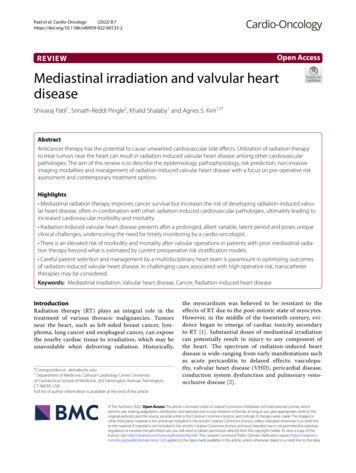
Patil et al. Cardio-Oncology(2022) 8:7Page 5 of 11coronary angiography and/or MRI, can facilitate incorporation of cardiac substructures (i.e., valves, coronaries, cardiac chambers) into radiation treatment planningand delivery, allowing better estimation of radiation doseto specific substructures of the heart and mitigation ofcardiac injury [40–42]. The selection of techniques andmodalities is a complex process and influenced by severalfactors, particularly the availability of technology, patientanatomy, and target volumes. An interdisciplinary discussion involving the oncologist, radiation oncologistand cardio-oncologist is key to determine optimal radiation therapy in improving patient survival, especially inpatients with established cardiovascular disease or riskfactors [43, 44]. The impact of modern cardiac-sparingradiation therapy on VHD is unknown, largely due toits delayed manifestation, and is an important area forfuture research.Multi‑modality non‑invasive imagingTransthoracic echocardiography is the first-line imagingmodality for detecting VHD (Table 2). Echocardiographicabnormalities include diffuse valve thickening due tofibrosis and focal or contiguous calcification in the valvular apparatus resulting in restricted motion of leaflets,initially causing regurgitation with eventual progressionto stenosis. Left-sided valves are more often affected thanright-sided valves. The aortic root, aortic valve annulus,aortic valve leaflets, aortic-mitral inter-valvular fibrosa,mitral valve annulus, and the base and mid portions ofthe mitral valve leaflets are typically affected with sparingof mitral valve tips and commissures [45]. Special focusshould be given to the measurement of aorto-mitralcurtain (AMC) thickness due to its prognostic importance (Fig. 1). An AMC thickness of greater than 6 mmhas been shown to be independently associated withincreased mortality [46]. The severity of valvular dysfunction should be graded based on the guidelines fromthe European Association of Cardiovascular Imagingand the American Society of Echocardiography. In addition, echocardiography can provide valuable informationregarding ventricular systolic and diastolic dysfunctionas well as pericardial pathology, importantly constrictivepericarditis.Multidetector cardiac computed tomography (MDCT)and cardiac magnetic resonance (CMR) are usefuladjunct imaging modalities. They provide complimentary information for pre-procedural planning, includingassessment of annular shape and size for aortic, mitral,and tricuspid valves, and ileo-femoral vasculature. Presence of significant calcification of the valvular apparatusmay preclude valvular repair. Extreme radiation-inducedcalcification of the ascending aorta (“porcelain aorta”)can increase the perioperative risk of embolic stroke dueto its manipulation. Therefore, its detection is importantto determine the suitability for aortic cross-clampingand cannulation in patients undergoing cardiac surgery.For patients undergoing redo surgeries, MDCT providescritical information regarding anatomical relationship ofvarious cardiovascular structures to the sternum and theextent of mediastinal fibrosis, which may preclude theuse of open cardiac surgery and favor minimally invasivetechniques for treatment [47]. Among individuals withinadequate information by transthoracic or transesophageal echocardiography, CMR can provide both structuraland functional data regarding valve dysfunction, as wellas information on ventricular function, mass, volume,regional wall motion abnormalities, and pericardialthickening, effusions, and features of constrictive physiology [48]. Coronary vasculopathy is common in patientswith radiation-induced VHD and commonly affects theostia or proximal coronary arteries. This can be detectednon-invasively by stress echocardiography, stress CMR,coronary CTA, radionuclide myocardial perfusion imaging with single photon emission CT (SPECT), or positronemission tomography (PET).ManagementAnnual follow-up with a cardiologist or cardio-oncologist for history and physical examination and utilizationof multimodality cardiac imaging when appropriate areTable 2 Common Echocardiographic Findings of Radiation-induced Valvular Heart Disease1Diffuse valve thickening due to fibrosis2Focal or contiguous calcification of valvular apparatus with restricted motion of leaflets3Initial regurgitation with eventual progression to stenosis4Fibrosis/calcification of aortic root, aortic valve annulus, aortic valve leaflets, aorticmitral inter-valvular fibrosa, mitral valve annulus, and the base and mid portions of themitral valve leaflets with typical sparing of mitral valve tips and commissures5Aorto-mitral curtain (AMC) thickness6Ventricular systolic and diastolic dysfunction7Pericardial pathology, importantly constrictive pericarditis
Patil et al. Cardio-Oncology(2022) 8:7Page 6 of 11Fig. 1 Transthoracic echocardiography measuring aorto-mitral curtain thickness. Example TTE image of aorto-mitral curtain thickness in a55-year-old man with a history of mediastinal radiation therapy for non-Hodgkin lymphoma at the age of 30. He underwent aortic and mitral valvereplacement for symptomatic severe valvular stenosisinvaluable for the early detection and timely intervention of radiation-induced VHD (Table 3). Development of new cardiopulmonary symptoms or physicalexam findings, particularly murmur, should promptimmediate diagnostic evaluation with echocardiography regardless of the time from MRT. The risk factors for developing VHD include anterior or left-sidedchest wall irradiation, exposure to a high cumulativedose of radiation ( 30 Gy) or a high daily fraction ofradiation 2 Gy, lack of shielding, young age at RT( 50 years), concomitant chemotherapy, presence ofpre-existing cardiovascular disease, or presence of cardiovascular risk factors (diabetes mellitus, hypertension, hyperlipidemia, obesity, and smoking). Apart fromthese traditionally described risk factors, there is growing evidence to include the radiation dose distributionto cardiac substructures to better identify high-riskindividuals [49]. An asymptomatic individual is considered high risk if a cardiac structure was in the radiationfield, typically observed in anterior or left-sided chestTable 3 Management Recommendations for the Prevention and Detection of Radiation-induced Valvular Heart Disease1Annual follow-up with a cardiologist or cardio-oncologist for history and physical examination2Assess risk factors for developing VHD: anterior or left-sided chest wall irradiation, exposure to a high cumula‑tive dose of radiation ( 30 Gy) or a high daily fraction of radiation 2 Gy, lack of shielding, young age at radio‑therapy ( 50 years), concomitant chemotherapy, presence of pre-existing cardiovascular disease, or presenceof cardiovascular risk factors (diabetes mellitus, hypertension, hyperlipidemia, obesity, and smoking)3For individuals with symptoms or murmur, check echocardiography4For low-risk asymptomatic individuals, screening for VHD with echocardiography is recommended at 10 years5For high-risk asymptomatic individuals, surveillance imaging for VHD should begin sooner, typically at 5 years6Asymptomatic individuals should undergo surveillance imaging every 5 years if initial screening echo is normal7Optimal management of underlying cardiovascular risk factors (hypertension, diabetes, hyperlipidemia, smok‑ing, obesity, sedentary lifestyle, obstructive sleep apnea) is imperative
Patil et al. Cardio-Oncology(2022) 8:7irradiation, with at least one other traditional risk factor for radiation-induced heart disease.For low-risk asymptomatic individuals, screening for VHD with echocardiography is recommendedat 10 years. For high-risk asymptomatic individuals,surveillance imaging for VHD should begin sooner,typically at 5 years. Asymptomatic individuals shouldundergo surveillance imaging every 5 years if initial screening echocardiogram was normal [45]. Morerecently, the international cardio-oncology society recommends screening with echocardiography as early as6–12 months in high-risk individuals and at least oneechocardiogram within 5 years of RT in all individuals inwhom the heart was in the radiation field [50]. Detectionof valvular and/or ventricular dysfunction should promptcloser interval follow-up depending on the severity ofthe abnormality detected. Radiation-induced coronaryartery disease often develops prior to significant valvular dysfunction and warrants earlier detection, typically5 years after completion of RT and repeated surveillanceat 5-year intervals. Patients with abnormal stress testsor those being planned for valvular intervention shouldundergo left heart catheterization to assess the coronaryanatomy and to confirm the findings of noninvasive stresstesting. The extent and severity of underlying atherosclerotic and/or radiation-induced coronary vasculopathymay have significant implications in planning for cardiacsurgery [45, 48, 50].Optimal management of underlying cardiovascular riskfactors (hypertension, diabetes, hyperlipidemia, smoking, obesity, sedentary lifestyle, obstructive sleep apnea)is imperative in these patients. Radiation-induced VHDprogresses over time and ultimately will require invasive structural interventions to relieve significant lesions.Patients with exposure to MRT can develop complexcardiac disease involving valvular, coronary, ventricular,and conduction system abnormalities of the heart as wellas simultaneous disease of the surrounding structures.There are no specific guidelines on the timing of intervention for patients with radiation-induced VHD. Interventions should be performed according to current existingnational and international guidelines [51, 52]. However,due to the presence of multiple structural abnormalitiesin these patients, a delayed surgical approach with an aimto perform a complete operation at the first surgery ispreferred to avoid the risk of cardiac reoperation, whichis associated with significant morbidity and mortality [48,53]. A comprehensive pre-operative evaluation should beperformed including echocardiography, coronary angiography, MDCT, and pulmonary function testing.Surgical risk stratification with current pre-operative risk stratification tools for cardiac valve surgery donot account for the adverse effects and complicationsPage 7 of 11related to prior MRT and may underestimate the truerisk. A retrospective analysis by Wu et al. of 173 patients(mean age, 63 14 years, 75% women, mean EuroSCORE7.8 3) with radiation-induced heart disease undergoingcardiac surgery matched to 305 controls based on age,gender, and type of procedure revealed a higher proportion of death in the RT group than in the comparisongroup (55% versus 28%; p 0.001) over a mean follow-upof 7.6 3 years, despite similar EuroSCOREs [10]. A retrospective analysis by Ejiofor et al. of 261 patients (meanage 62.6 12.1 years; 67% women) with prior MRT whounderwent valvular operations
to treat tumors near the heart can result in radiation‑induced valvular heart disease among other cardiovascular pathologies. The aim of this review is to describe the epidemiology, pathophysiology, risk prediction, non‑invasive imaging modalities and management of radiation‑induced valvular heart disease with a focus on pre‑operative risk