Transcription
Molecular cloning using polymerase chainreaction, an educational guide for cellularengineeringHoseini and SauerHoseini and Sauer Journal of Biological Engineering 2015, 9:2http://www.jbioleng.org/content/9/1/2
Hoseini and Sauer Journal of Biological Engineering 2015, YOpen AccessMolecular cloning using polymerase chainreaction, an educational guide for cellularengineeringSayed Shahabuddin Hoseini1,2 and Martin G Sauer1,2,3*AbstractBackground: Over the last decades, molecular cloning has transformed biological sciences. Having profoundlyimpacted various areas such as basic science, clinical, pharmaceutical, and environmental fields, the use ofrecombinant DNA has successfully started to enter the field of cellular engineering. Here, the polymerase chainreaction (PCR) represents one of the most essential tools. Due to the emergence of novel and efficient PCRreagents, cloning kits, and software, there is a need for a concise and comprehensive protocol that explains allsteps of PCR cloning starting from the primer design, performing PCR, sequencing PCR products, analysis of thesequencing data, and finally the assessment of gene expression. It is the aim of this methodology paper to providea comprehensive protocol with a viable example for applying PCR in gene cloning.Results: Exemplarily the sequence of the tdTomato fluorescent gene was amplified with PCR primers whereinproper restriction enzyme sites were embedded. Practical criteria for the selection of restriction enzymes and thedesign of PCR primers are explained. Efficient cloning of PCR products into a plasmid for sequencing and freeweb-based software for the consecutive analysis of sequencing data is introduced. Finally, confirmation of successfulcloning is explained using a fluorescent gene of interest and murine target cells.Conclusions: Using a practical example, comprehensive PCR-based protocol with important tips was introduced. Thismethodology paper can serve as a roadmap for researchers who want to quickly exploit the power of PCR-cloning buthave their main focus on functional in vitro and in vivo aspects of cellular engineering.Keywords: Cloning, Polymerase chain reaction (PCR), Recombinant DNA, Biological engineering, Educational guide,Transduction, TransfectionBackgroundVarious techniques were introduced for assembling newDNA sequences [1-3], yet the use of restriction endonuclease enzymes is the most widely used technique inmolecular cloning. Whenever compatible restriction enzyme sites are available on both, insert and vector DNAsequences, cloning is straightforward; however, if restriction sites are incompatible or if there is even no restriction site available in the vicinity of the insert cassette,cloning might become more complex. The use of PCRprimers, in which compatible restriction enzyme sites* Correspondence: sauer.martin@mh-hannover.de1Departments of Pediatric Hematology, Oncology and Blood Stem CellTransplantation, Hannover, Germany2Hannover Center for Transplantation Research, Hannover, GermanyFull list of author information is available at the end of the articleare embedded, can effectively solve this problem and facilitate multistep cloning procedures.Although PCR cloning has been vastly used in biological engineering [4-8], practical guides explaining allnecessary steps and tips in a consecutive order arescarce. Furthermore, the emergence of new high-fidelityDNA polymerases, kits, and powerful software makesthe process of PCR cloning extremely fast and efficient.Here we sequentially explain PCR cloning from the analysis of the respective gene sequence, the design of PCRprimers, performing the PCR procedure itself, sequencing the resulting PCR products, analysis of sequencingdata, and finally the cloning of the PCR product into thefinal vector. 2015 Hoseini and Sauer; licensee BioMed Central. This is an Open Access article distributed under the terms of the CreativeCommons Attribution License (http://creativecommons.org/licenses/by/4.0), which permits unrestricted use, distribution, andreproduction in any medium, provided the original work is properly credited. The Creative Commons Public DomainDedication waiver ) applies to the data made available in this article,unless otherwise stated.
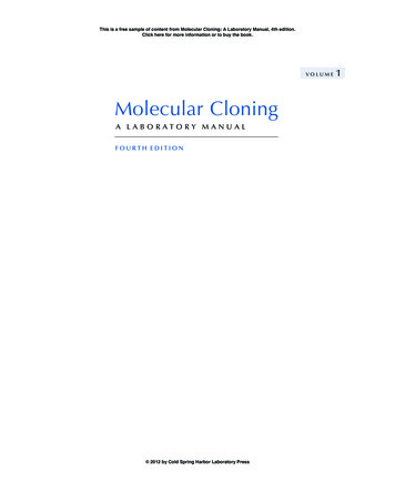
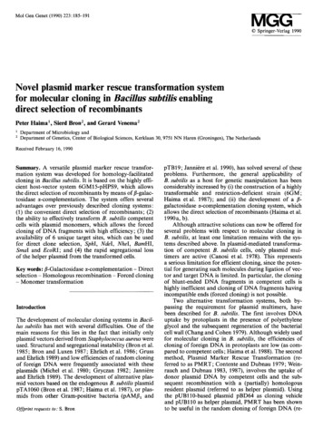
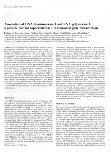
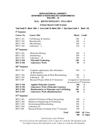
Hoseini and Sauer Journal of Biological Engineering 2015, 9:2http://www.jbioleng.org/content/9/1/2Results and discussionChoosing proper restriction enzymes based on definedcriteriaIn order to proceed with a concise example, tdTomatofluorescent protein was cloned into an alpharetroviralvector. Consecutively, a murine leukemia cell line expressing tdTomato was generated. This cell line will beused to track tumor cells upon injection into mice inpreclinical immunotherapy studies. However, this cloning method is applicable to any other gene. To begin thecloning project, the gene of interest (GOI) should be analyzed. First, we check whether our annotated sequencehas a start codon (ATG, the most common start codon)and one of the three stop codons (TAA, TAG, TGA). Incase the gene was previously manipulated or fused toanother gene (e.g. via a 2A sequence), it happens that agene of interest might not have a stop codon [9]. In suchcases, a stop codon needs to be added to the end of yourannotated sequence. It is also beneficial to investigatewhether your GOI contains an open reading frame(ORF). This is important since frequent manipulation ofsequences either by software or via cloning might erroneously add or delete nucleotides. We use Clone Managersoftware (SciEd) to find ORFs in our plasmid sequences;however, there are several free websites you can use tofind ORFs including the NCBI open reading frame .The tdTomato gene contains ATG start codon andTAA stop codon (Figure 1). The size of the tdTomatogene is 716 bp.In a next step, PCR primers that include proper restriction enzyme sites need to be designed for the amplification of the GOI. Several criteria should be considered inorder to choose the optimal restriction enzymes. First,binding sites for restriction enzymes should be ideallyPage 2 of 12available at a multiple cloning site within the vector. Alternatively they can be located downstream of the promoterin your vector sequence. Restriction enzymes should besingle cutters (single cutters target one restriction site onlywithin a DNA sequence) (Figure 2A). If they are double ormultiple cutters, they should cut within a sequence that isnot necessary for proper functioning of the vector plasmidand will finally be removed (Figure 2B). It is also possibleto choose one double cutter or multiple cutter enzymescutting the vector downstream of the promoter and alsonot within a vital sequence of the plasmid (Figure 2C).Double cutter or multiple cutter enzymes have two ormore restriction sites on a DNA sequence, respectively.Cutting the vector with double or multiple cutters wouldgive rise to two identical ends. In such a case, the insertcassette should also contain the same restriction enzymesites on both of its ends. Therefore, when the insert andvector fragments are mixed in a ligation experiment, theinsert can fuse to the vector in either the right orientation(from start codon to stop codon) or reversely (from stopcodon to start codon). A third scenario can occur, if thevector fragment forms a self-ligating circle omitting theinsert at all. Once the DNA has been incubated with restriction enzymes, dephosphorylation of the 5′ and 3′ends of the vector plasmid using an alkaline phosphataseenzyme will greatly reduce the risk of self-ligation [10]. Itis therefore important to screen a cloning product forthose three products (right orientation, reverse orientation, self-ligation) after fragment ligation.Second, due to higher cloning efficiency using stickyend DNA fragments, it is desirable that at least one(better both) of the restriction enzymes is a so-calledsticky-end cutter. Sticky end cutters cleave DNA asymmetrically generating complementary cohesive ends. Incontrast, blunt end cutters cut the sequence symmetricallyFigure 1 Overview of the start and the end of the gene of interest. (A) The nucleotide sequences at the start and the end of the tdTomatogene are shown. The coding strand nucleotides are specified in bold (B) The nucleotide sequences of the forward and reverse primers containingproper restriction enzyme sites and the Kozak sequence are shown.
Hoseini and Sauer Journal of Biological Engineering 2015, 9:2http://www.jbioleng.org/content/9/1/2Page 3 of 12Figure 2 Choosing proper restriction enzymes based on defined criteria for PCR cloning. (A) Two single-cutter restriction enzymes (E1 andE2) are located downstream of the promoter. (B) E1 and E2 restriction enzymes cut the plasmid downstream of the promoter several (here twotimes for each enzyme) times. (C) The E1 restriction enzyme cuts the plasmid downstream of the promoter more than once. (D) The PCR product,which contains the tdTomato gene and the restriction enzyme sites, was run on a gel before being extracted for downstream applications.leaving no overhangs. Cloning blunt-end fragments ismore difficult. Nevertheless, choosing a higher insert/vector molar ratio (5 or more) and the use 10% polyethylene glycol (PEG) can improve ligation of bluntend fragments [11].Third, some restriction enzymes do not cut methylatedDNA. Most of the strains of E. coli contain Dam or Dcmmethylases that methylate DNA sequences. This makesthem resistant to methylation-sensitive restriction enzymes[12]. Since vector DNA is mostly prepared in E. coli, it willbe methylated. Therefore avoiding methylation-sensitiverestriction enzymes is desirable; however, sometimes theisoschizomer of a methylation-sensitive restriction enzymeis resistant to methylation. For example, the Acc65I enzyme is sensitive while its isoschizomer kpnI is resistant tomethylation [13]. Isoschizomers are restriction enzymesthat recognize the same nucleotide sequences. If there remains no other option than using methylation-sensitive restriction enzymes, the vector DNA needs to be prepared indam dcm E. coli strains. A list of these strains and alsocommon E. coli host strains for molecular cloning is summarized in Table 1. Information regarding the methylationsensitivity of restriction enzymes is usually provided by themanufacturer.Fourth, it makes cloning easier if the buffer necessaryfor the full functionality of restriction enzymes is the samebecause one can perform double restriction digest. Thissaves time and reduces the DNA loss during purification.It may happen that one of the restriction enzymes is activein one buffer and the second enzyme is active in twice theconcentration of the same buffer. For example the NheIenzyme from Thermo Scientific is active in Tango 1X buffer (Thermo Scientific) and EcoR1 enzyme is active inTango 2X buffer (Thermo Scientific). In such cases, theplasmid DNA needs to be first digested by the enzyme requiring the higher buffer concentration (here EcoR1). Thiswill be followed by diluting the buffer for the next enzyme(requiring a lower concentration (here NheI)) in the samebuffer. However, the emergence of universal buffers hassimplified the double digest of DNA sequences [15]. Inour example the vector contains the AgeI and SalI restriction sites. These enzyme sites were used for designingPCR primers (Figure 1). It is essential for proper restriction enzyme digestion that the plasmid purity is high.DNA absorbance as measured by a spectrophotometercan be used to determine the purity after purification.DNA, proteins, and solvents absorb at 260 nm, 280 nm,and 230 nm, respectively. An OD 260/280 ratio of 1.8and an OD 260/230 ratio of 2 to 2.2 is considered to bepure for DNA samples [16]. The OD 260/280 and 260/230 ratios of our exemplary plasmid preparations were1.89 and 2.22, respectively. We observed that the purity ofthe gel-extracted vector and insert DNA fragments werelower after restriction digest; ligation works even in suchcases, however, better results can be expected using highpurity fragments.
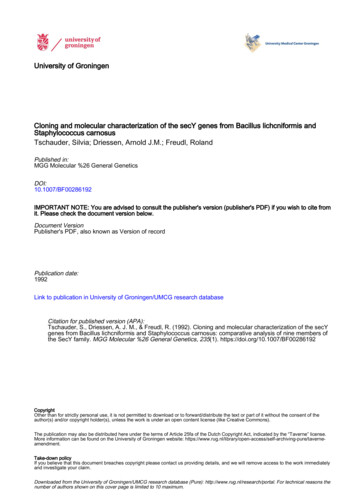
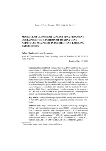
Hoseini and Sauer Journal of Biological Engineering 2015, 9:2http://www.jbioleng.org/content/9/1/2Page 4 of 12Table 1 Common E. coli host strains in gene cloningApplicationE. coli strainTransformation of large plasmidsGeneHogs, XL10 Gold, STBL4, SUREGeneration of single-stranded (ssDNA)INV110, JM109, JS5, NM522, SCS110, SURE, STBL4, XL10 Gold, XL1 Blue, TG1For storage of plasmids that tend to recombineDH10B, DH5α, STBL2, STBL3, STBL4, SURE, GeneHogs, Hb101, JM109, JS5, XL10 Gold,XL1 Blue, GC5, GC10Rapid cloning (fast cell growth)Mach1Cloning of unstable plasmidsSURE, STBL2, STBL3, STBL4High efficiency cloning for library constructionXL10-Gold, MegaX, DH10BBlue/white screeningDH10B, DH5α, MC1061, TOP10, XL1 Blue, Hb101, NM522, SCS110, STBL4, SURE, XL10Gold, GeneHogs, INV110, JM109, JS5, GC5, GC10For site-directed mutagenesisXL-mutS, BMH 71–18 mutS, ES1301 mutSFor random mutagenesisXL1-RedFor expression of toxic genesABLE C, ABLE KGeneral cloning and storage of plasmidsDH10B, DH5α, MC1061, TOP10, XL1-Blue, Hb101For proliferation of plasmids encoding the ccdB toxicgene (important in Gateway cloning)ccdB survival, DB3.1, XL1 Blue, JM109, DH5αFor generation of unmethylated DNA to be cut withmethylation-sensitive restriction enzymesJM110, ER2925, INV110, DM1, SCS110, E4109SData are derived from enzyme providers’ data sheets, the following website, and this reference [14]. http://openwetware.org/wiki/E. coli genotypes.The following plasmid repository website can be usefulfor the selection of different vectors (viral expressionand packaging, empty backbones, fluorescent proteins,inducible vectors, epitope tags, fusion proteins, reportergenes, species-specific expression systems, selection markers,promoters, shRNA expression and genome engineering):http://www.addgene.org/browse/.A collection of cloning vectors of E. coli is availableunder the following website: vectorExplanation.jsp.Designing cloning primers based on defined criteriaFor PCR primer design, check the start and stop codonsof your GOI. Find the sequence of the desired restrictionenzymes (available on the manufacturers’ websites) forthe forward primer (Figure 3A). It needs to be locatedbefore the GOI (Figure 1B). The so-called Kozak sequence is found in eukaryotic mRNAs and improves theinitiation of translation [17]. It is beneficial to add theKozak sequence (GCCACC) before the ATG start codonsince it increases translation and expression of the protein of interest in eukaryotes [18]. Therefore, we insertedGCCACC immediately after the restriction enzyme sequence AgeI and before the ATG start codon. Then, the first18 to 30 nucleotides of the GOI starting from the ATG startcodon are added to the forward primer sequence. Theseoverlapping nucleotides binding to the template DNA determine the annealing temperature (Tm). The latter is usuallyFigure 3 Designing primers based on defined criteria for PCR cloning. (A-B) Sequences of the forward and the reverse primer are depicted.The end of the coding strand is to be converted into the reverse complement format for the reverse primer design. For more information, pleasesee the text.
Hoseini and Sauer Journal of Biological Engineering 2015, 9:2http://www.jbioleng.org/content/9/1/2higher than 60 C. Here, we use Phusion high-fidelity DNApolymerase (Thermo Scientific). You can use the followingwebsites for determination of the optimal Tm: -tools/tm-calculator.The Tm of our forward primer is 66 C.Choose the last 18 to 30 nucleotides including the stopcodon of your GOI for designing the reverse primer(Figure 3B). Then calculate the Tm for this sequencewhich should be above 60 C and close to the Tm of theforward primer. Tm of the overlapping sequence of ourreverse primer was 68 C. Then, add the target sequenceof the second restriction enzyme site (in this case SalI)immediately after the stop codon. Finally, convert thisassembled sequence to a reverse-complement sequence.The following websites can be used to determine the sequence of the reverse oinformatics.org/sms/rev comp.htmlThis is important since the reverse primer binds thecoding strand and therefore its sequence (5′ 3′) mustbe reverse-complementary to the sequence of the codingstrand (Figure 1A).Performing PCR using proofreading polymerasesSince the PCR reaction follows logarithmic amplificationof the target sequence, any replication error during thisPage 5 of 12process will be amplified. The error rate of nonproofreading DNA polymerases, such as the Taq polymerase, is about 8 10 6 errors/bp/PCR cycle [19]; however, proofreading enzymes such as Phusion polymerasehave a reported error rate of 4.4 10 7 errors/bp/PCRcycle. Due to its superior fidelity and processivity [20-22],the Phusion DNA polymerase was used in this example. Itshould be noted that Phusion has different temperaturerequirements than other DNA polymerases. The primerTm for Phusion is calculated based on the Breslauermethod [23] and is higher than the Tm using Taq or pfupolymerases. To have optimal results, the Tm should becalculated based on information found on the website ofthe enzyme providers. Furthermore, due to the higherspeed of Phusion, 15 to 30 seconds are usually enough forthe amplification of each kb of the sequence of interest.After the PCR, the product needs to be loaded on agel (Figure 2D). The corresponding band needs to be cutand the DNA extracted. It is essential to sequence thePCR product since the PCR product might include mutations. There are several PCR cloning kits availablesome of which are shown in Table 2. We used thepJET1.2/blunt cloning vector (Thermo Scientific, patentpublication: US 2009/0042249 A1, Genbank accessionnumber EF694056.1) and cloned the PCR product intothe linearized vector. This vector contains a lethal gene(eco47IR) that is activated in case the vector becomescircularized. However, if the PCR product is cloned intoTable 2 Common vectors in gene cloningPlasmid nameAdvantagesDisadvantagesReferencespBR322Small size (4.4 kb), variety of cloning sites, medium copynumber (15–20)pUC18 and pUC19Small size (2.7 kb), high copy number (500–700), multiplecloning site, sequencing using M13 primerspLG338General purpose plasmid vector, size 7.3 kb, low copynumber (6–8), genes coding for membrane and regulatoryproteins which cannot be cloned into high-copy-numberplasmids[27]pMiniTInserted PCR product disrupts a toxic minigene, noblue/white selection required, works for both bluntend and single-base overhang-containing PCR productsNEBpCR 4Blunt-TOPO Inserted PCR product disrupts ccdB toxic gene [28], worksbased on topoisomerase I [29], no blue/white selectionrequiredNeeds blunt-end PCR productsLife TechnologiespDriveSize 3.8 kbNeeds single A overhangs, not usable forproofreading DNA polymerases, needsblue/white screeningQIAGEN[24,25]Not good if target protein is toxic or formembrane proteins, needs blue/whitescreen[24,26]StrataClone Blunt PCR Uses the DNA topoisomerase I and the DNA recombination Needs blunt-end PCR products, needsAgilent TechnologiesCloning Vectoractivity of Cre recombinase, up to 9 kb PCR product sizespecial competent cells for transformationpSC-B-amp/kan(cells expressing Cre recombinase)pJET1.2/bluntInserted PCR product disrupts the toxic gene eco47IR, noneed for blue/white screen, works for both blunt end andsingle-base overhang-containing PCR products, up to 10 kbPCR product sizeThermo ScientificData are derived from vector providers or the cited references. pBR322, pUC18, pUC19, and pLG338 are cloning vectors and the rest are PCR sequencing vectors.
Hoseini and Sauer Journal of Biological Engineering 2015, 9:2http://www.jbioleng.org/content/9/1/2the cloning site within the lethal gene, the latter is disrupted allowing bacteria to grow colonies upon transformation. Circularized vectors not containing the PCRproduct express the toxic gene, which therefore killsbacteria precluding the formation of colonies. Bacterialclones are then to be cultured, plasmid DNA consecutively isolated and sequenced. The quality of isolatedplasmid is essential for optimal sequencing results. Weisolated the plasmid DNA from a total of 1.5 ml culturedbacteria (yield 6 μg DNA; OD 260/280 1.86; OD 260/230 2.17) using a plasmid mini-preparation kit (QIAGEN).The whole process of PCR, including cloning of the PCRproduct into the sequencing vector and transfection ofbacteria with the sequencing vector can be done in oneday. The next day, bacterial clones will be culturedovernight before being sent for sequencing.Analysis of sequencing dataSequencing companies normally report sequencing dataas a FASTA file and also as ready nucleotide sequencesvia email. For sequence analysis, the following websitescan be /xylian.igh.cnrs.fr/bin/align-guess.cgiPage 6 of 12Here we will focus on the first website. On this websitepage, click on the “nucleotide blast” option (Figure 4A).A new window opens. By default, the “blastn” (blast nucleotide sequences) option is marked (Figure 4B). Thencheck the box behind “Align two or more sequences”.Now two boxes will appear. In the “Enter Query Sequence”box (the upper box), insert the desired sequence of yourgene of interest, which is flanked by the restriction sitesyou have already designed for your PCR primers. In the“Enter Subject Sequence” box (the lower box), enter the sequence or upload the FASTA file you have received fromthe sequencing company. Then click the “BLAST” buttonat the bottom of the page. After a couple of seconds, theresults will be shown on another page. A part of the alignment data is shown in Figure 4C. For interpretation, thefollowing points should be considered: 1) the number ofidentical nucleotides (shown under the “Identities” item)must be equal to the nucleotide number of your gene ofinterest. In our example, the number of nucleotides of thetdTomato gene together with those of the restriction enzyme sites and the Kozak sequence was 735. This equalsthe reported number (Figure 4C). 2) The sequence identity(under the “Identities” item) should be 100%. Occasionally,the sequence identity is 100% but the number of identicalFigure 4 Sequence analysis of the PCR product using the NCBI BLAST platform. (A) On the NCBI BLAST webpage, the “nucleotide blast”option is chosen (marked by the oval line). (B) The “blastn” option appears by default (marked by the circle). The sequence of the gene ofinterest (flanked by the restriction sites as previously designed for the PCR primers) and the PCR product are to be inserted to the “Enter QuerySequence” and “Enter Subject Sequence” boxes. Sequences can also be uploaded as FASTA files. (C) Nucleotide alignment of the first 60nucleotides is shown. Two important items for sequence analysis are marked by oval lines.
Hoseini and Sauer Journal of Biological Engineering 2015, s is lower than expected. This can happen if oneor more of the initial nucleotides are absent. Remember, allsequencing technologies have an error rate. For Sanger sequencing, this error rate is reported to range from 0.001%to 1% [30-33]. Nucleotide substitution, deletion or insertion can be identified by analyzing the sequencing results[34]. Therefore, if the sequence identity does not reach100%, the plasmid should be resequenced in order to differentiate errors of the PCR from simple sequencing errors.3) Gaps (under the “Gaps” item) should not be present. Ifgaps occur, the plasmid should be resequenced.The average length of a read, or read length, is at least800 to 900 nucleotides for Sanger sequencing [35]. Forthe pJET vector one forward and one reverse primerneed to be used for sequencing the complete gene.These primers can normally cover a gene size rangingup to 1800 bp. If the size of a gene is larger than 1800, anextra primer should be designed for each 800 extra nucleotides. Since reliable base calling does not start immediately after the primer, but about 45 to 55 nucleotidesdownstream of the primer [36], the next forward primershould be designed to start after about 700 nucleotidesfrom the beginning of the gene. Different websites, including the following, can be used to design these http://www.genscript.com/cgi-bin/tools/sequencing primer designBeing 735 bp in length, the size of the PCR product inthis example was well within the range of the pJET sequencing primers.After choosing the sequence-verified clone, vector andinsert plasmids were digested by the AgeI and SalI restriction enzymes (Figure 5). This was followed by gelpurification and ligation of the fragments. Transformation of competent E. coli with the ligation mixtureyielded several clones that were screened by restrictionenzymes. We assessed eight clones, all of which contained the tdTomato insert (Figure 6). It is important topick clones that are large. Satellite clones might not havethe right construct. We used a fast plasmid minipreparation kit (Zymo Research) to extract the plasmidfrom 0.6 ml bacterial suspension. The yield and puritywere satisfying for restriction enzyme-based screening(2.3 μg DNA; OD 260/280 1.82; OD 260/230 1.41).For large-scale plasmid purification, a maxi-preparationkit (QIAGEN) was used to extract the plasmid from450 ml of bacterial culture (yield 787 μg DNA; OD 260/280 1.89; OD 260/230 2.22). The expected yield of apBR322-derived plasmid isolation from 1.5 ml and 500 mlbacterial culture is about 2-5 μg and 500-4000 μg of DNA,respectively [37].Some plasmids tend to recombine inside the bacterialhost creating insertions, deletions and recombinationsPage 7 of 12[38]. In these cases, using a recA-deficient E. coli can beuseful (Table 1). Furthermore, if the GOI is toxic, incubation of bacteria at lower temperatures (25-30 C) andusing ABLE C or ABLE K strains might circumvent theproblem.Viral production and transduction of target cellsTo investigate the in vitro expression of the cloned gene,HEK293T cells were transfected with plasmids encodingthe tdTomato gene, alpharetroviral Gag/Pol, and thevesicular stomatitis virus glycoprotein (VSVG) envelope.These cells, which are derived from human embryonickidney, are easily cultured and readily transfected [39].Therefore they are extensively used in biotechnologyand gene therapy to generate viral particles. HEK293Tcells require splitting every other day using warmmedium. They should not reach 100% confluency foroptimal results. To have good transfection efficiency,these cells need to be cultured for at least one week tohave them in log phase. Transfection efficiency was22%, as determined based on the expression of tdTomatoby fluorescence microscopy 24 hours later (Figure 7A-B).To generate a murine leukemia cell line expressing thetdTomato gene for immunotherapy studies, C1498leukemic cells were transduced with freshly harvestedvirus (36 hours of transfection). Imaging studies (Figure 7C)and flow cytometric analysis (Figure 7D) four days aftertransduction confirmed the expression of tdTomato in themajority of the cells.ConclusionsIn this manuscript, we describe a simple and step-bystep protocol explaining how to exploit the power ofPCR to clone a GOI into a vector for genetic engineering. Several PCR-based creative methods have been developed being extremely helpful for the generation ofnew nucleotide sequences. This includes equimolar expression of several proteins by linking their genes via aself-cleaving 2A sequence [40,41], engineering fusionproteins, as well as the use of linkers for the design ofchimeric proteins [42-44]. Furthermore, protein tags[45,46] and mutagenesis (site-directed, deletions, insertions) [47] have widened the applications of biologicalengineering. The protocol explained in this manuscriptcovers for most situations of PCR-assisted cloning; however, alternative PCR-based methods are available beingrestriction enzyme and ligation independent [6,48-51].They are of special interest in applications where restriction enzyme sites are lacking; nevertheless, these methodsmight need several rounds of PCR or occasionally a wholeplasmid needs to be amplified. In such cases, the chanceof PCR errors increases and necessitates sequencing ofmultiple clones. In conclusion, this guideline assembles asimple and straightforward protocol using resources that
Hoseini and Sauer Journal of Biological Engineering 2015, 9:2http://www.jbioleng.org/content/9/1/2Page 8 of 12Figure 5 Vector and insert plasmid maps. (A) Illustration of the CloneJET plasmid containing the PCR product. Insertion of the PCR product inthe cloning site of the plasmid disrupts the integrity of the toxic gene eco47IR and allows the growth of transgene positive clones. The plasmidwas cut with the AgeI and SalI enzymes generating two fragments of 3 kb and 0.7 kb in size. The 0.7 kb fragment (tdTomato gene) was used asthe insert for cloning. (B) Illustration of the vector plasmid. The plasmid was cut with the AgeI and SalI enzymes generating two fragments of4.9 kb and 0.7 kb in size. The 4.9 kb fragment was used as the vector for cloning. AMP: Ampicillin resistance gene; PRE: posttranscriptionalregulatory element; MPSV: myeloproliferative sarcoma virus promoter.are tedious to collect on an individual basis thereby tryingto minimize errors and pitfalls from the beginning.leukemia cell line C1498 [52], was cultured in RoswellPark Memorial Institute (RPMI) 1640 medium supplemented with the same reagents used for DMEM. Cellswere split every other day to keep them on
proper restriction enzyme sites were embedded. Practical criteria for the selection of restriction enzymes and the design of PCR primers are explained. Efficient cloning of PCR products into a plasmid for sequencing and free web-based software for the consecutive analysis of sequencing data is introduced. Finally, confirmation of successful