Transcription
Staff Nurse Anesthetist, The Johns Hopkins HospitalAssistant Professor/Assistant Program DirectorColumbia University School of NursingProgram in Nurse AnesthesiaMaribeth Massie, CRNA, MSAnesthesia for Trauma
“It’s not the speed whichkills, it’s the sudden stop”OVERVIEW
8% worldwide death rate Leading cause of death in Americansfrom 1- 45 years of age MVC’s leading cause of death Blunt penetrating Often drug abusers, acutely intoxicated,HIV and Hepatitis carriersEpidemiology of Trauma
– First hour after injury– 50% of patients die within the first secondsto minutesÆ extent of injuries– 30% of patients die in next few hoursÆmajor hemorrhage– Rest may die in weeks Æ sepsis, MOSF “Golden Hour”Epidemiology of Trauma
Resuscitation Reduction of fractures Extrication of trapped victims Amputation Uncooperative patients– Initial assessment and BLS in trauma– GO TEAM: role of CRNA’s at MarylandShock Trauma Center ABC’SPre-hospital Care
Airway maintenance with cervical spineprotection Breathing: ventilation and oxygenation Circulation with hemorrhage control Disability ExposureInitial Management Plan
ALWAYS ASSUME A CERVICAL SPINE INJURYEXISTS UNTIL PROVEN OTHERWISE Provide MANUAL IN-LINE NECKSTABILIZATION Jaw-thrust maneuver– AIRWAY Primary Survey:Initial Assessment
Cross table lateral and swimmer’s view Xray Need to see all seven cervical vertebrae Only negative CT scan R/O injury– Cervical spine evaluation Airway cont’d:Initial Assessment
BicepsAbduct shoulder,Flex elbowC6 Wrist extensorsCock wristC7 TricepsExtend elbowC8 Finger flexorsGrasp finger in palmT1 Finger abductorsSpread fingers C5– Functional assessment of cervical level Cervical spine cont’d:Initial Assessment
––––––Airway obstructionPaO2 80 mmHg or SpO2 90% with O2Shock with SBP 90 mmHgSevere head injury or unconscious (GCS 9)Anticipated surgery with multisystem injuryCombativeness Indications for intubation:ALWAYS ASSUME FULL STOMACHPRECAUTIONSÆ RAPID SEQUENCEINTUBATIONInitial Assessment
Succinylcholine: 1-2 mg/kg Zemuron: 1.2 mg/kg Vecuronium: .2 mg/kg– “Pent, Sux, Tube”– May have to decrease amount of sedative drugsand give appropriate dose of RSI muscle relaxants Use slow inspiratory flow rates (1-1.5 sec inspiratorytime) Avoid stomach distentionÆgastric inflation occurs wheninspiratory pressure exceeds EOP ( 15-18 cm H2O)– Preox Rapid sequence intubation (or modified)Initial Assessment
– MAC vs. Miller debate 10 # pressure required to seal esophagus– Remove front of C-collar and maintain inline stabilization– Cricoid pressure (Sellick’s maneuver) afterPent given Airway cont’d:Initial Assessment
Awake intubation: local, topicalsuperior laryngeal nerve blocks Awake fiberoptic: may be too bloody Awake cricothyrotomy/tracheostomy Gum elastic bougie/LMA Know your difficult airway algorithm!Initial Assessment
– Always verify correct position of ETT, evenif arrive intubated !!– 100 % O2– May have Combitube in; change to ETT– Nasal intubation: watch with basilar skullfractures BREATHINGInitial Assessment
– Control hemorrhage first!– Crystalloids vs. colloids vs. blood products?– Alot or alittle?– Early or later? CirculationInitial Assessment
– Allergies, PMH//PSH, meds, ETOH/druguse, weight, last meal After primary survey complete, attemptto complete a head-to-toe assessment Ask pertinent questions if patient ableto answerSecondary survey
Cardiac: S/S shock, EKG changes Respiratory: Breath sounds, crepitus,respiratory patterns/distress, CXR Neurologic: GCS, LOC; assume C-spineinjury until ruled outÆ Lateral C-spineXray, palpate neck Renal: monitor urine output, amountand colorTrauma/preop assessment
Endocrine: release of stress hormones(catecholamines and glucose) Hematologic: hypovolemic shock;coagulopathies– Gastric emptying slows or stops at time oftrauma Gastrointestional: FULL STOMACH!!!!Trauma/preop assessment
CBC, electrolytes, urinalysis, PT/PTT,lactate, baseline ABG (as conditionpermits); T&C for at least 4 units CXR, lateral C-spine, CT/MRI 12 lead EKG FAST: focused abdominal sonographyfor trauma DPL: diagnostic peritoneal lavageLaboratory/diagnostic tests
– Versed in small doses (.5-1 mg IV)– Bicitra 30 cc preop Preop: Sedation rarely necessaryAnesthetic management of traumapatient
Standard monitors Preoxygenation Basic airway and difficult airwayadjuncts RSI with cricoid pressure Invasive monitors as indicatedInduction
Thiopental 3-4 mg/kg; reduce doses inunstable patients; most commonly used intrauma Ketamine 0.5-1 mg/kg; useful for burn andhypovolemic patients; avoid with headinjuried Etomidate 0.1-0.3 mg/kg; reduce doses withhypovolemia; ?myoclonus effects Propofol 1-2 mg/kg in stable patients; reducedoses in hypovolemiaInduction agents
– May give nondepolarizing dose prior to Suxto inhibit fasciculations (esp. with SCI) Succinylcholine:1-2 mg/kg; useful forRSI/emergency; contraindicated inburns, spinal cord injury and crushinjuries 24-48 hours after injuryMuscle relaxants
– Vecuronium .28 mg/kg (250-300mcg/kg)high dose; onset in 80 secs;duration 75-90 min; good cardiovascularstability without histamine release– Rocuronium 1.2 mg/kg high dose; onset45-60 secs; duration 67 minutes NondepolarizersMuscle relaxants
O2/air/Forane mixture Avoid N2O if any question ofpneumothorax, pnuemocephalus,pneumomediastinum, bowel injury Fentanyl 1- 10 mcg/kg/hr Monitor fluids and administer carefully Prepare to give blood products ifnecessaryMaintenance
Forced air warming systems Heat moisture exchangers– Level 1: warms IVF and blood to 42*C anddelivers at 75-30,000 ml/hr– Rapid infusion system (RIS): warms to 42*C andcan deliver products as bolus and various rates,up to 3000ml/min; cell saver can be attached tosystem Common with traumatic injuries and relatedprocedures Warm all IV fluidsHypothermia
Normal extubation criteria Hemodynamically unstable, elderly withrib and long bone fractures, those whohave received massive fluid and bloodresuscitation, severe burns, and thosewith coagulopathies should remainintubatedEmergence
– Hypothermia, atelectasis, V/Q mismatch,coagulopathyLong-acting opioidsEpidural infusionsIntercostal blocksComplications:– Correct acid-base imbalances and electrolytedisturbances Monitored and labs followed closelyPostop
Blunt trauma: caused by high-velocity orlow-velocity impact from generally dullobjects Penetrating trauma: result of sharp objectspiercing through tissue, such as stab woundsproduced by knives or bullet woundsproduced by gunfire Impalement injuries: combination of bluntand penetrating trauma Falls: vertical high-velocity injuries Burns: thermal, electrical or chemicalMechanism of injury
Airway burns and smoke inhalationinjuries: associated with carbon dioxidepoisoning Environmental injuries: poisonousinsects and snakes, animals orconsequences of nature Biological, chemical or nuclear warfareMechanism of injury cont’d
Result of direct impact, deceleration,continuous pressure, shearing, and rotaryforces Associated with injuries from high-speedcollisions and falls from heights Motor vehicle crashs (MVC) are classified ashead on, rear impact, side impact, rotationalimpact, and rollover Injuries commonly much more severe thanpenetratingBlunt trauma
– Type of wounding instrument– Velocity of instrument at time of impact– Type of tissue that instrument passesthrough (organs, vessels, nervous tissue,muscle, fat, bone) Often requires surgical intervention Damage depends on 3 factors:Penetrating trauma
Blunt or penetrating trauma Most ominous sign: hypoxia fromtension pneumothorax, hemothorax,flail chest, hypovolemia, cardiactamponade Chest wall trauma can result in aboveThoracic injuries
–––– Treatment:: needle decompression second intercostalspace midclavicular lineÆ chest tube 4th or 5th ICS,midaxillary linechest wall hyperresonant to percussionBreath sounds decreased or absent unilaterallySubcutaneous emphysemaCXR confirms Accumulation of air between parietal andvisceral pleura Results in severe V/Q mismatch and hypoxia S/S:Pneumothorax
Can be caused from bleeding of heartand great vessels Fluid load before chest tube placement Differentiated from pneumothorax bydullness to percussion with absentbreath soundsHemothorax
Develops from air entering pleural cavitythrough a one way valve in lung or chest wall With each inspiration, more air becomestrapped in thorax, increasing intrapleuralpressure Eventually the ipsilateral lung collpases andthe mediastinum and trachea shift tocontralateral sideTension pneumothorax
Hyperresonance to percussion of chest wallIpsilateral absence of breath soundsContralateral tracheal shiftDistended neck veins?Differentiated from cardiac tamponade byhyperresonance to percussion over tensionpneumo– 14 gauge catheter 2nd ICS midclavicular lineÆchest tube Treatment––––– S/STension pneumothorax cont’d
– Paradoxical chest wall movement and/or splintingdue to intense pain Results from comminuted fractures of at leastthree adjacent ribs with associatedcostochondral separation or sternal fracture Accompanied by hemothorax or pulmonarycontusion Patients with 3 or more rib fractures havegreater likelihood of hepatic or splenic injury S/SFlail chest
IV thoracic epidural intercostal blocks– O2 with humidification– Pain meds: Chest Xray and ABG confirm diagnosis TreatmentFlail chest cont’d
– If worsening respiratory failure, intubation withPEEP, frequent suctioning to avoid bronchialplugging and atelectasis, and careful volumeresuscitation Intra-alveolar hemorrhage and edemaresulting from sudden increase in intraalveolar pressure and rupture of alveolarcapillary interface Difficult to diagnosis; high index os suspicionwith thoracic injuries TreatmentPulmonary contusion
Later pulmonary complication Attributed to direct thoracic injury,sepsis, aspiration, head injury, massivetransfusion, oxygen toxicity, and fatembolism Mortality rate reaching 50%ARDS
– Dysrhythmias: heart block to Vfib; ST segmentelevation– Elevated CPK-MB; ? troponin elevation– CHF– Anginal pain which may or may not respond tonitrates Associated with blunt trauma Contusion most often right ventricle since liesdirectly posterior to sternum S/SMyocardial contusion
– Management of dysrhythmias– Increase CVP to optimize right ventricularoutput TreatmentMyocardial contusion cont’d
– Dyspnea– Orthopnea– tachycardia Life-threatening emergency Bleeding into pericardial space, whichrestricts cardiac filling during diastoleand creates a low cardiac output state Initial symptomsCardiac tamponade
If advanced too far, will see ectopyRequires thoracotomyFluid load and treat with pressors if necessaryAvoid bradycardia; Ketamine useful agent– Pericardiocentesis: 16 g catheter inserted at thexiphochondral junction toward left scapula at 45*angle Treatment May not be evident in hypovolemia– Beck’s triadÆ neck vein distention, hypotension,muffled heart sounds– Pulsus paradoxus: 10 mmHg decrease in bloodpressure during spontaneous inspiration Classic symptomsTamponade cont’d
Aortic ruptureValvular ruptureSeptal ruptureDiaphragmatic herniationEsophageal ruptureAssociated thoracic injuries
High risk for exsanguinating hemorrhage andperitonitis Results from blunt and penetrating trauma Retroperitoneal injuries can damageabdominal aorta, IVC, kidneys, pancreas,duodenum Intraperitoneal injuries can injure spleen,liver, stomach, small bowel, colon, rectumAbdominal and Pelvic trauma
Intraabdominal injuries associated withparalytic ileus and peritoneal irritation(muscle guarding, tenderness to percussion,abdominal distention) 1-3 liters of blood can sequester inabdomen/retroperitoneal space with minimalsigns Diagnosis confirmed with free air on Xray orFAST or CT or by bloody DPLAbdominal and pelvic injuries cont’d
Performed when abdominal injurysuspected from mechanism of injury Not performed routinely now that FASTavailable FAST and DPL can prevent unnecessaryexploratory lap Can use local with sedationDiagnostic peritoneal lavage (DPL)
100,000 RBC’s/ml 500,000 WBC’s/mlAmylase 200 unitsBacteria– False positive results 2% – Positive finding: 10 cc gross blood Peritoneum lavaged with fluid that is thendrained by gravity and examined for presenceof RBC’s, bile, amylase, and WBC’sDPL cont’d
– Decreases incidence of sepsis– Can take to angiography to embolize lac Most common injury in blunt abdominaltrauma and with penetrating wounds ofleft lower thorax and upper abdomen Routine splenectomy rare Splenorrhaphy (repairing the spleen)more commonSplenic laceration
Second most common injury associatedwith abdominal trauma Exsanguniating hemorrhage can occur Majority of liver injuries (85-90%) healspontaneously and may only requiresurgical drainageLiver laceration
Urethrogram should be performed before foley inserted– Arterial bleeding can be embolized– Bladder injuries often associated with pelvicfracture Result in major hemorrhage 25% of time Exsanguination 1% of time Bleeding results from disruption of veins frombone fragments Emergent or elective external fixation can befollowed by angiographyPelvic fractures
– The pelvis can hold up to 3 liters Anesthetic concerns revolve aroundhemorrhage, hypothermia, sepsis/peritonitisand impairment of ventilation Warming measure are crucial since large heatloss from open mesentery and shock Avoid N20 to prevent bowel distention Fluid resuscitation imperativeAbdominal and pelvic trauma
Usually not immediately life-threateningand part of secondary survey Can be associated with vascular injuriescausing hemorrhage, shock, sepsis, fatemboli, and thromboembolic hypoxicrespiratory failureExtremity trauma
Ideal to repair in first few hours postinjury so full stomach precautions Should repair within 6 hours to lessenincidence of sepsis If obvious hemorrhage, hold pressuremanually; can have MAST pants appliedwhile in fieldOpen fractures
– Pain– Pulselessness– Pallor– Paresthesias– Paresis– Confirmed with angiography S/SVascular trauma
Emergency fasciotomy must be done toprevent irreversible muscle and nervedamage Diagnosis confirmed by compartmentpressures 40 cm H20– Calf pain on dorsiflexion of foot Characterized by severe pain in affectedextremityCompartment syndrome
–––––FeverPetechaieDysrhythmiasFat globules in urine, plasma, retinal vesselsMental deterioration 1-3 days post trauma Commonly lead to thromboembolic hypoxicrespiratory failure due to fat globules orfracture debris reaching pulmonary vascularbed Fat embolism syndrome:Long bone fractures
– Aggressive cardiovascular and pulmonarysupport Diagnosis: elevated serum lipase, fat inurine, and thrombocytopenia Treatment: early fracture stabilizationis key to preventionFat embolism syndrome cont’d
Positioning Associated injuries TourniquetsAnesthetic concerns with extremitytrauma
– Need to hydrate, osmotic diuretics, alkalinize urineto protect kidneys– Follow lactate; 2 can be sign of underresuscitation Can occur with blunt and penetrating trauma Increased risk of myoglobinuria, leading torhabdomyolisis Always check urine and document color withtrauma patients; inform surgeon immediatelyif becoming bloodyCrush injuries
Goal is prevention of secondary brain damageresulting from intracranial bleeding, increasedICP, edema Management should include early control ofairway, cardiovascular stability, andavoidance of increased ICP Patients with suspected head injury should beplaced head up position to promote venousdrainage and decrease ICP; moderatehyperventilation to 30 mmHgHead injury
– C collar– Inline stabilization with intubation– RSI/airway adjuncts High index of suspicion related tomechanism of injury Always treat as suspected C-spineinjury unless proven otherwiseSpinal cord injury
Paralysis Pain Position: patient holding head upright withboth hands may indicate Jefferson (hangman) fracture C1; “hold-up” position witharms above head may indicate C4-5 fracture;“prayer position” with arms folded acrosschest possible C5-6 fractureSigns and symptoms related to SCI
Paresthesias Ptosis PriapismS/S of SCI cont’d
– C7 most common site of injury Leading cause of death at scene:aspiration Most injuries occur in males in 20’s-30’s Occur from falls, MVC’s, diving injuries,penetrating missiles, sports injuries Must obtain lateral C-spine XraySCI
Oral intubation: induce patient then removefront of C collar and hold in-linestabilization/RSI– Topical anesthesia– Avoid transtracheal block due to increased risk ofaspiration and movement of neck with coughing Nasal intubation method of choice if patientdoes not have associated basilar skullfracture/LeFort 2-3 fracturesAnesthetic management with SCI
– Acutely may want to avoid secondary tofasciculations that may exacerbate SCI– \Can give curarizing dose of NDMR– High dose Vec or Roc good alternative Succinylcholine: do not give to patients 24 hours post massive muscle ordenervation injuries, SCI’s, crushinjuries or burnsMuscle relaxants with SCI
Hypotension Bradycardia Hypothermia/poikilothermia (bodytemperature migrates towardenvironmental level) Results from sympathectomy in SCIpatients More intensified at T6 level and higherSpinal shock
– Unable to maintain adequate cardiac filling pressuresbut overaggressive fluid administration canprecipitate pulmonary edema (neurogenic) Careful volume resuscitation– Hemmorrhagic shock tend to behypotensive, tachycardiac with cold,clammy skin– Treatment: Patients present with hypotension,bradycardia and warm, pink extremitiesSpinal shock
– If arm embolizes, patient at severedisadvantage May require pressorsÆ Dopamine 4-5mcg/kg/min Avoid using radial arteries for arterialline if paraplegicSpinal shock cont’d
HypertensionBradycardiaDysrhythmiasCutaneous vasodilation above andvasoconstriction below injury– Severe headaches– Seizures– Loss of consciousness–––– Seen in 85% of patients with injuries aboveT5 S/SAutonomic hypereflexia
Occurs after spinal shock passed Usually seen 24 hours post injury andwhen patients return to OR forsubsequent operations Caused by stimulation below level oflesion Treatment: stop stimulus; deepenanesthesia; cardiovascular supportAutonomic hypereflexia
2 million patients will be brought to traumacenters for burns and associated injuries Majority are thermal injuries in children 5years Electrical burns cause tissue damage bythermal injury and injury to underlyingstructures and heart Chemical burns depend on chemical,concentration, and duration of exposureThermal injury
First-degree burn: superficial involvingupper layers of epidermis; skin red andedematous and painful like sunburn Second-degree burn: partial thicknessburns extend damage through dermis;regeneration can occur; blisters developand have white or red areas that arepainfulDegree of burn
Third-degree burn: full thickness burncharacterized by destruction of all layers ofskin, including nerve endings; skin will notregenerate and healing does not occur unlessdead tissue debrided and skin grafts placed;skin charred and not painful Fourth-degree burn: involve destruction ofall layers of skin and extend intosubcutaneous tissue, fascia, muscle, andboneDegree of burn cont’d
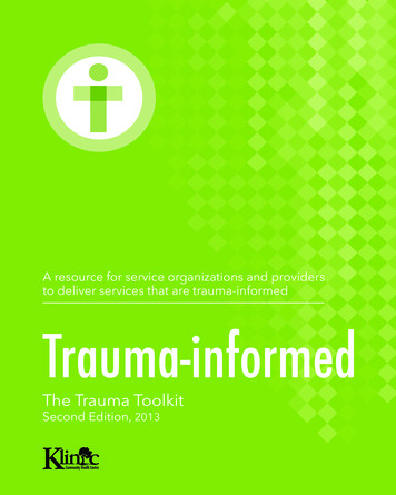
First degree burn (epidermal burn)
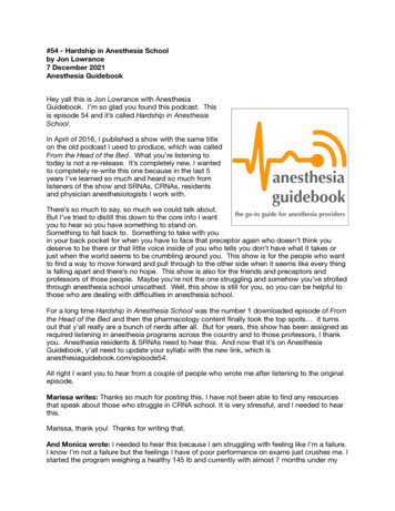
Second degree burn (superficial dermalburn)
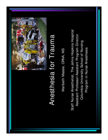
Third degree burn (sub-dermal burn)
Fourth degree burn
Size of burn estimation to assess total BSAburned Body divided into regions that represent 9%or multiples of 9% of total BSA Adults: head/neck 9%; arms/hands 9% eachextremity; thighs/legs 18% each extremity;anterior/posterior trunk 18% each side;perineum 1% Children calculated slightly different due tolarge head Size of hand roughly equal to 1% BSARule of Nines
– First 24 hours– Includes airway management and treatingany circulatory and associated injuries– Suspicion of upper and lower airway injuryis increased with singedeyebrows/eyelashes and black soot aroundnose and mouth Resuscitative phaseThree phases of burn inury
– Needle electrodesEarly intubationMultiple large bore IV accessAggressive fluid resuscitationStandard and invasive monitors placedearly Temperature regulation Anesthetic management of burnpatient
– Albumin concentration decreased after 48 hoursÆalbumin-bound drugs (such as benzos andanticonvulsants) have an increased free fractionand prolonged effect– Cardiovascular support– Require higher than normal doses of NDMR (2-5times normal dose)– Ketamine for dressing changes and escharotomies Varied drug responsesManagement cont’d
–––––––Respiratory irritation (coughing)Sore throatDysphagiaHemoptysisCarbon-colored sputumTachypnea, use of accessory muscles, wheezingCrepitus High index of suspicion if loss ofconsciousness at scene and if fire occurred inclosed space S/S of inhalation injuryAirway injury
Hoarseness demands immediateattentionÆ means airway becomingedematous and can quickly obstructglottis Diagnosis made withcarboxyhemboglobin levels Should be intubated immediately if anysuspicion of injuryInhalation injury
– Tachypnea– Cherry red color of blood (only when COHgB 40%) Results from inhalation of CO produced byfires, exhaust from internal combustionengines and cooking and charcoal stoves Produces tissue hypoxia by its 200 timesaffinity for Hgb compared to oxygen COHgB formedÆ pulse oximeter may displayhigher than actual O2 saturation S/SCarbon monoxide (CO) poisoning
NoneMild H/A, confusionSevere H/A, blurred visionDisorientation, N/V,irritability, syncopeTachycardia, tachypnea,agitationDeath0-55-1011-2021-40 6041-60ManifestationsCO HgB level (%)Clinical manifestation of CO exposure
Treatment: 100% O2 immediately Hyperbaric oxygen therapy (HBO) maybe initiated if symptoms not abatingCO poisoning
4ml/kg LR per percent BSA burned½ given over first 8 hoursRest over next 16 hoursIn addition to maintenance– 3ml/kg per percent BSA burned– ½ over first 8 hours– Rest over next 16 hours Brooke formula–––– Parkland formulaFluid resuscitation
Occurs following rhabdomyolisis andhemoglobinuria due to hemolysis; affectsrenal blood flow via damage to renalparenchyma FFP may protect renal function since itcontains haptoglobin, which binds freehemoglobin Aggressive fluid resuscitation Maintenance of urine output with osmoticdiuretics and sodium bicarb to protect kidneysMyoglobinuria
Multiple skin omiesMay still be hemodynamically unstable inthis phase Debridement and grafting phase
May continue for rest of life Release of contractures Multiple plastic surgeryReconstructive phase
Skin grafting
Anesthesia for Trauma Maribeth M a s s ie, C R N A, M S Staff Nurse A n estheti s t, Th e Joh n s Hopkins Hospital As si stant Prof e s sor/A s si sta n t Program Director Columbia University School of Nursing Program in Nurse A n esth . MAC vs. Miller deb a te. Initial Assessment Awake intubation: local,