Transcription
The Spine Center at SinaiPatient GuideAnterior Lumbar Interbody Fusion (ALIF)Anterior Cerivical Diskectomy and Fusion (ACDF)Lumbar DiskectomyPosterior Lumbar Instrumented Fusion2401 West Belvedere AvenueBaltimore, Maryland om
Table of ContentsIntroduction . . . . . . . . . . . . . . . . . . . . . . . . . . . . . . . . . . . . . . . . . . . . . . . . . . . . . . . . . . . . . . . . . . . . .1Anterior Lumbar Interbody Fusion (ALIF) . . . . . . . . . . . . . . . . . . . . . . . . . . . . . . . . . . . . . . . . . . . . . . .2IntroductionWelcome to The Spine Center at Sinai. Our team of physicians and nurses is dedicated to providing expertcare for your spine throughout your treatment process. To help guide you throughout your stay we havecustomized this patient education booklet to your condition. We recommend you keep it with you and useit as a reference guide.Anatomy, Symptoms, Surgery . . . . . . . . . . . . . . . . . . . . . . . . . . . . . . . . . . . . . . . . . . . . . . . . . . . .2-7Anterior Cervical Diskectomy and Fusion . . . . . . . . . . . . . . . . . . . . . . . . . . . . . . . . . . . . . . . . . . . . . . .8Anatomy, Symptoms, Surgery . . . . . . . . . . . . . . . . . . . . . . . . . . . . . . . . . . . . . . . . . . . . . . . . . . .8-13Lumbar Diskectomy . . . . . . . . . . . . . . . . . . . . . . . . . . . . . . . . . . . . . . . . . . . . . . . . . . . . . . . . . . . . . .14Anatomy, Symptoms, Surgery . . . . . . . . . . . . . . . . . . . . . . . . . . . . . . . . . . . . . . . . . . . . . . . . . .14-17The Spine Center’s inpatient unit is located on the third floor of the main hospital and is staffed by speciallytrained nurses and orthopedic technicians. It includes eight private inpatient rooms and one semiprivate inpatientrooms plus a specially equipped physical therapy unit to enhance patient recovery. Three operating rooms onSinai’s fourth floor are dedicated solely to spine patients and procedures, and are equipped with the latesttechnology available.The staff at The Spine Center at Sinai looks foward to caring for you during your stay.Posterior Lumbar Instrumented Fusion . . . . . . . . . . . . . . . . . . . . . . . . . . . . . . . . . . . . . . . . . . . . . . .18Anatomy, Symptoms, Surgery . . . . . . . . . . . . . . . . . . . . . . . . . . . . . . . . . . . . . . . . . . . . . . . . . .18-24Before Surgery Information . . . . . . . . . . . . . . . . . . . . . . . . . . . . . . . . . . . . . . . . . . . . . . . . . . . . . . . .25After Surgery Information . . . . . . . . . . . . . . . . . . . . . . . . . . . . . . . . . . . . . . . . . . . . . . . . . . . . . . . . . .27Frequently asked questions related to surgery . . . . . . . . . . . . . . . . . . . . . . . . . . . . . . . . . . . . . . . . . .28Glossary . . . . . . . . . . . . . . . . . . . . . . . . . . . . . . . . . . . . . . . . . . . . . . . . . . . . . . . . . . . . . . . . . . . . . . .30 2003 Copyrighted MaterialPatient Handbook for Anterior Lumbar Interbody Fusion (ALIF); Anterior Cervical Diskectomy and Fusion; Lumbar Diskectomy; and Posterior LumbarInstrumented Fusion.This notice is to inform the user that all the materials found in this package are copyrighted. This material is to be used only as specified andcannot be copied or used in any other capacity without the expressed written consent of Sinai Hospital.1
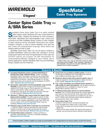
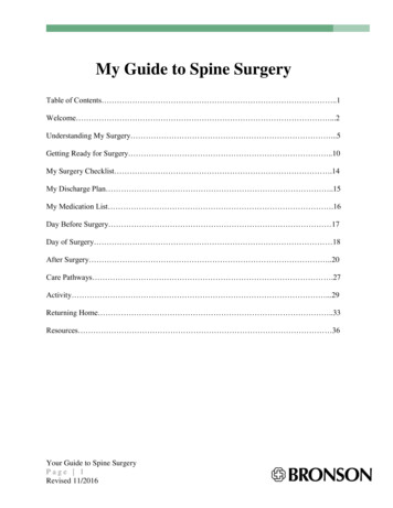
Anterior Lumbar Interbody Fusion (ALIF) is an inpatient procedure that requires a three to five dayhospital stay. The length of time for the procedure varies but typically lasts four to six hours. Theusual time for discharge is three days after surgery. Independent ambulation (walking without assistance) and adequate pain control are goals to be met prior to discharge. On rare occasions, patientswill be admitted to a rehabilitation unit prior to discharge home.The spinal cord passes through a canal in the spine and ends in the upper lumbar spine. It then continues down the spine as a bundle of nerve roots. These nerve roots exit through openings betweenthe vertebrae.Anatomy of the Lower BackThe lumbar spine consists of five vertebrae, which are separated by disks. The disks cushion thespine during movement similar to the way shock absorbers cushion movement on a car. The disksalso function as joints to allow movement in your spine.The center or nucleus of the disk is made of a spongy material and is surrounded by tough outer ringsknown as the annulus. The nucleus is the shock absorber, and the annulus holds the disk together.VertebraeSymptomsDegenerative disk disease can be caused by a number of factors including trauma, too much weighton the spine, aging, smoking and poor genes. Degenerative arthritis of the lower spine may, overtime, damage the disk and the vertebrae, which can irritate the nerve roots.Degenerative disk disease is a common cause oflow back pain. Bone spurs can irritate nerve rootsthat, in turn, can cause symptoms of leg pain.23
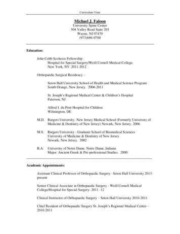
Surgery–Purpose and MethodPurpose of SurgeryThe purpose of anterior lumbar fusion is to take pressure off the nerves and to stabilize or strengthenthe lower spine. The fusion locks two or more vertebrae together and prevents abnormal or excessivemovement of the vertebrae to minimize back pain.The goal of surgery is to relieve the symptoms of pain, muscle weakness, numbness and tingling or,in some cases, to halt the progression of symptoms.Method of SurgerySurgery is performed in the supine position (lying on the back). An incision is made on the abdomento expose the vertebrae.Degenerative spondylolisthesis is a condition that develops when the disk weakens and flattens. Thevertebrae begin to slip back and forth. The facet joints and nerve roots become irritated, which canlead to symptoms of low back and leg pain.45
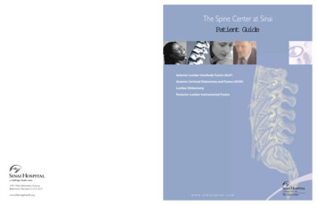
A bone graft (also known as an autograft) will be taken from the front of your pelvisthrough a small incision. The removed bone is placed at the surgical site to promote newbone growth.The placement of the cages restores disk height and relieves compression on the affected nerves.Bone morphogenetic protein (BMP) is a substance that is now available for general usein patients undergoing anterior fusion approaches. It eliminates the need for obtaining abone graft from the patient.The degenerated disk is removed and hollow “cages” containing either bone graft or theBMP substance are placed in the space.The underlying layers will be closed with absorbable sutures. The outer incision will be closed withmetal staples or absorbable sutures. Staples should be removed seven to 14 days after surgery.Over the next few months, bone growth within the cages will cause the adjacent vertebrae to fusetogether and function as one vertebra.67
Surgery for Anterior Cervical Diskectomy and Fusion (ACDF) requires at least an overnight stay tomonitor breathing and the level of pain due to the location of the incision. The length of time for theprocedure varies depending on the extent of surgery but typically lasts two hours. The earliest time fordischarge is the morning after surgery.The center or nucleus of the disk is made of a spongy material and is surrounded by tough outer rings (the annulus).Anatomy of the Cervical SpineThe cervical spine is the most flexible part of the spine and consists of seven vertebrae. Each vertebra is separatedby disks that cushion the spine during movement similar to the way shock absorbers cushion movement on a car.The spinal cord passes through the canal in the spine. A pair of spinal nerves exits the spinal cord at openings orforamen on either side of each vertebra.SymptomsIrritated nerve roots that exit the spine cause symptoms of neckand arm pain, numbness and weakness.89
Trauma or sudden movement can cause the weakened outer rings of the disk to tear. The material in the center ofthe disk squeezes through the tear in the outer rings and presses against the nerve roots. This is what is known asa herniated or ruptured disk.The disks in a healthy spine act as shock absorbers, protecting the vertebrae, nerves and spinal cord fromdamage or injury. Aging, too much weight on the spine, and/or trauma may contribute to degenerative diskdisease.Characteristic of this disease is that the disks wear down and become flat. The vertebrae above andbelow the disk grind, slip and lose stability. This may cause bone growths (bone spurs). Bone spurs cannarrow the path to nerve roots or narrow the spinal canal (stenosis). Pain is caused by pressure on thenerve roots or spinal cord.10Surgery – Purpose and MethodPurpose of SurgeryThe purpose of a cervical diskectomy is to take pressure off the nerves by removing portions of the disk or thebone spurs that are pressing on the nerve roots. The goal is to relieve symptoms of pain, muscle weakness,numbness or tingling. For some patients, the goal is to halt the progression of these symptoms.11
Method of SurgeryAnterior Cervical Diskectomy and Fusion (ACDF) is the surgical removal of a portion of the disk that ispressing on the nerve roots followed by a fusion with bone graft to ensure stability.A one to three inch incision is made in the front of the neck. The incision will be either on the right or leftside of the neck.Bone from your pelvis (autograft) or from abone bank (allograft) will be fused to the vertebrae above and below the removed disk.Bone morphogenetic protein (BMP) is a substance that is now available for general use inpatients undergoing anterior fusion approaches.It eliminates the need for obtaining a bone graftfrom the patient or a bone bank.In order to reach the disk, your throat and windpipe are moved, which may cause temporary hoarseness, sorethroat or difficulty swallowing. The disk that is pressing on the nerve is then removed.In some cases, a metal plate and screws will also beused to add stability and reduce the length of timein a brace.The fusion maintains the height between the vertebrae and provides stability to the spine. The bonegraft will eventually fuse the vertebrae together.The skin incision is closed with sutures just belowthe surface and will not be visible.1213
Lumbar diskectomy is typically performed as an outpatient surgery and takes 1 to 2 hours. Patients are usually discharged on the same day.Anatomy of the Lower BackThe lower spine consists of five vertebrae, which are separated by disks. The disks cushion the spine duringmovement similar to the way shock absorbers cushion movement on a car.The spinal cord passes through a canal in the spine and ends in the lumbar spine in a bundle of nerve roots.These nerve roots exit through openings in the side of the vertebrae.SymptomsDegenerative disk disease can be caused by trauma, too much weight on the spine or aging. Degenerative arthritis of the lower spine can also damage the disk.Degeneration of the disk causes it to weaken. As the disk weakens, the outer rings are unable to contain thespongy center. The material in the center of the disk presses against the nerve roots and is squeezed through atear in the outer rings. This is what is known as a herniated or ruptured disk.Degenerative spondylolisthesis is a condition that develops when the disk weakens and flattens and the vertebraebegin to slip back and forth. The facet joints and nerve roots become irritated, which again lead to the symptomsof low back and leg pain.The center or nucleus of the disk is made of a spongy material and is surrounded by tough outer rings knownas the annulus.VertebraePressure on the nerve roots maycause symptoms of low back painor numbness and weakness in thelegs. Pressure on the sciatic nerveor sciatica may cause shooting orradiating pain from the buttockdown the leg.1415
Surgery – Purpose and MethodPurpose of SurgeryThe purpose of a lumbar diskectomy is to take pressure off the nerves by removing the portion of the disk that ispressing on the nerve roots. The goal of surgery is to relieve the symptoms of pain, muscle weakness, numbness andtingling or, in some cases, to halt the progression of these symptoms.Under magnification, the surgeon removes the part of the disk that is pressing on the nerve root.The incision will be closed in layers, with either staples or absorbable sutures. Staples should beremoved seven to 10 days after surgery.Method of SurgeryA lumbar diskectomy is the surgical removal of a portionof the disk that is pressing on the nerve roots. A one tothree inch vertical incision will be made in the lower back.The surgeon will first move the soft tissueaside from the vertebrae. A small portion of thebone is removed to allow better visualizationof the disk (laminotomy).1617
Posterior Lumbar Instrumented Fusion is an inpatient procedure that requires a three to five day hospital stay.The length of time for the procedure varies depending on the extent of surgery but typically lasts four to sixhours. The usual time for discharge is three days after surgery or when your pain is well under control and youare able to ambulate independently. Rarely, patients will be admitted to a rehabilitation unit prior to dischargehome.The spinal cord passes through a canal in the spine and ends in the upper lumbar spine. The cord thencontinues in a bundle of nerve roots. These nerve roots exit through openings in the side of the vertebrae.Anatomy of the Lower BackThe lumbar spine consists of five vertebrae, which are separated by disks. The disks cushion the spine duringmovement similar to the way shock absorbers cushion movement on a car. The disks also act as joints to allowmotion.The center or nucleus of the disk is made of a spongy material and is surrounded by tough outer rings known asthe annulus. The ligaments allow and control movement of the spine.VertebraeSymptomsDegenerative disk disease can be caused by trauma, too much weight on the spine, aging, smoking or heredity.Degenerative arthritis of the lower spine can over time, damage the disk and the vertebrae that irritate the nerveroots. Bone spurs can form where the vertebrae rub against each other. This can cause the spinal canal to narrow(spinal stenosis) and press against nerve roots. Irritation of the nerve roots cause symptoms of pain and numbness and tingling of the legs. Degenerative disk disease and arthritis are typically the cause of low back pain.1819
Degenerative spondylolisthesis is a condition that develops when the disk weakens and flattens and the vertebraebegin to slip back and forth.The facet joints and nerve roots become irritated which again lead to the symptoms of low back and leg pain.2021
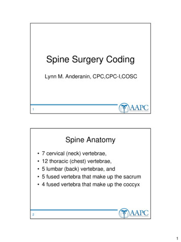
SurgeryPurpose of SurgeryThe purpose of a lumbar fusion is to take pressure off the nerves (decompression) and to stabilize the disk space.The fusion locks two or more vertebrae together, which prevents movement of the vertebrae. The goal ofsurgery is to relieve the symptoms of pain, muscle weakness, numbness and tingling, or in some cases, to haltprogression of these symptoms.Method of SurgerySurgery is performed in the prone position (lying on the stomach). A vertical incision will be made in thelower back.The soft tissue will be moved aside from the vertebrae. If there is too much pressure on a nerve, then a smallportion of bone is removed.Lumbar vertebrae will be fused together by placing a bone graft either between the transverse process("wings") or between the vertebrae. Bone from the pelvis (autograft) or from a bone bank (allograft) will beplaced next to the actual bone.Metal supports or instrumentation can be used if extra support is needed. Metal rods will be placed to holdthe spine straight. Metal screws are placed into the vertebrae to hold the metal rods. The underlying layerswill then be closed with absorbable sutures. The outer incision may be closed with metal staples. Staplesshould be removed seven to 14 days after surgery.Over the next several months, the graft will grow to join the two separate bones.2223
24
What You Need to Know Before SurgeryPreoperative EvaluationMake an appointment with your primary care physician for a preoperative evaluation within 30 days of surgery.The evaluation will include the following:- A physical examination- A health history- EKG (if over 40 years of age or with history of heart disease)- Chest X-ray (with history of lung disease)- Review of current medications- Bloodwork including type and crossmatchNutritionA balanced diet is an important part of surgical preparation to promote healing and prevent infection. Eat a healthy balanced diet prior to and after surgery. An iron supplement may be recommended before surgery if you donate blood.Discuss the need for other vitamin or mineral supplements with your surgeon.Post Anesthesia Care UnitAfter surgery, you will be cared for in the Post Anesthesia Care Unit (PACU) or Recovery Room for approximately onehour. Your head will be elevated and medications will be given to manage pain.Normal sensations after surgery include sore throat, raspy voice and difficulty swallowing. These minor irritations willsubside a few weeks following surgery. The use of lozenges and eating soft foods will minimize throat irritation.Medications to Avoid Before Surgery:Medications to discontinue or continue will be discussed with your primary care physician. Ten to 14 days beforesurgery, all medications that may cause "thinning" of the blood should be stopped. This includes prescription andnon-prescription drugs such as:Day of SurgeryYOU ARE TO HAVE NOTHING TO EAT OR DRINK AFTER MIDNIGHT THE NIGHT BEFORE SURGERY AND INTHE MORNING THE DAY OF SURGERY. If you have been instructed to take any medication, do so with only sips ofwater.Coumadin ng Your Hospital StayFor the first day or two after surgery, pain will be managed intravenously. The brace will be applied the day after surgery.Gingko BilobaVitamin E (tocopherol)All drugs containing aspirinAll non-steroidal anti-inflammatory drugs (NSAID's)Herbal stimulantsTrentalYou will be encouraged to spend time out of bed. Goals to be met for discharge will be to get out of bed, use the bathroomand walk independently. Fluids and soft foods will be offered gradually.Do not expect to be pain free. The goal of post-operative pain management is to make you comfortable enough to breatheadequately and to walk. While rest is an important part of recovery from surgery, activity will speed the healing process.Tylenol (acetaminophen) can be continued.SmokingIt is imperative that smoking and the consumption of nicotine is stopped prior to and for at least three months followingsurgery. If unable to stop “cold turkey,” speak with your primary care physician about medications that aid in smokingcessation. Any aid used must not contain nicotine. Smoking and specifically nicotine will significantly increase thefailure rate of bone fusion.Blood DonationBecause of the potential for blood loss during surgery, it will be necessary to donate one to two units of your own blood.If this is not possible, family members or friends may donate units for you. The American Red Cross will contact you toschedule appointments for donation. Three to four weeks must be allowed for blood donations.Nursing staff will help you with bathing and personal hygiene. Wear the back brace at all times unless otherwise instructedby your doctor. Typically discharge will be scheduled for three to five days after surgery, depending on your individualprogress.Prior to discharge home, the nurse will ensure that pain is controlled. It is often a good idea to request a pain tablet beforedischarge to ensure a more comfortable drive home. The following goals should be met beforedischarge to home: Walking independently Eating soft foods Control of pain, nausea and vomitingBrace FittingIf you are required to wear a brace following surgery, you will be fitted prior to surgery. Brace fitting will take approximately one to two hours of your time excluding travel time. Please allow at least three weeks to a month for yourbrace to be created and fitted. Generally speaking the brace will extend from just below your rib cage to the bend atyour hips. If your sacral spine is involved, an attached thigh cuff will be needed to reduce movement.The purpose of the brace is to immobilize your spine during the recovery period to give the fusion the best environment inwhich to heal. It is important that you wear the brace snugly as it is intended to limit your turning and bending movements.2526
At Home After SurgeryAfter surgery you will experience some pain around the incision and stiffness. The majority of patients experience areduction in back pain very soon after surgery. In some cases, discomfort may gradually decrease with time. Numbnessand tingling will be the slowest symptoms to improve and may not completely go away.ActivityRest at home for the first few days. Walk as much as you feel comfortable and work up to at least 30 minutes per day.Limit the use of stairs and walking on hills for the first one to two weeks. Do not lift anything greater than five pounds forthree months. Usually you may drive eight to 12 weeks after surgery. Clarify the time for return to driving with your surgeon.Showering or BathingClarify showering instructions with your surgeon.Frequently Asked QuestionsHow much pain should I experience after surgery?Naturally, you will have pain associated with your incision. You may or may not notice an immediateimprovement in your symptoms the first few days following surgery. However, in time, the pain shoulddecrease. If you have concerns, call your surgeon. A new onset, persistent or worsening pain of the lowerextremities should be reported to your surgeon immediately for evaluation.Why is my throat sore? How long will it last?Sore throat, hoarseness, and difficulty swallowing are common side effects that are experienced the first fewdays following surgery. Occasionally, hoarseness may be prolonged as a consequence of the type of anteriorsurgical technique used to access the cervical disk. Hoarseness should improve over time. Swallowing willimprove as inflammation decreases. Eat a diet consisting of soft foods for the first week or two following surgery to avoid difficulty.How long will I have to wear the brace after my lumbar or thoracic fusion?The normal length of time is four months after which time you will be weaned from the brace.Dressing ChangesThe incision should be kept clean and dry for at least three to five days after surgery or as directed by your surgeon.Change the dressing as needed or at least every other day during this time. Staples should be removed approximatelyseven to 14 days after surgery.Pain ManagementPain medicine will be prescribed for the first few weeks after surgery. Take pain medication as needed to keep pain at amanageable level. Gradually taper to non-prescription medication. Non-steroidal anti-inflammatory drugs (NSAIDs) mustbe avoided for the first few months after surgery.Emotional RecoverySurgery places a stress on the body’s reserves. Feelings of tiredness and discouragement are normal as pre-operativesymptoms slowly subside. Prescription pain medications can alter sleep patterns, elimination and emotional responses. Apositive attitude and patience are needed for successful recovery from any surgery. Speak with your surgeon or primarycare physician about any emotional difficulties that you experience.Follow-Up CareKeep all appointments with your surgeon after surgery. Continued follow up is critical for a complete and successfulrecovery.Report any of the following symptoms to your surgeon Drainage, bleeding, redness or swelling of the wound Opening of the incision Temperature greater than 100.4 F or chills New, persistent or worsening pain or numbness Difficulty with urination or bowel movementsHow long will I have to wear the collar after my cervical fusion?This will depend upon the surgical procedure performed, but generally a minimum of two weeks.When will I be allowed to drive after my cervical fusion?You may drive only after your surgeon has instructed you to remove your neck brace and you have regainedan adequate range of motion in your neck.When will I be allowed to drive after my lumbar or thoracic fusion?You may drive only after your surgeon released you to do so. It is important that you test your ability on avery quiet side street or in an empty parking lot prior to venturing into traffic. Pain, stiffness and the bracemay impair your ability to drive safely. Limit time spent sitting in the car to 45 minutes. If a trip is longer than45 minutes, take a break to walk and stretch your legs for a few minutes.When can I return to work after my cervical fusion?The date for return to work will depend on your specific job activities and usually ranges from three weeks tothree months following surgery. Speak with your surgeon regarding the specifics of your occupation and yourrate of healing.When can I return to work after my lumbar or thoracic fusion?The date for return to work will depend on your specific job activities and usually ranges from eight weeks toeight months following surgery. Speak with your surgeon regarding the specifics of your occupation and yourrate of healing.When can I resume normal activities? (sex, sports, exercising)Sex - As long as you are lying on your back, you may resume sexual activity as soon as you feel comfortable.Remember that you must leave your collar on unless otherwise instructed by your doctor.Sports - Again this will depend on the specific procedure performed but for most it is safe to resume fullactivities after six months.2728
Exercise – Walking is encouraged during the healing process as is any low impact aerobic or cardiovascularactivity. For more strenuous activities such as weight lifting, check with your surgeon.Glossary of TermsAnnulus – The outer rings of rigid fibrous tissue surrounding the nucleus in the disk.What activities should be avoided?Generally speaking lifting is to be avoided while the fusion is healing. Please speak with your surgeon forspecific weight limitations. Prolonged sitting or standing should also be avoided as this places the mostpounds of pressure on your spine. If you have access to the Internet, you will find useful information at thefollowing sites and their associated euniverse.comwww.srs.orgPlease understand that information on these Web sites is geared to the general population. To address issues specific toyour condition and symptoms, please speak with your spine surgeon or a member of the spine team.Anterior – A relative term indicating the front of the body.Bone Spur – An abnormal growth of bone, usually present in degenerative arthritis or degenerative diskdisease.Cervical Spine – The group of seven bones, vertebrae, that form the upper and most flexible part of spinalcolumn. It is located between the skull and the thoracic spine.Degenerative Arthritis – The inflammatory process that causes gradual impairment and loss of use of a joint.Degenerative Disk Disease – The loss of water from the disks that reduces elasticity and causes flattening ofthe disks.Disk– The complex of fibrous and gelatinous connective tissues that separate the vertebrae in the spine. Theyact as shock absorbers to limit trauma to the bony vertebrae.Herniated Disk – The abnormal protrusion of soft disk material that may impinge on nerve roots. Alsoreferred to as ruptured disk, protruding disk.Laminotomy – The removal of a small portion of the lamina, the bone that lies posterior to the vertebra.Lumbar – The portion of the spine lying below the thoracic spine and above the pelvis.Lumbar Diskectomy – The removal of a protruding or free fragment of intravertebral disk material.Nerve Root – The portion of a spinal nerve that lies closest to its origin from the spinal cord.Nucleus Pulposis or Nucleus – The relatively soft center of the disk that is protected by the rigid fibrousouter rings.Posterior – A relative term indicating that an object is to the rear of or behind the body.Spine – The flexible column of 24 vertebrae, disks, ligaments and muscle that lie between the head and pelvisand behind the rib cage. Also referred to as the spinal column.Vertebra (e) – The bone or bones that form the spine.2930
The Spine Center at Sinai Patient Guide Anterior Lumbar Interbody Fusion (ALIF) Anterior Cerivical Diskectomy and Fusion (ACDF) Lumbar Diskectomy Posterior Lumbar Instrumented Fusion www.sinaispine.com 2401 West Belvedere Avenue Baltimore, Maryland 21215-5271 www.lifebridgehealth.org.