Transcription
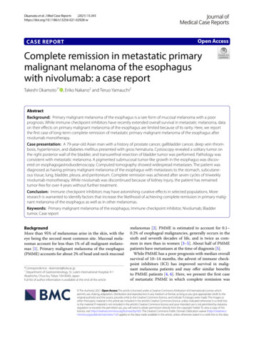
(2021) 15:345Okamoto et al. J Med Case pen AccessCASE REPORTComplete remission in metastatic primarymalignant melanoma of the esophaguswith nivolumab: a case reportTakeshi Okamoto1* , Eriko Nakano2 and Teruo Yamauchi2AbstractBackground: Primary malignant melanoma of the esophagus is a rare form of mucosal melanoma with a poorprognosis. While immune checkpoint inhibitors have recently extended overall survival in metastatic melanoma, dataon their effects on primary malignant melanoma of the esophagus are limited because of its rarity. Here, we reportthe first case of long-term complete remission of metastatic primary malignant melanoma of the esophagus afternivolumab monotherapy.Case presentation: A 79-year-old Asian man with a history of prostate cancer, gallbladder cancer, deep vein thrombosis, hypertension, and diabetes mellitus presented with gross hematuria. Cystoscopy revealed a solitary tumor onthe right posterior wall of the bladder, and transurethral resection of bladder tumor was performed. Pathology wasconsistent with metastatic melanoma. A pigmented submucosal tumor-like growth in the esophagus was discovered on esophagogastroduodenoscopy. Computed tomography showed widespread metastases. The patient wasdiagnosed as having primary malignant melanoma of the esophagus with metastases to the stomach, subcutaneous tissue, lung, bladder, pleura, and peritoneum. Complete remission was achieved after seven cycles of triweeklynivolumab monotherapy. While nivolumab was discontinued because of kidney injury, the patient has remainedtumor-free for over 4 years without further treatment.Conclusion: Immune checkpoint inhibitors may have astonishing curative effects in selected populations. Moreresearch is warranted to identify factors that increase the likelihood of achieving complete remission in primary malignant melanoma of the esophagus as well as in other melanomas.Keywords: Primary malignant melanoma of the esophagus, Immune checkpoint inhibitor, Nivolumab, Bladdertumor, Case reportBackgroundMore than 95% of melanomas arise in the skin, with theeye being the second most common site. Mucosal melanomas account for less than 1% of all malignant melanomas [1]. Primary malignant melanoma of the esophagus(PMME) accounts for about 2% of head and neck mucosal*Correspondence: okamotot@luke.ac.jp1Department of Gastroenterology, St. Luke’s International Hospital, 9‑1Akashicho, Chuo‑ku, Tokyo 104‑8560, JapanFull list of author information is available at the end of the articlemelanomas [2]. PMME is estimated to account for 0.1–0.2% of esophageal malignancies, generally occurs in thesixth and seventh decades of life, and is twice as common in men than in women [3–5]. About half of PMMEpatients have metastases at the time of diagnosis [5].While PMME has a poor prognosis with median overallsurvival of 10–14 months, the advent of immune checkpoint inhibitors (ICI) has improved survival in malignant melanoma patients and may offer similar benefitsto PMME patients [4, 6]. Here, we present the first caseof metastatic PMME in which complete remission was The Author(s) 2021. Open Access This article is licensed under a Creative Commons Attribution 4.0 International License, whichpermits use, sharing, adaptation, distribution and reproduction in any medium or format, as long as you give appropriate credit to theoriginal author(s) and the source, provide a link to the Creative Commons licence, and indicate if changes were made. The images orother third party material in this article are included in the article’s Creative Commons licence, unless indicated otherwise in a credit lineto the material. If material is not included in the article’s Creative Commons licence and your intended use is not permitted by statutoryregulation or exceeds the permitted use, you will need to obtain permission directly from the copyright holder. To view a copy of thislicence, visit http:// creat iveco mmons. org/ licen ses/ by/4. 0/. The Creative Commons Public Domain Dedication waiver (http:// creat iveco mmons. org/ publi cdoma in/ zero/1. 0/) applies to the data made available in this article, unless otherwise stated in a credit line to the data.
Okamoto et al. J Med Case Reports(2021) 15:345achieved after nivolumab monotherapy and sustained forover 4 years after discontinuing nivolumab.Case presentationA 79-year-old Asian man presented with gross hematuria that started 2 weeks prior. His morning urine wasbright red when he began to urinate, after which it grewprogressively lighter in color. His urine later in the daywas pink and sometimes yellow or colorless at night.He noted mild weight loss despite normal appetite andfood intake. He also complained of mild cough and slightdyspnea upon exertion.He had a history of open cholecystectomy for gallbladder cancer, radical prostatectomy with penile prosthesisimplantation for prostate cancer, inferior vena cava filterplacement for deep vein thrombosis, hypertension, anddiabetes mellitus. Medications included edoxaban, anangiotensin II receptor blocker, a dipeptidyl peptidase-4inhibitor, and a proton pump inhibitor. He was an occasional drinker and had quit smoking 40 years ago. No significant family history was noted. He had opted to receiveannual medical checkups including serum and urine testing with tumor markers, chest roentgenogram, abdominal ultrasound, and esophagogastroduodenoscopy(EGD) for over 30 consecutive years. No abnormalitiesPage 2 of 8were noted at the last checkup, which was conducted11 months prior to his presentation.Physical examination was significant only for abdominal surgical scars and decreased breath sounds on theleft side. Laboratory testing revealed an increase inwhite blood cells (10,200 mm3) and C-reactive protein(6.76 mg/dL). Neuron-specific enolase was elevated(30.7 ng/mL), while other tumor markers such as carcinoembryonic antigen, squamous cell carcinoma antigen,and prostate-specific antigen were within their normalranges. Urinalysis showed over 50 red blood cells perhigh-powered field, and urine cytology showed severeatypia, raising the suspicion of urothelial carcinoma.Contrast computed tomography (CT) showed a wellenhanced bladder tumor with no signs of deep invasion.Severely enlarged abdominal lymph nodes and lung,pleural, and peritoneal metastases were also observed.A small subcutaneous nodule in the left thorax wasalso suspected to be a metastatic lesion. While metastatic urothelial carcinoma was a possible explanation, acoexisting cancer of unknown origin was also suspected(Fig. 1).Cystoscopy revealed a 15-mm white mass on the rightposterior wall of the bladder (Fig. 2). Transurethral resection of bladder tumor (TUR-BT) was performed withno complications. While macroscopic findings of theFig. 1 Contrast computed tomography revealed tumors (white arrowheads) in the left lung (a), subcutaneous tissue (b), abdominal lymph nodes(c), and bladder (d) consistent with metastatic disease
Okamoto et al. J Med Case Reports(2021) 15:345Page 3 of 8after seven cycles. Pathology from renal biopsy was suggestive of both mild interstitial tubulonephritis and IgAnephropathy (Fig. 5). Partial recovery was achieved afterdiscontinuing nivolumab and introducing steroids at0.5 mg/kg, which were tapered over a 3 month period.While nivolumab therapy was not reintroduced, repeatedCT scans taken every 3–6 months also showed no signsof recurrence. Follow-up endoscopy 1 year later revealedthat all esophageal and gastric tumors had disappeared,leaving only slight melanosis with no remaining pathological evidence of malignancy (Fig. 6a). The melanosis also disappeared 6 months thereafter (Fig. 6b). Thepatient has remained tumor-free for over 5 years withoutfurther treatment.Fig. 2 A 15-mm white mass was observed in the right posterior wallof the bladder and removed by transurethral resection of bladdertumorbladder tumor appeared consistent with those of urothelial tumors, further testing was conducted to rule outconcomitant malignancies. EGD revealed a 20-mm flat,pigmented lesion with a nonpigmented nodule in themid-esophagus, a 10-mm pigmented submucosal tumorlike growth in the distal esophagus, and polypoid lesionswith central depression in the distal esophagus and stomach (Fig. 3). Endoscopic ultrasound-guided fine needleaspiration of an abdominal lymph node and thoracentesiswere also performed. Colonoscopy was unremarkable.Esophageal biopsy showed melanin granules withintumor cells and melanoblasts in the interstitium, withimmunohistochemistry positive for melan A (Fig. 4a–c).In situ lesions were also observed, suggesting that theesophagus was the primary site (Fig. 4d). Stomach biopsyrevealed similar proliferation of tumor cells. Pathology of the bladder tumor revealed no signs of muscularinvasion. Immunohistochemistry for both the bladdertumor and lymph node was positive for melan A, vimentin, S-100, HMB45, SOX10, and p53 and negative forAE1/AE3 (Fig. 4e–h). The MIB-1 proliferative index was80%. A negative test for the EWSR1 chimeric gene ruledout clear cell sarcoma. No BRAF mutation was found.Comprehensive dermal and ocular examinations werenegative for melanoma. The patient was diagnosed withPMME with metastases to the stomach, subcutaneoustissue, lung, bladder, pleura, and peritoneum.Nivolumab was started at 2 mg/kg every 3 weeks,which was the standard dosage at the time. CT followup after six cycles revealed complete remission of all visible tumors. However, acute kidney injury was observedDiscussion and conclusionsSince the first report by Baur in 1906, over 300 PMMEcases have been reported to date [6]. Risk factors forPMME have not been elucidated; those associated withcutaneous melanoma such as sun exposure have no connection with PMME. Over 90% are found in the middle and distal thirds of the esophagus. PMME lesionsare usually polypoid and pigmented but can take a variety of forms and are amelanotic in 10–25% of cases [7].Diagnosis is therefore based on histology, as defined byAllen and Spitz in 1953: a) typical melanoma histologyand melanin granules within tumor cells, b) originationfrom epithelium with junctional activity, and c) junctional activity with melanotic cells in adjacent epithelium[8, 9]. Diagnosis can present challenges even with the useof immunohistochemistry markers such as S-100 (themost sensitive), melan-A (the most specific), and HMB45 [10]. Metastatic melanoma to the esophagus is rarerthan PMME.Surgery has traditionally been the only option to prolong survival in melanoma, as efficacy of both chemotherapy and radiotherapy are limited. The introductionof programmed death 1 (PD-1) inhibitors nivolumab andpembrolizumab as well as the cytotoxic T-lymphocyteassociated antigen 4 inhibitor ipilimumab in combinationwith nivolumab significantly extended overall survivalin metastatic melanoma [11–13]. ICIs are positioned asfirst-line therapy for melanoma without BRAF mutationsand 3 year overall survival rates have reached 51%, 40%,and 56%, respectively [14].While nivolumab has been shown to be less effectivein mucosal melanomas than in cutaneous melanomas,the standard treatment and the efficacy of ICI in PMMEremains unclear due to its rarity [15]. The largest studyevaluating ICI treatment in PMME included a cohort of12 patients treated with PD-1 inhibitors in a retrospective analysis of 76 unresectable or metastatic PMMEs[16]. The study revealed promising results, with 75%
Okamoto et al. J Med Case Reports(2021) 15:345Page 4 of 8Fig. 3 Esophagogastroduodenoscopy revealed a 20-mm flat, pigmented lesion with a nonpigmented nodule in the mid-esophagus (a), a 10-mmpigmented submucosal tumor-like growth in the distal esophagus (b), and polypoid lesions with central depression in the distal esophagus (c) andstomach (d). All were confirmed to be malignant melanoma lesions on pathologyFig. 4 Low-power (a) and high-power (b) magnification of esophageal biopsies showed melanin granules within tumor cells and melanoblastsin the interstitium. Immunohistochemistry was positive for melan A (c). In situ lesions were also observed, suggesting that the esophagus wasthe primary site (d). Immunohistochemistry for the resected bladder specimen was positive for melan A (e), SOX10 (f), and p53 (g). The MIB-1proliferation index was 80% (h)
Okamoto et al. J Med Case Reports(2021) 15:345Page 5 of 8Fig. 5 Periodic acid–Schiff staining (a) and IgA immunofluorescence microscopy (b) of the right kidney, suggestive of mild tubulointerstitialnephritis and IgA nephropathyFig. 6 All esophageal and gastric tumors had disappeared 1 year after nivolumab treatment, leaving only slight melanosis with no remainingpathological evidence of malignancy (a). The melanosis also disappeared 6 months thereafter (b)achieving partial response and the other 25% maintainingstable disease for at least 4 months. The mean progression-free survival was 15.6 months and severe toxicity was only seen in 1 patient. While case reports of ICItreatment for PMME are scarce, one case of unresectablePMME maintained good partial response after 30 coursesof nivolumab monotherapy, while another had stable disease for 11 cycles [17, 18]. Another PMME case refractory to chemoradiotherapy showed good partial responseafter 3 cycles of nivolumab but discontinued treatmentafter 7 cycles due to irAEs, ultimately resulting in tumordeath [19]. Our case is the first report of complete remission in metastatic PMME with nivolumab monotherapy.Despite a large representation of the elderly in thecancer population, they are almost always underrepresented in clinical trials. ICI treatment was expectedto have less efficacy on elderly patients due to immunosenescence, or reduced immune function due tocontinuous remodeling of lymphoid organs whichincrease the incidence of infections, neoplasia, andautoimmune disease [20]. Similarly, the elderly wereexpected to experience more immune-related adverseevents (irAEs). However, meta-analyses of ICI treatment showed no significant difference in efficacybetween the younger and older groups with cut-offsof 65, 70, and 75 years of age [21, 22]. Frequency andseverity of irAEs were also similar between the youngerand elderly groups [23]. Efficacy and irAE profile independent of age were also seen in various ICI studieslimited to melanoma patients [24–26]. In fact, a largestudy with 538 metastatic melanoma subjects showedthat patients above the age of 60 experienced higherefficacy with anti-PD1 therapy than their youngercounterparts, with the chance of disease progressiondecreasing by 13% for every decade of life [27]. Theauthors reproduced the result in mice and proposed
Okamoto et al. J Med Case Reports(2021) 15:345that a change in the regulatory T cell composition withage may help explain this higher efficacy. While immunosenescence may lead to decreased immune function,the elderly also have increased chronic inflammationwhich suggests an augmented immune function. Agemay have played a role in the complete remission of our79 year-old patient.It is also possible that spontaneous regression, wherehistologically-proven cancer regresses without sufficienttherapy to explain it, also played a role. Melanomas,renal cell carcinomas, and neuroblastomas are amongthe most reported malignancies which regress spontaneously [28]. In malignant melanomas, complete regressionis observed in 3–15% of primary lesions and 0.08–0.71%of metastatic lesions. Most reports of regression involvecutaneous lesions, and there are no reports of spontaneous regression of PMME. While a combination of immunologic, endocrine, inflammatory and metastatic tumornutritional factors have been implicated, the underlyingmechanisms remain to be elucidated.Despite its benefits, ICI treatment also has the potential to cause irAEs. The two types of irAEs causing kidney injury - tubulointerstitial nephritis and glomerulardisease – have been reported to coexist after nivolumabtherapy for other cancers and after ICI treatment formelanoma [29]. Renal pathology in our case also showedsigns of both mild tubulointerstitial nephritis and immunoglobulin A (IgA) nephropathy. Full or partial recoveryis generally achieved after discontinuation of ICIs andconcomitant medications, as in our case [30].Our patient had metastases to the stomach and bladder, both of which are rare in melanoma patients. Mostmelanoma lesions arising in the gastrointestinal (GI)tract are metastases. As melanoblasts are only presentin the oral cavity, esophagus, and anorectum, the histogenesis of primary melanomas arising in other GIsites remains to be elucidated [31]. Melanoma metastasizing to GI organs is underrecognized, despite autopsies showing melanoma metastases to the esophagusin 4%, stomach in 20%, duodenum in 12%, small bowelin 58%, colon in 22%, rectum in 5%, and liver in 68%of cases [32]. A study of CT scans for metastatic melanoma showed metastases to the esophagus in 7%, stomach in 24%, duodenum in 19%, small bowel in 48%,colon in 4%, liver in 15%, gallbladder in 3%, biliary treeand pancreas in 6%, and mesentery and omentum in18% of cases [33]. Sensitivity of the CT for small bowelmetastases is estimated at 60–70%, and endoscopy maybe warranted in suspicious cases. On the other hand,symptoms such as GI bleeding, abdominal pain, andbowel obstruction may only manifest in a small minority of patients with advanced GI metastases [34]. Thus,Page 6 of 8endoscopic evaluations to rule out metastases shouldbe considered even in the absence of GI symptoms.Stomach and small bowel metastases commonly present as submucosal nodules with central depressionpresenting a characteristic “bull’s eye” appearance [35].One case of PMME with gastric metastasis has beenreported [36]. A few case reports suggest limited efficacy of immunotherapy for cutaneous melanoma withgastric metastases [37, 38].Urothelial tumors account for 95% of bladder tumors,while metastases only account for 2.3% [39, 40]. Directinvasion from colorectal, prostate, and cervical cancersare the most common metastatic tumors, with rare distant metastases seen mainly from the stomach, skin,lung, and breast. Metastases almost always present assolitary tumors, and the majority are adenocarcinomas [40]. Both primary and metastatic melanoma ofthe bladder are extremely rare, accounting for less than0.1% of bladder tumors and with only about 30 reportsof each [40–42]. On the other hand, an autopsy studyfound bladder metastases in 18% of melanoma patients,suggesting that the latter is more common [32]. Bothare generally asymptomatic but may present with painless hematuria [43]. Most metastases come from cutaneous melanoma; there is only one report of PMMEmetastasizing to the bladder in the English literature[42, 44]. While generally found in the context of widespread metastasis, surgery was performed in 15 cases(including 8 TUR-BT cases) in a review of 24 melanoma cases with metastases to the bladder [42].One interesting aspect of the bladder metastasis inour case is that it appeared amelanotic despite the primary tumor being melanotic. A Japanese case reportalso demonstrated an amelanotic bladder metastasis ofa melanotic melanoma [45]. In that case as well as ours,histology revealed melanin pigments in the metastaticbladder lesion, and immunohistochemistry was positive for both S-100 and HMB-45. The pathophysiologybehind this phenomenon has not been described in theliterature and remains to be elucidated.In conclusion, we report a case of long-term completeremission of metastatic PMME after nivolumab monotherapy. The patient remains tumor-free 55 monthsafter diagnosis and 50 months after the last nivolumabinjection. This case sheds light on the prognosis of thisrare and dismal disease. More research is warranted toidentify factors that increase the likelihood of achieving complete remission with ICIs in PMME as well in asother melanomas.AbbreviationsCT: Computed tomography; EGD: Esophagogastroduodenoscopy; GI: Gastrointestinal; ICI: Immune checkpoint inhibitor; IrAE: Immune-related adverse
Okamoto et al. J Med Case Reports(2021) 15:345Page 7 of 8effects; PD-1: Programmed death 1; PMME: Primary malignant melanoma ofthe esophagus; TUR-BT: Transurethral resection of bladder tumor.AcknowledgementsNoneAuthors’ contributionsTO wrote the manuscript. EN and TY critically reviewed and edited the manuscript. All authors read and approved the final manuscript.FundingThe authors declare that they have received no funding for the publication ofthis article.Availability of data and materialsData sharing is not applicable to this article as no datasets were generated oranalyzed during the current study.13.14.15.16.17.18.Declarations19.Ethics approval and consent to participateNot applicable20.Consent for publicationWritten informed consent was obtained from the patient for publication ofthis case report and any accompanying images. A copy of the written consentis available for review by the Editor-in-Chief of this journal.21.22.Competing interestsThe authors declare that they have no competing interests.Author details1Department of Gastroenterology, St. Luke’s International Hospital, 9‑1 Akashicho, Chuo‑ku, Tokyo 104‑8560, Japan. 2 Department of Oncology, St. Luke’sInternational Hospital, 9‑1 Akashicho, Chuo‑ku, Tokyo 104‑8560, Japan.Received: 10 June 2020 Accepted: 24 May 202123.24.25.26.References1. Trap TK, Fu Y, Calcaterra TC. Melanoma of the nasal and paranasal sinusmucosa. Arch Otolaryngol. 1987;113:1086–9.2. Nandapalan V, Roland NJ, Helliwell TR, Williams EM, Hamilton JW, JonesAS. Mucosal melanoma of the head and neck. Clin Otolaryngol Allied Sci.1998;23:107–16.3. Suzuki H, Nagayo T. Primary tumors of the esophagus other than squamous cell carcinoma. Histologic classification and statistics in the surgicaland autopsied materials in Japan. Int Adv Surg Oncol. 1980;3:73–109.4. Sabanathan S, Eng J, Pradhan GN. Primary malignant melanoma of theesophagus. Am J Gastroenterol. 1989;84:1475–81.5. Hussein MR. Extracutaneous malignant melanomas. Cancer Invest.2008;26:516–34.6. Sabanathan S, Eng J. Primary malignant melanoma of the esophagus.Scand J Thorac Cardiovasc Surg. 1990;24:83–5.7. Bisceglia M, Perri F, Tucci A, et al. Primary malignant melanoma of theesophagus: a clinicopathologic study of a case with comprehensiveliterature review. Adv Anat Pathol. 2011;18:235–52.8. Allen AC, Spitz S. Malignant melanoma; a clinicopathological analysis ofcriteria for diagnosis and prognosis. Cancer. 1953;6:1–45.9. Stringa O, Valdez R, Beguerie JR, et al. Primary amelanotic melanoma ofthe esophagus. Int J Dermatol. 2006;45:1207–10.10. Kobayashi J, Fujimoto D, Murakami M, Hirono Y, Goi T. A report ofamelanotic malignant melanoma of the esophagus diagnosed appropriately with novel markers: a case report. Oncol Lett. 2018;15:9087–92.11. Robert C, Long GV, Brady B, et al. Nivolumab in previously untreatedmelanoma without BRAF mutation. N Engl J Med. 2015;372:320–30.12. Schachter J, Ribas A, Long GV, et al. Pembrolizumab versus ipilimumabfor advanced melanoma: final overall survival results of a ed, open-label phase 3 study (KEYNOTE-006). Lancet.2017;390:1853–62.Larkin J, Chiarion-Sileni V, Gonzalez R. Combined nivolumab andipilimumab or monotherapy in untreated melanoma. N Engl J Med.2015;373:23–34.Corrie PG, Chao D, Board R, et al. Metastatic melanoma patient outcomessince introduction of immune checkpoint inhibitors in England between2014 and 2018. J Clin Oncol. 2020;38:55.Kaunitz GJ, Cottrell TR, Lilo M, et al. Melanoma subtypes demonstratedistinct PD-L1 expression profiles. Lab Invest. 2017;97:1063–71.Wang X, Kong Y, Chi Z, et al. Primary malignant melanoma of theesophagus: a retrospective analysis of clinical features, management, andsurvival of 76 patients. Thorac Cancer. 2019;10:950–6.Ito S, Tachimori Y, Terado Y, Sakon R, Narita K, Goto M. Primary malignantmelanoma of the esophagus successfully treated with nivolumab: a casereport. J Med Case Rep. 2021;15:237.Rochefort P, Roussel J, la Fouchardière A, et al. Primary malignant melanoma of the esophagus, treated with immunotherapy: a case report.Immunotherapy. 2018;10:831–5.Inadomi K, Kumagai H, Arita S, et al. Bi-cytopenia possibly induced byanti-PD-1 antibody for primary malignant melanoma of the esophagus: acase report. Medicine (Baltimore). 2016;95:e4283.Pawelec G. Immunosenescence: impact in the young as well as the old?Mech Ageing Dev. 1999;108:1–7.Kasherman L, Siu DHW, Lee KWC, et al. Efficacy of immune checkpointinhibitors in older adults with advanced stage cancers: a meta-analysis. JGeriatr Oncol. 2020;11:508–14.Nishijima TF, Muss HB, Shachar SS, Moschos SJ. Comparison of efficacyof immune checkpoint inhibitors (ICIs) between younger and olderpatients: a systematic review and meta-analysis. Cancer Treat Rev.2016;45:30–7.Corbaux P, Maillet D, Boespflug A, et al. Older and younger patientstreated with immune checkpoint inhibitors have similar outcomes inreal-life setting. Eur J Cancer. 2019;121:192–201.Joshi K, Atwal D, Ravilla R, et al. Immunotherapy outcomes in advancedmelanoma in relation to age. Perm J. 2020. https:// doi. org/ 10. 7812/ TPP/ 19. 093.Betof AS, Nipp RD, Giobbie-Hurder A, et al. Impact of age on outcomes with immunotherapy for patients with melanoma. Oncologist.2017;22:963–71.Archibald WJ, Victor AI, Strawderman MS, Maggiore RJ. Immunecheckpoint inhibitors in older adults with melanoma or cutaneousmalignancies: the Wilmot Cancer Institute experience. J Geriatr Oncol.2020;11:496–502.Kugel CH 3rd, Douglass SM, Webster MR, et al. Age correlates withresponse to anti-PD1, reflecting age-related differences in intratumoral effector and regulatory T-cell populations. Clin Cancer Res.2018;24:5347–56.Kalialis LV, Drzewiecki KT, Klyver H. Spontaneous regression of metastasesfrom melanoma: review of the literature. Melanoma Res. 2019;19:275–82.Mamlouk O, Selamet U, Machado S, et al. Nephrotoxicity of immunecheckpoint inhibitors beyond tubulointerstitial nephritis: single-centerexperience. J Immunother Cancer. 2019;7:2.Koda R, Watanabe H, Tsuchida M, et al. Immune checkpoint inhibitor(nivolumab)-associated kidney injury and the importance of recognizing concomitant medications known to cause acute tubulointerstitialnephritis: a case report. BMC Nephrol. 2018;19:48.Backman H. Metastases of malignant melanoma in the gastrointestinaltract. Geriatrics. 1969;24:112–20.Dasgupta T, Brasfield R. Metastatic melanoma. a clinicopathological study.Cancer. 1964;17:1323–39.Kawashima A, Fishman EK, Kuhlman JE, Schuchter LM. CT of malignantmelanoma: patterns of small bowel and mesenteric involvement. J Comput Assist Tomogr. 1991;15:570–1.Patel K, Ward ST, Packer T, et al. Malignant melanoma of the gastro-intestinal tract: a case series. Int J Surg. 2014;12:523–7.Pomerantz H, Margolin HN. Metastases to the gastrointestinal tractfrom malignant melanoma. Am J Roentgenol Radium Ther Nucl Med.1962;88:712–7.Wu PR, Yen HH, Chen CJ. Gastrointestinal: primary esophageal melanomawith gastric metastasis. J Gastroenterol Hepatol. 2011;26:1338.
Okamoto et al. J Med Case Reports(2021) 15:34537. Farshad S, Keeney S, Halalau A, Ghaith G. A case of gastric metastaticmelanoma 15 years after the initial diagnosis of cutaneous melanoma.Case Rep Gastrointest Med. 2018. https:// doi. org/ 10. 1155/ 2018/ 76849 64.38. Genova P, Sorce M, Cabibi D, et al. Gastric and rectal metastases frommalignant melanoma presenting with hypochromic anemia and treatedwith immunotherapy. Case Rep Oncol Med. 2017. https:// doi. org/ 10. 1155/ 2017/ 20790 68.39. Dahm P, Gschwend JE. Malignant non-urothelial neoplasms of the urinarybladder: a review. Eur Urol. 2003;44:672–81.40. Bates AW, Baithun SI. Secondary neoplasms of the bladder are histological mimics of nontransitional cell primary tumours: clinicopathologicaland histological features of 282 cases. Histopathology. 2000;36:32–40.41. Bumbu GA, Berechet MC, Pop OL, et al. Primary malignant melanomaof the bladder—case report and literature overview. Rom J MorpholEmbryol. 2019;60:287–92.42. Meunier R, Pareek G, Amin A. Metastasis of malignant melanoma to urinary bladder: a case report and review of the literature. Case Rep Pathol.2015. https:// doi. org/ 10. 1155/ 2015/ 173870.Page 8 of 843. Efesoy O, Cayan S. Bladder metastasis of malignant melanoma: a casereport and review of literature. Med Oncol. 2011. https:// doi. org/ 10. 1007/ s12032- 010- 9730-x.44. Charfi S, Ellouze S, Mnif H, et al. Plasmacytoid melanoma of the urinarybladder and lymph nodes with immunohistochemical expression ofplasma cell markers revealing primary esophageal melanoma. Case RepPathol. 2012. https:// doi. org/ 10. 1155/ 2012/ 916256.45. Maeda T, Uchida Y, Kouda T, et al. Metastatic malignant melanoma of theurinary bladder presenting with hematuria: a case report. Acta Urol Jap.2008;54:787–90 (in Japanese).Publisher’s NoteSpringer Nature remains neutral with regard to jurisdictional claims in published maps and institutional affiliations.Ready to submit your research ? Choose BMC and benefit from: fast, convenient online submission thorough peer review by experienced researchers in your field rapid publication on acceptance support for research data, including large and complex data types gold Open Access which fosters wider collaboration and increased citations maximum visibility for your research: over 100M website views per yearAt BMC, research is always in progress.Learn more biomedcentral.com/submissions
Follow-up endoscopy 1 year later revealed that all esophageal and gastric tumors had disappeared, leaving only slight melanosis with no remaining patho- . melan-A (the most specic), and HMB-45 [10]. Metastatic melanoma to the esophagus is rarer than PMME. Surgery has traditionally been the only option to pro-long survival in melanoma, as .