Transcription
Clinical Outcomes of Osteochondral AllograftTransplantation for Secondary Treatment ofOsteochondritis Dissecans of the Knee in SkeletallyMature PatientsEric J. Cotter, B.S., Rachel M. Frank, M.D., Kevin C. Wang, B.S., Trifon Totlis, M.D., Ph.D.,Sarah Poland, B.A., Maximillian A. Meyer, M.D., and Brian J. Cole, M.D., M.B.A.Purpose: To report clinical outcomes of osteochondral allograft transplantation (OCA) for skeletally mature patients withosteochondritis dissecans (OCD) lesions of the knee in whom prior surgical intervention has failed, and to describe returnto-sport (RTS) rates and any predictive demographic or preoperative variables associated with a higher likelihood to RTS.Methods: Patients who underwent OCA by a single surgeon (B.J.C.) between October 1, 2002, and November 30, 2014,for a diagnosis of OCD with a minimum of 2 years’ follow-up were included. Demographic, surgical history, operative, andpostoperative data points including RTS were analyzed for all patients collectively, with subanalysis to identify anydemographic and operative variables associated with the ability to RTS. Failure was defined as revision OCA, grossappearance of graft failure on second-look arthroscopy, or conversion to arthroplasty. Results: We identified 41consecutive patients (43 knees), with 37 patients (39 knees, comprising 26 male and 13 female knees) available for clinicalfollow-up at an average of 7.29 3.30 years. There was significant improvement (P .05) in all patient-reported outcomescores, with the exception of the Short Form 12 mental subscale (P ¼ .910). Most patients (81.6%) reported being eithermostly satisfied or completely satisfied at final follow-up. Of patients self-identifying as athletes preoperatively (n ¼ 22), 18(81.8%) achieved RTS at an average of 14.0 8.7 months. Athletes with failure to RTS had a significantly greater body massindex (RTS, 24.68 3.67; no RTS, 27.82 0.69; P ¼ .005). Subsequent surgery was performed in 14 patients (35.9%), andprimary OCA failed in 2 patients at an average of 6.2 3.8 years. Conclusions: OCA is a successful secondary surgicaltreatment for OCD of the knee in skeletally mature patients and leads to clinically meaningful improvements in patientreported outcome scores and high patient satisfaction and RTS rates in low-level athletes at an average of 7.29 years’follow-up. Athletes with failure to RTS were found to have a significantly higher body mass index. Although reoperation maybe common after OCA (35.9%), the failure rate is low (5.1%) in this series. Level of Evidence: Level IV, case series.From the Department of Orthopaedic Surgery, Rush University MedicalCenter (E.J.C., R.M.F., K.C.W., S.P., M.A.M., B.J.C.), Chicago, Illinois,U.S.A.; and Department of Anatomy and Surgical Anatomy, Aristotle University of Thessaloniki (T.T.), Thessaloniki, Greece.The authors report the following potential conflict of interest or source offunding: B.J.C. receives support from Arthrex, Regentis. Consultation over past36 months. Arthrex, Saunders/Mosby-Elsevier, Elsevier Publishing. IP royalties. Regentis. Stock ownership. Aesculap/B. Braun, Arthrex, Athletico,National Institutes of Health (NIAMS, NICHD). Previous research support.Smith & Nephew, Tornier, Ossur. Other financial or material support. FullICMJE author disclosure forms are available for this article online, assupplementary material.Received July 3, 2017; accepted October 20, 2017.Address correspondence to Brian J. Cole, M.D., M.B.A., Department ofOrthopaedic Surgery, Rush University Medical Center, 1611 W Harrison St,Ste 300, Chicago, IL 60612, U.S.A. E-mail: bcole@rushortho.comÓ 2017 by the Arthroscopy Association of North America0749-8063/17813/ dult osteochondritis dissecans (OCD) lesions arefocal lesions of subchondral bone thought torepresent juvenile lesions that went unrecognized;however, authors have noted de novo cases.1,2 OCDlesions are most commonly seen in the knee and occurmost often in the medial femoral condyle (70%),followed by the lateral femoral condyle (15%-20%),patella (5%-10%), and trochlea ( 1%).3-5 Furthermore, these lesions can be debilitating, especially inactive individuals who regularly place high stress loadsacross the knee joint, and may lead to an inability toperform activities of daily living or to return to sport(RTS).6 Age and skeletal maturity are important variables influencing clinical decision making becauseolder, skeletally mature individuals are less likely tosucceed with nonoperative treatment and more likelyto progress to surgery owing to a higher likelihood ofArthroscopy: The Journal of Arthroscopic and Related Surgery, Vol-,No-(Month), 2017: pp 1-81
2E. J. COTTER ET AL.unstable OCD fragments in the setting of an unfavorable healing environment.3,5,7 Adult forms of OCDlesions, particularly advanced lesions in stage 3 (unstable but not dislocated fragment) or stage 4 (presenceof loose body) according to the classification systemproposed by Clanton and DeLee,8 are commonlytreated surgically because of the inferior healingpotential compared with juvenile OCD.2,9,10Surgical options for OCD have been classified aspalliative, reparative, and restorative.2,9,10 Palliativetechniques such as loose body removal or debridementprovide pain relief and improvement of mechanicalsymptoms but do not address underlying articularcartilage disease, and degenerative changes are oftennoted later on, especially in adult patients.11 For patients in whom prior palliative or reparative interventions failed, restorative options includingosteochondral allograft transplantation (OCA) haveshown encouraging clinical outcomes.12,13 OCA affordsseveral biological advantages over marrow stimulationtechniques including restoration of the articular surfacewith hyaline cartilage and has been shown to effectively treat lesions larger than 2 cm2 in a single-stageprocedure.14,15 Until recently, there has been apaucity of literature regarding athletes’ ability to RTSafter OCA.6,16-19 Although patients with OCD lesionstreated with OCA have been included in RTS investigations, no study has reported RTS and return torecreational activities exclusively for this etiology usingonly a dowel technique for treatment of femoralcondyle lesions.The purposes of this study were to report midtermclinical outcomes of OCA as a secondary procedure forskeletally mature patients with OCD lesions and todescribe RTS rates and any predictive demographic orpreoperative variables associated with a higher likelihood to RTS. We hypothesized that patients wouldshow significant clinical improvement after OCA, aswell as high rates of RTS, with no significant associations between demographic or operative variables andthe ability to RTS.MethodsAfter institutional review board approval (No.15050301), prospectively collected data from our electronic health record database were searched for allskeletally mature patients undergoing OCA for OCD bya single surgeon (B.J.C.) from October 1, 2002, throughNovember 30, 2014, using Current Procedural Terminology codes 27415 (osteochondral allograft, knee,open) and 29867 (arthroscopy, knee, surgical; osteochondral allograft) and International Classification ofDiseases, Ninth Revision code 732.7 (OCD) based onphysical examination, radiographic, and/or magneticresonance imaging findings. Indications for OCAincluded focal, symptomatic articular cartilage defectsand failure of prior ipsilateral knee surgery. Weexcluded patients who underwent OCA without anOCD lesion, had an inaccurate diagnosis (i.e., not OCDdespite being coded as such), underwent bone graftingat the time of allograft transplantation, receivedDeNovo NT (Zimmer, Warsaw, IN), or underwentallograft transplantation in a joint other than the knee.Demographic information, mechanism of injuryincluding traumatic versus insidious, medical and/orsurgical history, intraoperative findings, and postoperative data were collected, as described in Table 1. Asports medicine fellow (R.M.F.) reviewed all preoperative radiographs for patients younger than 21 years todetermine skeletal maturity. Preoperative and finalfollow-up patient-reported outcome (PRO) metricswere analyzed, including the following: InternationalKnee Documentation Committee (IKDC) score, subscores of the Knee Injury and Osteoarthritis OutcomeScore (KOOS), Western Ontario and McMaster Universities Osteoarthritis Index (WOMAC) subscores andoverall score, and Short Form 12 (SF-12) mental andphysical subscale scores. Patients who preoperativelyself-identified as athletes, either recreational orcompetitive (high school or college), were askedwhether they returned to sporting activities, at whatlevel they returned to sporting activities, and how longit took them to RTS; if they did not RTS, they wereTable 1. Data Collected From All Patients Undergoing OCA for iveDataAge at date of surgery, sex, BMI, and insurance status (including Workers’ Compensation status)Type and level of athlete before symptom onset, symptom duration, and number and type of prior ipsilateral kneesurgical proceduresLaterality; compartment involved; size of defect; depth of defect; and concomitant procedures including MAT, ligamentrepair or reconstruction, and/or osteotomyComplications, reoperations, return to sport, satisfaction, whether patient would undergo procedure again, andvalidated PRO scores at minimum of 2 yr after surgeryBMI, body mass index; MAT, meniscal allograft transplantation; OCA, osteochondral allograft transplantation; OCD, osteochondritis dissecans;PRO, patient-reported outcome.
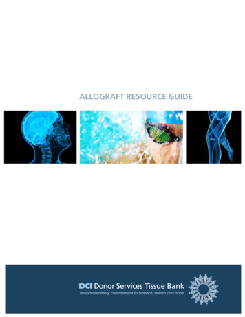
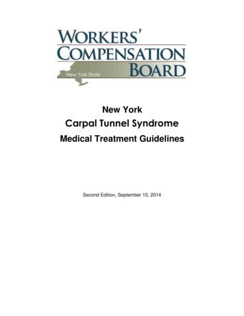
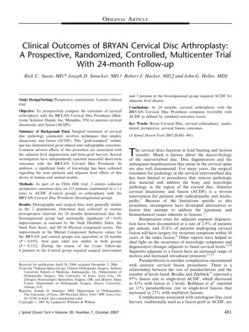
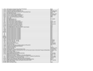
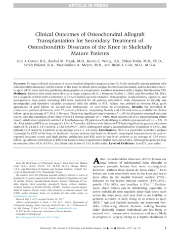
OSTEOCHONDRITIS DISSECANS TREATMENTFig 1. Intraoperative photograph of the right knee of a22-year-old male patient showing an osteochondritis dissecans lesion of the lateral femoral condyle.asked whether their knee was the primary reason.There were only 2 college athletes and no professionalathletes in this study. In addition, all patients wereasked how satisfied they were with their surgicaloutcome using a 4-point Likert scale (1, dissatisfied; 2,somewhat satisfied; 3, mostly satisfied; and 4,completely satisfied) and whether they would choose toundergo the procedure again. Demographic, medicaland/or surgical history, intraoperative, and postoperative variables were analyzed first for all patients inthis study; a subanalysis was then performed betweenthose patients who did not RTS and those who did.Age-related differences in clinical outcomes wereevaluated using regression analysis with age as acontinuous variable. Failure was defined as revisionOCA, gross appearance of graft failure on second-lookarthroscopy, or conversion to arthroplasty.Surgical TechniqueThe surgical technique for OCA of the senior author(B.J.C.) has been previously described.20,21 The procedure is performed with the patient in the supineposition. A diagnostic arthroscopy is performed toconfirm that the focal chondral lesion is amenable toOCA and to evaluate for concomitant pathology. Ifnecessary, concomitant procedures are performedbefore OCA. Next, a minieparapatellar arthrotomy isperformed to visualize and prepare the medial or lateralfemoral condyle defect site (Figs 1 and 2). A cannulated, cylindrical sizing guide (Arthrex, Naples, FL) isplaced flush over the defect to determine appropriatesizing of the graft. A 2.4-mm guide pin is then driventhrough the cannulated sizing guide into the base of thedefect. The guide is removed and used to size theallograft at the appropriate topographic location. A3cannulated cutting reamer of the same size is used toream to a depth of approximately 6 to 8 mm. Next, aruler is used to measure the depth of the defect socketat the 3-, 6-, 9-, and 12-o’clock positions. The donorcondyle is then prepared using the same sizing guide,and the 12-o’clock position is marked with a markingpen. A donor harvester is passed through a metalbushing of the appropriate size corresponding to thedefect and drilled through the entire depth of the donortissue. The graft plug is trimmed to the previouslymeasured depths using a sagittal saw. Pulsatile lavage isused on the allograft plug to remove any remainingmarrow elements. The graft is carefully put into placeby hand and gently impacted with an oversized tamp tosecure a press fit. If fixation is required, the seniorsurgeon prefers resorbable compression screws(Arthrex). The wound is irrigated, and all layers areclosed in standard fashion.Rehabilitation ProtocolThe preferred rehabilitation protocol of the seniorauthor (B.J.C.) has been previously described.20,21 Patients are placed in a hinged knee brace that is locked inextension for the first 2 weeks and is used for a total of4 to 6 weeks with touch-down weight bearing. Use ofthe brace is discontinued once patients show thequadriceps strength necessary to support ambulationand perform a straight leg raise without extension lag.At 6 weeks, partial weight bearing on the surgical kneeis allowed, with progression to full weight bearing astolerated. At approximately 6 to 8 months after surgery,patients are permitted to begin higher-impact exercises,with progression to sport-specific activities after8 months.Fig 2. Intraoperative photograph of the right knee of thesame 22-year-old male patient shown in Figure 1 showing theplacement of an osteochondral allograft within the lateralfemoral condyle lesion.
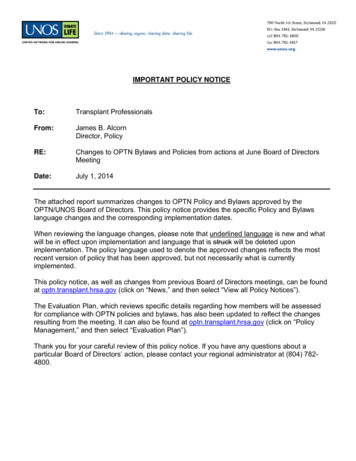
4E. J. COTTER ET AL.Statistical AnalysisStatistical analysis was performed using descriptivestatistics; the c2 or Fisher exact test for comparison ofcategorical variables; and the Student t test and MannWhitney U test for comparison of parametric andnonparametric continuous variables, respectively.Binomial logistic regression analysis was used tocompare patient age as a continuous variable with clinical outcome metrics including complications, reoperations, satisfaction, willingness to undergo the procedureagain, RTS, RTS at the same level of sporting activitybefore the initial presentation of symptoms or higher,and failure. The Wilcoxon signed rank test was used foranalysis of change in PROs from baseline to final followup. A Kaplan-Meier curve was generated to show graftsurvivorship. All reported P values are 2 tailed, with an alevel of .05 detecting significant differences (SPSSStatistics, version 23.0; IBM, Armonk, NY).ResultsThe retrospective review over the period from October1, 2002, to November 30, 2014, yielded 67 patients (70knees). A total of 25 patients (26 knees) were excludedfor the following reasons: different treatment (DeNovoNT, 19 knee); iliac crest bone graft (1 knee); incorrectdiagnosis (avascular necrosis due to medication, 1 knee);incorrect location of OCD lesion (ankle, 1 knee); skeletally immature on preoperative radiographs (1 knee);and/or concomitant patellofemoral compartment procedures including patellofemoral anteromedialization,medial patellofemoral ligament reconstruction, and/ormicrofracture (4 knees). We identified 43 knees in 41patients for inclusion; 4 patients (4 knees) were lost tofollow-up. Thus 39 knees (90.9%) in 37 patients (26male and 13 female knees) were analyzed at a minimum2-year follow-up. The average time to final follow-upwas 7.29 3.30 years. All patients underwent at least1 previous ipsilateral surgery (mean, 2.3 1.3 procedures). Of the knees, 32 (82.1%) underwent priorarticular cartilage procedures whereas the remainderunderwent previous knee surgery unrelated to the OCDlesion (meniscectomy and ligament reconstruction).Complete demographic information is provided inTable 2.There were 21 lateral femoral condyle OCA and 24medial femoral condyle OCA cases. Of note, 4 patientsunderwent OCA on both condyles concomitantly and 2patients received 2 grafts (snowman technique), each tothe medial femoral condyle for elongated or obliquelesions. The average defect size was 460.87 168.12 mm2. Major concomitant procedures wereperformed in 11 patients, including 6 meniscal allografttransplantations, 3 high tibial osteotomies, and 4 distalfemoral osteotomies.Table 2. Demographic and Surgical History of All PatientsNo.SexAge, yrBMILateralityWorkers’ Compensation casesNo. of previous surgicalproceduresDuration of symptoms, yrAthlete (recreational, competitive,professional)Follow-up, yrNo. of knees with failed articularcartilage proceduresFailed OCD ORIFFailed loose body removalFailed microfracture and/ordrillingFailed ACIFailed OATSData37 patients, 39 knees26 male and 13 female knees26.01 9.96 (range,15.78-49.25)26.54 4.7720 right (51.3%) and 19 left(48.7%) knees3 (7.5%)2.3 1.34.54 4.3824 (60.0%)7.29 3.3032 (82.1%)17 (43.5%)13 (32.5%)21 (52.5%)1 (2.5%)1 (2.5%)NOTE. All percentages are based on the total number of knees. Dataare presented as mean standard deviation or number.ACI, autologous chondrocyte implantation; BMI, body mass index;OATS, osteochondral autograft transplantation; OCD, osteochondritisdissecans; ORIF, open reductioneinternal fixation.Clinical OutcomesOne patient did not respond to follow-up regardingsatisfaction with the clinical outcome and whether hewould choose to undergo the operation again. For 31 ofthe remaining 38 knees (81.6%), the patient stated thathe or she was either mostly satisfied (19) or completelysatisfied (12) at final follow-up. In addition, for 35knees (92.1%), the patient stated that he or she wouldundergo the procedure again. There was a statisticallysignificant improvement in all PROs (P .05), with theexception of the SF-12 mental subscale (P ¼ .910), frombaseline to final follow-up (Fig 3). An important findingwas that the minimal clinically importance difference(MCID) in improvement from the PRO preoperativelyto the PRO at a minimum 2-year follow-up previouslypublished for the IKDC score22 was achieved(improvement by 25.54). In addition, MCID improvements were achieved for all KOOS subscores, specifically pain (20.68), symptom (14.28), activities of dailyliving (20.88), sport (25.96), and quality of life (30.56).For WOMAC subscores, the MCID was not achieved forthe WOMAC pain subscore (3.74) or WOMACsymptom subscore (14.73).22Return to SportThere were 7 basketball athletes (29.2%), 4 footballathletes (16.7%), 3 running athletes (12.5%), 3 cyclingathletes (12.5%), 2 skiers (8.3%), 2 soccer athletes(8.3%), 2 wrestling athletes (8.3%), and 1 hockey
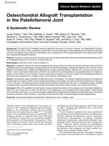
5OSTEOCHONDRITIS DISSECANS TREATMENTFig 3. Summary of patient-reported outcome metrics for patients undergoing osteochondral allograft transplantation fortreatment of osteochondritis dissecans lesions in the knee at an average follow-up of 7.36 3.29 years. Significant differences(asterisks) were noted for the following: International Knee Documentation Committee (IKDC) score (P .0001), all subscoresof the Knee Injury and Osteoarthritis Outcome Score (KOOS) (P .0001 for each), Western Ontario and McMaster UniversitiesOsteoarthritis Index (WOMAC) pain score (P .0001), WOMAC stiffness score (P ¼ .002), WOMAC function score (P .0001),WOMAC overall score (P .0001), and Short Form 12 (SF-12) physical subscale score (P ¼ .002). (ADL, activities of daily living;QOL, quality of life.)athlete (4.2%). Two patients were unable to be reachedto ask whether they achieved RTS; however, 18 of theremaining 22 patients (81.8%) who preoperatively selfidentified as athletes achieved RTS at an average of14.0 8.7 months, with 13 of 18 (72.2%) returning atthe same level or higher. On subanalysis between thosepatients who returned to sport and those who did not,only body mass index (BMI) was shown to be significantly different between groups, with those with failureto RTS having a higher BMI (RTS, 24.68 3.67; noRTS, 27.82 0.69; P ¼ .005) (Table 3). Of the 4 athleteswho did not RTS, 3 were recreational athletes whereas1 was a college hockey player. All 4 identifiedcontinued discomfort and/or lack of confidence in theirknee as primary reasons for not returning to sport.Age AnalysisShapiro-Wilk testing confirmed a normal distributionof patient age (mean, 26.01 9.96 years; range,15.78-49.25 years). There was no significant differencein age between patients who self-identified as athletes(24 knees; mean, 23.94 8.12 years) at preoperativeoffice visits and non-athletes (15 knees; mean,30.01 12.02 years; P ¼ .095). On logistic regressionanalysis of age (continuous) with postoperative outcomes, no significant differences were noted in anyoutcome variable. Specifically, no significant relationswith complications (odds ratio [OR], 1.010; P ¼ .852),reoperation (OR, 1.028; P ¼ .414), patient satisfaction(OR, 1.022; P ¼ .957), willingness to undergo the procedure again (OR, 1.022; P ¼ .977), RTS (OR, 0.990;P ¼ .875), RTS at the same level or higher (OR, 1.227;P ¼ .150), and failure (OR, 1.121; P ¼ .159) were shown.Complete analysis with reported 95% confidence intervals can be found in Table 4.ComplicationsComplications occurred in 4 patients, including alow-grade incisional infection (n ¼ 1), superficial skinTable 3. Demographic, Preoperative, Operative, andOutcome Variable Comparisons Between Athletes WhoReturned to Sport and Those Who Did NotVariableRTSAge, yr24.26 8.19BMI24.68 3.67Symptom duration, yr5.94 5.17Failed previous cartilage15 (83.3%)surgeryFailed OCD ORIF8 (44.4%)Failed microfracture8 (44.4%)Major concomitant surgery6 (33.3%)Reoperation6 (33.3%)Complications3 (16.7%)Mostly or complete satisfied 16 (88.9%)No RTSP Value24.96 10.29 .88327.82 0.69.005*1.25 1.06.2354 99.541.264.999.135NOTE. All percentages are based on the total number of knees. Dataare presented as mean standard deviation or number.BMI, body mass index; OCD, osteochondritis dissecans; ORIF, openreductioneinternal fixation; RTS, return to sport.*Statistically significant.
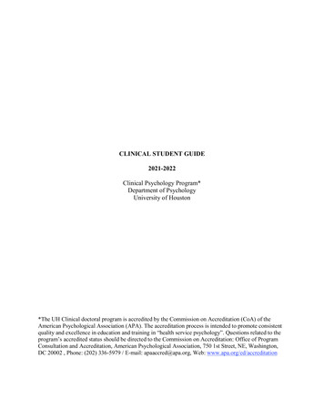
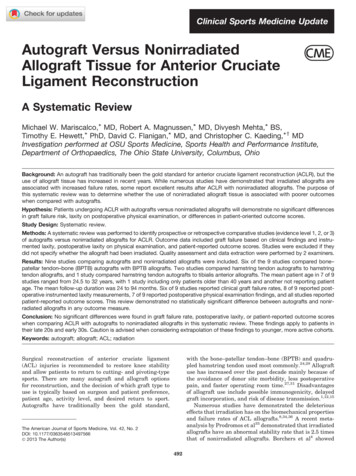
6E. J. COTTER ET AL.Table 4. Binomial Logistic Regression for DemographicCharacteristics and Outcomes Based on Patient Age(Continuous)VariableRTSRTS at same level or higherComplicationsSatisfactionWilling to undergo procedure 1.0281.12195% 9-1.1290.962-1.0980.956-1.313P Value.875.150.852.957.977.414.159NOTE. Higher ORs signify that the outcome variable was more likelyto occur as patient age increased.CI, confidence interval; OR, odds ratio; RTS, return to sport.dehiscence (n ¼ 1), an acute hematoma that requiredevacuation and washout (n ¼ 1), and a transientperoneal nerve palsy (n ¼ 1) that resolved after7 months.ReoperationsIn 14 patients (35.9%), subsequent surgery was performed at an average of 2.93 2.47 years after indexOCA. The reoperations included hardware removalfrom a distal femoral osteotomy (n ¼ 1), partial lateralmeniscectomy (n ¼ 1), debridement and plica excisionprocedures (n ¼ 11), and separate-site microfracture(n ¼ 1); all patients were found to have intact allograftsat the time of secondary surgery.Graft SurvivorshipIn 2 patients (5.1%), progression to failure occurredat an average of 6.2 3.8 years. Specifically, a 32-yearold male patient underwent revision OCA at 3.57 yearsand a 41-year-old female patient underwent a unicompartmental knee arthroplasty at 8.97 years after theindex procedure. Both had an insidious onset ofsymptoms and had previous failure of microfracture atthe site of eventual OCA, in each case involving themedial femoral condyle. The 32-year-old man waspreviously active running and playing basketball,whereas the 41-year-old woman did not identify as anathlete. Neither patient had postoperative complications, and both underwent a subsequent second-lookarthroscopy 2.55 and 4.3 years after the index procedure, respectively, showing mild, clinically insignificantgraft degeneration. A Kaplan-Meier curve showed97.0% graft survivorship at 5 years (Fig 4).all PRO metrics except the SF-12 mental subscale, and92.1% of patients stating they were willing to undergothe procedure again. MCID improvements from previously published literature22 were achieved for the IKDCscore, all KOOS subscores, the WOMAC function score,and the WOMAC total score, indicating clinicallysignificant improvement in outcomes. Furthermore, theresults show that OCA for OCD has high utility inreturning athletes (predominantly recreational andhigh school athletes) to sport (81.8%). Athletes withfailure to RTS were shown to have a significantly higherBMI (P ¼ .005), with all other analyzed demographicand operative variables found to have no associationwith the ability to RTS. In addition, regression analysisbased on patient age did not identify any patient-,injury-, or surgery-specific variables associated withinferior clinical outcomes. This information can be usedto counsel patients with OCD lesions of the knee,especially low-level athletes, in whom previous surgicalintervention has failed regarding OCA as a viable kneejoint preservation procedure regardless of patient age.An interesting finding was that both patients withfailure had undergone previous marrow stimulationprocedures at the site of OCA. Recently, Gracitelliet al.23 reported no significant differences in graft survival or clinical outcomes between patients undergoingprimary OCA and patients undergoing OCA after afailed marrow stimulation procedure in a controlmatched analysis of 46 patients, 42 of whom had apreoperative diagnosis of OCD. Furthermore, in arecent study of 180 patients who underwent OCA, theauthors performed a Kaplan-Meier analysis to evaluatewhat effect, if any, previously failed microfracture hadon graft survivorship.20 They reported no significantDiscussionThe principal findings of this study suggest that OCAis an effective procedure for secondary treatment ofOCD of the knee in skeletally mature patients, as shownby the 82.1% of patients who were mostly orcompletely satisfied with their clinical outcome, significant improvements from baseline to final follow-up inFig 4. Kaplan-Meier survival analysis curve showing survivalat an average follow-up of 7.29 3.3 years. There were 2graft failures, with 1 patient undergoing revision osteochondral allograft transplantation at 3.57 years and another undergoing unicompartmental knee arthroplasty at 8.97 years.At 5 years, the graft survival rate was 97%.
7OSTEOCHONDRITIS DISSECANS TREATMENTdifference (P ¼ .370). In our study, there was aninsignificant number of patients who underwent previous marrow stimulation to reach a safe, accurateconclusion on whether this may have had a significanteffect on failure for these 2 patients.In this study, 81.8% of athletes were able to RTS and72.2% were able to RTS at the same level. There was nosignificant difference in RTS (OR, 0.990; P ¼ .875) orsame-level RTS (OR, 1.227; P ¼ .150) based on patientage. Other authors have reported slightly higher rates ofRTS after OCA. Krych et al.16 noted an 88% RTS rate atan average of 9.6 months, with 79% achieving thepreinjury activity level, in a study of 43 patients treatedwith OCA not specific to only OCD. However, theyshowed that age of 25 years or older and a preoperativeduration of symptoms greater than 12 months were riskfactors for an inability to RTS, neither of which wasseen in our study.More recently, Nielsen et al.6 conducted the largest RTSstudy regarding treatment of focal cartilage lesions of theknee of any etiology with OCA. Of the 149 knees included,112 (75.2%) were able to RTS or return to recreationalactivity after OCA, with 91% of athletes reporting beingsatisfied or extremely satisfied with their clinical outcome.None of the aforementioned RTS investigations reported arelation between a higher BMI and inability to RTS asshown in our investigation. Although the 4 athletes whodid not RTS had a mean BMI of 27.82, this may be anincidental finding given the relatively small number ofathletes who did not RTS or may be indicative of the effectsof added load across the knee joint. Furthermore, the results of our study suggest that lower-level athletes treatedwith OCA for OCD lesions of the knee have a comparable,if not slightly improved, opportunity to RTS whencompared with OCA for any etiology.Sadr et al.13 recently published the largest investigation to date of clinical outcomes after OCA for treatment of OCD lesions in the knee. In a population of 149knees with a minimum 2-year follow-up, they reportedhigh patient satisfaction (95% satisfied or extremelysatisfied), excellent graft survival (95% at 5 years and93% at 10 years), and significantly improved clinicaloutcome scores at final follow-up. However, theirinvestigation used both dowel and shell techniques andincluded patellofemoral grafts, differing from our study,in which only a dowel technique was used and patellofemoral lesions were excluded because of inherentlyunique biomechanical and concomitant pathologyconsiderations. Similarly to the results of Sadr et al.,13previous investigations have reported good to excellent clinical outcomes and graft survival with OCA forthe treatment of OCD. Garrett24 reported graft survivalin 16 of 17 patients at a 2- to 9-year follow-up inpatients who had undergone at least 1 prior surgicalprocedure for OCD. In addition, Emmerson et al.25 reported excellent outcomes in 47 of 65 knees (72%) thatunderwent OCA for the treatment of OCD, with 95%survivorship at 5 years and 76% survivorship at10 years. Although 11 patients (27.5%) in our studyunderwent subsequent second-look arthroscopy anddebridement indicative of the presence of symptoms,this is not uncommon for this procedure and all wereshown to have intact grafts without degeneration.Other authors have reported good clinical outcomesof OCA in investigations that included but were notexclusive to patients with OCD lesions of the knee.Levy et al.26 reported an 82% 10-year survival rate ina long-term follow-up study of 129 knees, with a meanage of 33 years, after OCA treatment of femoralcondyle lesions. They found a significant associationbetween age older than 30 years at the time of surgeryand allograft failure. This finding differs from the result
nology codes 27415 (osteochondral allograft, knee, open) and 29867 (arthroscopy, knee, surgical; osteo-chondral allograft) and International Classification of Diseases, Ninth Revision code 732.7 (OCD) based on physical examination, radiographic, and/or magnetic resonance imaging findings. Indications for OCA