Transcription
Clinical Sports Medicine UpdateOsteochondral Allograft Transplantationin the Patellofemoral JointA Systematic ReviewJorge Chahla,*y MD, PhD, Matthew C. Sweet,z MD, Kelechi R. Okoroha,y MD,Benedict U. Nwachukwu,y MD, MBA, Betina Hinckel,§ MD, Jack Farr,k MD,Adam B. Yanke,y MD, PhD, William D. Bugbee,{ MD, and Brian J. Cole,y MD, MBAInvestigation performed at Rush University Hospital, Chicago, Illinois, USABackground: The initial focus of cartilage restoration algorithms has been on the femur; however, the patellofemoral compartment accounts for 20% to 30% of significant symptomatic chondral pathologies. While patellofemoral compartment treatmentinvolves a completely unique subset of comorbidities, with a comprehensive and thoughtful approach many patients may benefitfrom osteochondral allograft treatment.Purpose: To perform a systematic review of clinical outcomes and failure rates after osteochondral allograft transplantation (OCA)of the patellofemoral joint at a minimum 18-month follow-up.Study Design: Systematic review; Level of evidence, 4.Methods: A systematic review of the literature regarding the existing evidence for clinical outcomes and failure rates of OCA forpatellofemoral joint chondral defects was performed with the Cochrane Database of Systematic Reviews, the Cochrane CentralRegister of Controlled Trials, PubMed, and MEDLINE from studies published between 1990 and 2017. Inclusion criteria were asfollows: clinical outcomes and failure rates of OCA for the treatment of chondral defects in the patellofemoral joint, English language, minimum follow-up of 18 months, minimum study size of 5 patients, and human studies. The methodological quality ofeach study was assessed with a modified version of the Coleman methodology score.Results: The systematic search identified 8 studies with a total of 129 patients. The methods of graft procurement and storage timeincluded fresh (121 patients, 93.8%), and cryopreserved (8 patients, 6.2%) grafts. The mean survival rate was 87.9% at 5 years and77.2% at 10 years. The following outcome scores showed significant improvement from pre- to postoperative status: modifiedd’Aubigné-Postel, International Knee Documentation Committee, Knee Society Score–Function, and Lysholm Knee Score.Conclusion: OCA of the patellofemoral joint results in improved patient-reported outcome measures with high patient satisfactionrates. Five- and 10-year survival rates of 87.9% and 77.2%, respectively, can be expected after this procedure. These findingsshould be taken with caution, as a high percentage of patellofemoral osteochondral allografts were associated with concomitantprocedures; therefore, further research is warranted to determine the effect of isolated osteochondral transplantations.Keywords: osteochondral allograft transplantation; patellofemoral, knee; cartilage; repairChondral lesions of the patellofemoral compartment arenoted in .33% of patients undergoing knee arthroscopy,making it the second-most common site for cartilagedefects after the medial femoral condyle.6,9,11,24 Chondrallesions of the patellofemoral joint are particularly challenging to manage, as treatment of the lesion itself mustbe performed in concert with any concomitant patellofemoral anatomic and biomechanical derangements. Theseabnormalities can include excessive lateral tilt, malalignment, maltracking, patella alta, limb coronal and axialmalalignment, and trochlear dysplasia, all of which shouldbe taken, cataloged, and addressed with a risk:rewardratio approach.3,6,11,13,24Several treatment modalities have been described forthe management of patellofemoral lesions, including osteochondral autograft transplantation, marrow stimulation(microfracture, abrasionplasty, or drilling), autologouschondrocyte implantation, osteochondral allograft transplantation (OCA), and particulated juvenile cartilage allograft.6,17 However, inferior outcomes for patellofemoralcartilage procedures have been consistently reported inthe literature when compared with other locations irrespective of the surgical technique used.6,10OCA procedures offer numerous advantages, includingthe presence of metabolically active chondrocytes withoutThe American Journal of Sports Medicine1–9DOI: 10.1177/0363546518814236Ó 2018 The Author(s)1
2Chahla et alconcurrent donor site morbidity,29 natural hyaline cartilagewith structural osseous bed that can accept full loading assoon as the bone base has healed,21 and immunoprivilegedcharacteristics.1 While extensive literature exists evaluatingosteochondral allograft in the knee, the majority of outcomesare reported for OCAs of the femoral condyles.2,8,10,20,27There are fewer and less-powered studies that assess outcomes of OCA in the patellofemoral joint. Therefore, the purpose of this study was to perform a systematic review ofclinical outcomes and failure rates after OCA transplantationof the patellofemoral joint at a minimum follow-up of 18months. It was hypothesized that improved functional andobjective outcomes would be obtained after a patellofemoralOCA with high mid- to long-term survival rates.METHODSArticle Identification and SelectionThis study was conducted in accordance with the 2009PRISMA (Preferred Reporting Items for SystematicReviews and Meta-Analyses) guidelines.26 A systematicreview of the literature regarding the existing evidence forclinical outcomes and failure rates of OCA transplantationof the patellofemoral joint was performed with the CochraneDatabase of Systematic Reviews, the Cochrane Central Register of Controlled Trials, PubMed (1990-2017), and MEDLINE (1990-2017). The queries were performed in May2018. The literature search strategy included the following:Search 1: osteochondral[All Fields] AND (‘‘allografts’’[MeSH Terms] OR ‘‘allografts’’[All Fields] OR ‘‘allograft’’[All Fields]) AND patellofemoral[All Fields]Search 2: osteochondral[All Fields] AND (‘‘allografts’’[MeSH Terms] OR ‘‘allografts’’[All Fields] OR ‘‘allograft’’[All Fields]) AND (‘‘patella’’[MeSH Terms] OR‘‘patella’’[All Fields])Search 3: osteochondral[All Fields] AND (‘‘allografts’’[MeSH Terms] OR ‘‘allografts’’[All Fields] OR ‘‘allograft’’[All Fields]) AND trochlea[All Fields]The American Journal of Sports MedicineSystematic review registration was performed in May 2018with the PROSPERO international prospective register ofsystematic reviews (registration CRD42018094608).Inclusion criteria were as follows: original researchreporting clinical outcomes and failure rates of OCA forthe treatment of chondral defects in the patellofemoraljoint, English language, minimum follow-up of 18 months,minimum of 5 patients, and human studies. We excludedcadaveric studies, animal studies, biomechanical reports,basic science articles, editorial articles, case reports, literature reviews, surgical technique descriptions, instructional courses, OCA for tumor, and studies in which OCAof the patellofemoral joint were not available independentof combined OCA groups.Two independent reviewers (J.C. and M.C.S.) performeda review of the abstracts from all identified articles. Fulltext articles were obtained for review if necessary to allowfor further assessment of inclusion and exclusion criteria.Additionally, all references from the included studieswere reviewed and reconciled to verify that no relevantarticles were missing from the systematic review. If datawere not specifically reported for patellofemoral OCA instudies that otherwise met inclusion criteria, authorswere contacted to provide missing data points.23Data Collection and ProcessingThe level of evidence of each study was assigned according to the classification system specified by Wrightet al.35 Data were abstracted from the full text or directlyfrom the authors of all eligible articles via standardizeddata collection forms. Abstracted and recorded dataincluded patient demographics, follow-up period, surgicaltechniques, postoperative imaging results (if reported),and objective and subjective outcomes. For continuousvariables (eg, age, follow-up, outcome scores), the means,SDs, interquartile ranges, and ranges were collected (ifreported). Data were recorded into a custom spreadsheetwith a modified information extraction table.15*Address correspondence to Jorge Chahla, MD, PhD, Rush University Medical Center, 1611 W Harrison, Suite 300, Chicago, IL 60612, USA (email:jachahla@msn.com).yRush University Medical Center, Chicago, Illinois, USA.zWayne State University School of Medicine, Detroit, Michigan, USA.§Kaiser Permanente Southern California, San Diego, California, USA.kCartilage Restoration Center of Indiana, OrthoIndy, Indianapolis, Indiana, USA.{Scripps Clinic, La Jolla, California, USA.One or more of the authors has declared the following potential conflict of interest or source of funding: J.F. reports personal fees from Advanced Biosurfaces, Arthrex Inc, Arthrocare, Exatech, BioRegeneration Technologies, Vericel, Ceterix Orthopaedics, DePuy, Genzyme, Knee Creations LLC, MedShape Inc, Mitek, Moximed Inc, NuOrtho Surgical Inc, NuTech Medical, Osiris Therapeutics Inc, RTI Biologics Inc, Schwartz Biomedical LLC, Scienceand Biomaterials Inc, Springer, Stryker, Thieme Medical Publishers Inc, and ZimmerBiomet and nonfinancial support from Arthrocare, DePuy, DePuy/Mitek,Genzyme, Histogenics, Knee Creations Inc, Moximed Inc, NuTech Medical, RTI Biologics Inc, and ZimmerBiomet. W.D.B. is a consultant for and hasreceived research support from the Joint Restoration Foundation, a nonprofit tissue bank that receives, processes, and distributes osteochondral allografts;is a consultant for and receives royalties from DePuy Synthes; receives royalties from Smith & Nephew and Zimmer Biomet; and holds investment interest inOrthAlign. A.B.Y. has received research support from Arthrex Inc, NuTech, and Smith & Nephew. B.J.C. holds stock options in Aqua Boom, Biomerix, Giteliscope, Ossio, and Regentis; is a paid consultant for Arthrex, Flexion, Regentis, Smith & Nephew, and Zimmer; receives research support from Aesculap/B.Braun, Arthrex, Geistlich, Medipost, the National Institutes of Health (National Institute of Arthritis and Musculoskeletal and Skin Diseases and National Institute of Child Health and Human Development), Novartis, Sanofi-Aventis, and Zimmer; receives intellectual property royalties from Arthrex, DJ Orthopaedics,and Elsevier; and receives other financial or material support from Athletico, JRF Ortho, Smith & Nephew, and Tornier. AOSSM checks author disclosuresagainst the Open Payments Database (OPD). AOSSM has not conducted an independent investigation on the OPD and disclaims any liability or responsibility relating thereto.
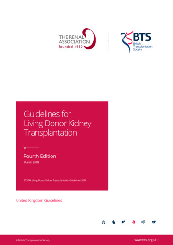
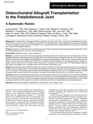
AJSM Vol. XX, No. X, XXXXPatellofemoral OCA Transplantation3Figure 1. Study search strategy according to the PRISMA (Preferred Reporting Items for Systematic Meta-Analyses) guidelines.25 OCA, osteochondral allograft transplantation; PF, patellofemoral.Based on a preliminary survey of the most commonlyused outcome scales, outcome scores were recorded forthe following: modified d’Aubigné-Postel score, LysholmKnee Score, International Knee Documentation Committee (IKDC) knee form, Knee injury and Osteoarthritis Outcome Score, and Knee Society Score–Function (KSS-F)score. If none of these scales were used, results were documented for the primary functional scale used in the study.Literature Quality EvaluationTwo reviewers (J.C. and M.C.S.) used a modified version ofthe Coleman methodology score (mCMS) to assess themethodological quality of each study.19 The 2-part mCMSgrades cartilage-related studies based on 10 criteria. PartA includes study size, mean follow-up, number of surgicalprocedures, type of study, description of surgical procedure,postoperative rehabilitation, included patients’ magneticresonance imaging outcome, and inclusion patients histological outcome. Part B includes outcome criteria, procedurefor assessing clinical outcomes, and description of patientselection process. The maximum score of the mCMS is100, which indicates that a study largely avoids chance,biases, and confounding factors.RESULTSStudy SelectionThe systematic search performed with the previously mentioned keywords identified 8 studies after removal of duplicates and application of exclusion criteria. Followingreview of all references from the included studies, no additional studies met the inclusion criteria (Figure 1). Twoindependent reviewers (J.C. and M.C.S.) performed a methodological quality assessment of included articles. Themean Kon-Verdonk mCMS score of the included studieswas 41.9 (range, 25-51) out of 100 points.19Study Characteristics and DemographicsThree prospective and 5 retrospective case series met theinclusion criteria for this systematic review. Two studiesevaluated OCA isolated to the femoral trochlea; 2 studiesevaluated OCA isolated to the patella; and 4 studies evaluated OCA of the patellofemoral joint or OCA for bipolarchondral lesions (femoral trochlea and patella). The methods of procurement and storage time included fresh (121patients, 93.8%) and cryopreserved (8 patients, 6.2%)grafts. The 8 studies included in the analysis reported ona total of 129 patients (range, 8-28 patients per study).The weighted mean patient age was 36.9 years (range, 1264 years), and the weighted mean follow-up was 6.6 years(range, 1.8-30.1 years). Importantly, 91.2% of the patientshad previous operations (mean, 2.85 procedures beforeOCA transplantation). The patient demographics, indications for OCA transplantation, concomitant procedures,and locations of the allograft transplant are described indetail in Table 1. Details regarding lesion size, plug size,and prior surgical treatment are outlined in Table 2.Functional OutcomesPre- and postoperative patient-reported outcome scores forpatellofemoral OCA were assessed in 7 studies (Table3).4,7,14,18,32-34 Sixteen outcome measures were reported inthese 7 studies, of which the most commonly used were theIKDC form (4 studies),7,14,33,34 modified d’Aubigné-Postelform (3 studies),7,14,18 and KSS-F form (3 studies).7,14,32All studies reported significant improvement in at least 1clinical score. All 4 studies that utilized IKDC scores reporteda significant improvement from baseline to postoperativefollow-up (P \ .001), with an aggregate preoperative IKDCscore of 41.8 and postoperative IKDC score of 68.1.7,14,33,34The aggregate mean improvement in total IKDC score frompreoperative to final follow-up was 26.3. Studies with themodified d’Aubigné-Postel scores reported significant postoperative improvement (P \ .001), with mean preoperative12.2, and mean postoperative of 15.9.7,14,18 The aggregate
4Chahla et alThe American Journal of Sports MedicineTABLE 1Characteristics of Included StudiesaFirstAuthorbJournal, Year,Study TypebAge, y, Mean 6 SD MalePatients(Range)Sex, n (%) (Knees), nLocationof DefectBipolar(Knees, n)Cameron7 AJSM, 2016,retrospective30.2 6 10.6 (12-47) 20 (72.4)28 (29)TrochleaNoneGracitelli14 AJSM, 2015,retrospective33.7 (14-64)13 (46.4)27 (28)PatellaNoneTorgaSpak32CORR, 2006,retrospective37 (24-56)0 (0)11 (14)Patella (14),trochlea (12)12Jamali18CORR, 2005,retrospectiveCartilage, 2018,prospectiveAJSM, 2018,prospectiveAJSM, 2017,prospective42 (19-64)7 (39)18 (20)128 (80)10 (10)Patella (20),trochlea (12)Trochlea47.0 (40-53)13 (68.4)19 (19)31.1 (24-48)3 (37.5)8 (8)Wang34,d33,dWangFrank12,dBakay4Int Orthop, 1998,retrospective37.1 6 8.2 (23-52)NANA8 (8)Patella (7),trochlea (16)Patella (2),trochlea (6)PatellaNoneTechnique(Knees, n)cDowel (25),shell (4)EtiologyConcomitantProceduresOCD, DCL,OA, traumaLateral retinaculumrelease (38%),diagnostic arthroscopy,hardware removal,debridement, PF softtissue realignment,trochleoplasty, loosebody removal,meniscus repair, lysisof adhesions,manipulationDowel, shellOCD, DCL, OA,Lateral retinaculum2(.10 cm ortrauma, AVNrelease (37%),75% of patella)realignment surgeryfor the extensormechanism (1 vastusmedialis imbrication,1 tibial tubercleosteotomy with medialPF ligamentreconstruction, 3 tibialtubercle osteotomiesisolated)Femoral (32) and tibialShellTrauma, OA, bonyosteotomies 1 to 3 ymalalignment,patellar instability, before OCAiatrogeniccartilage shavingDowel, shellTraumaLateral retinaculumrelease (45%)DCL, AVN, trauma Realignment osteotomyFollow-up,Mean (Range), y7.0 (2.1-19.9)9.7 (1.8-30.1)10.0 (2.5-17.5)7.8 (2.0-18)3.9 (2.0-10.7)5DowelOCD, DCL, OARealignment osteotomy3.3 (2.0-6.8)NoneDowelGSW, traumaNoneDoweleTraumaTTT anteromedialization4.4 (2.1-7.7)(4), microfracture toarea other than OATsite, partialmeniscectomyLateral retinaculumNArelease (75%)aAJSM, American Journal of Sports Medicine; AVN, avascular necrosis; CORR, Clinical Orthopaedics and Related Research; DCL, degenerative chondral lesion; GSW, gun shot wound;Int Orthop, International Orthopaedics; NA, not available; OA, osteoarthritis; OAT, osteochondral autograft transplantation; OCA, osteochondral allograft transplantation; OCD, osteochondritis dissecans; PF, patellofemoral; TTT, tibial tubercle transfer.bLevel of evidence: 4 (all studies).cType of graft: fresh (all studies unless noted otherwise).dData obtained from respective authors of studies.eCryopreserved graft.mean preoperative KSS-F score was 53.4 and postoperativewas 80.2.7,14,32 All other patient reported outcomes are demonstrated in Table 3.Radiographic AnalysisPostoperative radiographic results were reported for 2 studies. Torga Spak and Teitge32 evaluated radiographs at finalfollow-up for graft incorporation, resorption, collapse, cystformation, and osteophyte formation. Radiographs wereused to measure the patellar thickness ratio; patellar heightwas measured with the Blackburne-Peel index and theCaton-Deschamps index. The patellar thickness ratio wasdefined as the length of the patella to the maximal thicknessof the patella, as measured on lateral radiographs. Thepatellar thickness ratios of the bipolar shell allografts (6 ofthe 8 surviving allografts) showed a mean 14% volumetricreduction of the patellar allograft-host bone unit. Themean preoperative patellar thickness ratio of these graftswas 0.46 (range, 0.40-0.56), and at follow-up (mean, 8.1years; range, 48-204 months) it was 0.40 (range, 0.130.56). At final follow-up, for the 8 patients with an intactallograft, radiographs demonstrated mild degenerativechanges for 6 patients and no degenerative changes for 2patients, as measured by the Fairbank grading scale.Jamali et al18 evaluated the radiographic outcomes of 12patients. The mean radiographic follow-up was 70 months(range, 18-183 months). Patellofemoral arthrosis and tibiofemoral arthrosis in the medial and lateral compartmentswere classified with the modified Fairbank and Ählbackcriteria as described by Lundberg and Messner. Radiographs were evaluated for visibility of the allograft-hostjunctions, allograft radiodensity when compared with the
AJSM Vol. XX, No. X, XXXXPatellofemoral OCA Transplantation5TABLE 2Characteristics of Patients Included in the StudiesaFirstAuthorLesionSize, cm2,Mean 6 SD (Range)PriorSurgery, %;Proceduresper Patient,n (Mean)6.1 6 3.6 (2.3-20.0)89.7; 2.4Cameron7Gracitelli14Torga Spak3210.1 (4-18)NA92.9; 3.2100; 4.4Jamali1813.2 (2.5-22.5)d100, 2.6Wang347.4 (1.0-15.3)80; 2.7Wang337.1 (0.8-15.3)79, 1.9Frank12Bakay44.1 6 1.8 (2.2-6.3)NA100; 2.75NAReoperations, n (%):Procedures Performed,b n6 (21.4): MUA for arthrofibrosis(1), AS/D and partialmeniscectomy (1), scar tissueremoval (1), chondral flapdebridement and loose bodyremoval (1), debridement,chondroplasty of patella/LFC,and synovectomy (1), TKA (1)17 (60.7): AS/D (9), HR (6), ACLR(1), PF realignment (1), MUA(1), loose body removal (1), TKA(4), PFA (2), patellectomy (1),revision OCA (1)12 (86): HR (12), MPFLR (3), TTT(3), AS/D (2), IT rotationalosteotomy (2), revision Maquet(1) FVO (1), TIR osteotomy (1)13 (65): AS/D (7), ASC (2), HR (2),TKA (2), revision allograft (2),arthrotomy and fat paddebridement (1), arthrodesis (1)3 (30): TKA (1), meniscectomy (1),HR, MUA, and opensynovectomy (1)11 (58): TKA (5), UKA (3), ASCand loose body removal (2), I&D(1)4 (50): trochlea (3), patella (1)eNAFailure, n (%)Mean Timeto Failure,mo (Range)TKA (1) for persistent pain(probable complex regionalpain syndrome)100 at 5 y, 91.7at 10 y1 (3.4)91.2TKA (4), PFA (2), revisionOCA (1), patellectomy (1)(reasons unknown)78.1 at 5 y, 78.1at 10 y, 55.8 at15 y8 (28.6)51.6 (6-165.6)Revision at 12 mo for graftfragmentation; conversionto TKA 20 mo after graftfragmentationscTKA (2), revision allograft (2),arthrodesis (1)86 at 5 y, 72 at10 y2 (14.3)NA67 at 10 y5 (25)32.2 (13-52)TKA (1)90 at 2 y1 (10)20.05 TKA, 3 UKA89.5 at 2 y8 (42.1)33.6 (11-72)Trochlea (2)eHyperpressure of PF joint (1)87.5 at 5 yNA2 (25)1 (12.5)40.4NAReason for FailuresAllograftSurvivorshipaACLR, anterior cruciate ligament reconstruction; ASC, arthroscopic chondroplasty; AS/D, diagnostic arthroscopy with debridement; FVO, femoral varus osteotomy; HR, hardwareremoval; I&D, irrigation and debridement; IT, intertrochanteric; LFC, lateral femoral condyle; MPFLR, medial patellofemoral ligament reconstruction; MUA, manipulation under anesthesia; NA, not available; OCA, osteochondral allograft; PF, patellofemoral; PFA, patellofemoral arthroplasty; TIR, tibial internal rotation; TKA, total knee arthroplasty; TTT, tibial tubercle transfer; UKA, unicompartmental knee arthroplasty.bSome patients had .1 procedure (n number of knees undergoing reoperation).cThe 2 grafts that did not survive were in the same patient: the first unipolar allograft (patella) required revision 12 months after implantation; the revised bipolar allograft was converted to a TKA at 20 months.dTrochlea (information only for 8 knees). Plug size (information for 3 knees): mean, 7.1 cm2 (range, 1.8-17.8 cm2).eReason for 2 failed trochlear grafts and reoperations not reported.surrounding bone (increased, decreased, or the same), andthe presence of subchondral cysts. Of the 12 knees evaluated radiographically, 4 had no evidence of patellofemoralarthrosis, 6 had mild arthrosis, and 2 had advanced arthrosis. The allograft-host interface was not visible radiographically in 9 patients and was identifiable in 3 patients. Theradiodensity of the grafts was identical to the host in 8patients and was increased in 4 patients. Although 4 graftshad subchondral lysis, 3 of them still had good or excellentclinical scores.SatisfactionFour studies reported patient satisfaction as a measuredoutcome.4,7,14,18 Two studies assessed patient satisfactionwith a 5-point scale (extremely satisfied to extremely dissatisfied), with an aggregate mean 89% of patients extremelysatisfied or satisfied with the allograft surgery.7,14 Jamaliet al18 assessed patient satisfaction with a similar 4-pointordinal scale and found that 87.5% of patients wereextremely satisfied or satisfied with the allograft surgery.Bakay et al4 used a modified Bentley score,5 reporting75% of patients with excellent or good results.Reoperation and Failure RatesThe weighted mean failure rate for all 8 studies was 20.1%(n 28 patients; range, 3.4%-42.1%) (Table 2). The definition of failure among the identified studies was highly variable; each study used a different definition for graft failure.Cameron et al7 and Wang et al33,34 defined failure as revision of the graft or conversion to arthroplasty. Gracitelliet al14 defined failure as any reoperation resulting inremoval of the allograft. Torga Spak and Teitge32 definedfailure as occurrence of any 1 of the following: (1) the clinical rating was \70 points on the KSS-F and LysholmKnee Score scales; (2) there was radiographic evidence ofresorption or collapse; or (3) the patient required totalknee arthroplasty, fusion, or allograft revision. Jamaliet al18 defined failure as a poor modified d’Aubigné-Postelscore (\11) with the need for revision allografting, patellectomy, arthrodesis, or total knee arthroplasty. Frank
6Chahla et alThe American Journal of Sports MedicineTABLE 3Outcomes MeasuresaFirst Author: Outcome MeasuresCameron7,bModified d’Aubigné-PostelIKDC totalIKDC painIKDC functionKSS-FUCLA activity scoreSatisfactionExtremely satisfiedSatisfiedSomewhat satisfiedSomewhat dissatisfiedDissatisfiedGracitelli14,bModified d’Aubigné-PostelIKDC totalIKDC painIKDC functionKSS-FSatisfaction, %Torga Spak32,d,eLysholm Knee ScoreKSS-FJamali18,eModified d’Aubigné-PostelSatisfaction (n 16), %Extremely satisfiedSatisfiedDissatisfiedImprovement (n 16), %Substantial improvementSomewhat improvementNo changeSubstantially worseWang34,fIKDCKOS-ADLMarx Activity ScaleOverall conditionSF-36General healthPainPhysical functioningRole limited per physical healthWang33,fIKDCKOS-ADLMarx Activity ScaleOverall conditionSF-36General healthPainPhysical functioningRole limited per physical 224.63.12.519.32.2P 49.378.6666616.523.814.330.482.069.583.065.06 22.06 28.8616.86 ed)
AJSM Vol. XX, No. X, XXXXPatellofemoral OCA Transplantation7TABLE 3(continued)First Author: Outcome MeasuresPreoperativePostoperativeFrank12: NABakay4Bentley score, %ExcellentGoodFairPoorP Value2550250aAll values are presented as mean 6 SD unless noted otherwise. Bold indicates statistically significant value (P \ .05). IKDC, International Knee Documentation Committee; KOS-ADL, Knee Outcome Survey–Activities of Daily Living; KSS-F, Knee Society Score–Function;NA, not available; SF-36, 36-Item Short Form Health Survey; UCLA, University of California, Los Angeles.bOverall satisfaction with allograft surgery was determined using a 5-point scale with descriptors from extremely satisfied to dissatisfied.cExtremely satisfied or satisfied with results.dStudy reported results only for surviving allografts.eOutcomes from survival cohort.fData provided upon request from study authors.et al12 defined failure as revision OCA, conversion to kneearthroplasty, or gross appearance of graft failure at second-look arthroscopy. Bakay et al4 defined failure as fragmentation of the graft or a fair/poor result as determinedby Bentley score.5 The most common treatment for failurewas conversion to total knee arthroplasty (data availablefor 6 studies only, 13 of 25 patients). The weighted meantime to failure was 44.8 months (range, 20-91.2 months)(data available for 6 studies only).Of the 8 articles reviewed, 7 reported reoperations afterpatellofemoral OCA. In these 7 studies, 66 of 128 kneesunderwent reoperation (Table 2), for a weighted meanreoperation rate of 51.6% (range, 21.4%-86%). The mostcommon reoperation performed was hardware removal(32% of all reoperations, n 21 knees),14,18,32,34 whichcould have been from any of the concomitant proceduresperformed (eg, realignment procedures), followed by diagnostic arthroscopies, with or without debridement (27%of all reoperations, n 18 knees).14,18,32No study reported any intraoperative complications,and 3 studies (38%) reported no postoperative complications within the documented follow-up period. TorgaSpak and Teitge32 reported on 4 patients with persistentpostoperative anterior knee pain, thought to be associatedwith unrecognized bony malalignment, as well as 1 patientwho developed a skin rash 2 weeks after allograft implantation, which resolved with a course of prednisone. Additionally, all patients had low-grade synovitis thatresolved spontaneously, with the worst case lasting 12months. Wang et al33,34 reported that 3 patients developedarthrofibrosis postoperatively, which was successfullytreated with lysis of adhesions and scar excision. Whileno patients developed superficial or deep infections afterOCA, 1 patient developed a septic joint after arthroscopiclysis of adhesions, which was treated with arthroscopicirrigation and debridement.33 This patient ultimatelyunderwent total knee arthroplasty in the same knee 45months after arthroscopic irrigation and debridement.Bakay et al4 reported that a single patient experienceda hyperalgesic reaction on the fifth postoperative daybut recovered within 2 days with steroid and calciumderivative therapy. Frank et al12 reported complications;however, the authors did not stratify complications basedon graft location (patellofemoral specific).Graft SurvivalSeven studies performed Kaplan-Meier survival analysisfor patellofemoral OCAs (Table 2). The weighted mean 5year survival rate reported was 87.9%.7,12,14,32 The mean10-year survival rate was 77.2%,7,14,18,32 and the 15-yearsurvival rate was 55.8% (reported in only 1 study).14DISCUSSIONThe compelling findings of this study were that OCA transplantation for the patellofemoral joint yielded improvedpostoperative outcomes with high patient satisfaction andsurvival rates at short- to medium- and long-term followup (mean 10-year survival rate reported, 77.2%). Additionally, although the mean reoperation rate was 51.6%, themost common reoperation performed was hardwareremoval (31.8%), which is common in all patellar realignment series.28 Finally, while heterogeneous definitionsfor failure were used among the reviewed studies, the overall mean failure rate was 20.1%, which is similar to variousother biologic cartilage–restorative procedures at the tibiofemoral compartments.10 Of note, all the studies includedin this review had concurrent procedures performed atthe time of the osteochondral transplantation (6 of the 8had a bony realignment procedure), which does not allowfor an exclusive analysis of OCA for the patellofemoraljoint.
8Chahla et alWhile there is extensiv
Results: The systematic search identified 8 studies with a total of 129 patients. The methods of graft procurement and storage time included fresh (121 patients, 93.8%), and cryopreserved (8 patients, 6.2%) grafts. The mean survival rate was 87.9% at 5 years and 77.2% at 10 years. The following outcome scores showed significant improvement from .