Transcription
Scheele et al. Journal of Orthopaedic Surgery and 2019) 14:331RESEARCH ARTICLEOpen AccessInfluence of bone density on morphologiccement penetration in minimally invasivetibial unicompartmental knee arthroplasty:an in vitro cadaver studyChristian B. Scheele1,2*, Matthias F. Pietschmann1, Christian Schröder1, Igor Lazic2, Thomas M. Grupp1,3 andPeter E. Müller1AbstractBackground: Unicompartmental knee arthroplasty is an established treatment option for anteromedialosteoarthritis. However, large registry studies report higher rates of aseptic loosening compared to total kneearthroplasty. The objective of this study was to assess the impact of bone density on morphological cementpenetration. Moreover, an alternative regional bone density measuring technique was validated against theestablished bone mineral density assessment.Methods: Components were implanted on the medial side of 18 fresh-frozen cadaver knees using a minimallyinvasive approach. Bone density has been quantified prior to implantation using Hounsfield units and bone mineraldensity. Morphological cement penetration has been assessed in different areas and was correlated with local bonedensity.Findings: A highly significant correlation between Hounsfield units and trabecular bone mineral density wasdetected (r 0.93; P 0.0001), and local bone density was significantly increased in the anterior and posterior area(P 0.0003). The mean cement penetration depth was 1.5 (SD 0.5 mm), and cement intrusion into trabecular bonewas interrupted in 31.8% (SD 23.7%) of the bone-cement interface. Bone density was correlated significantlynegative with penetration depth (r 0.31; P 0.023) and positive with interruptions of horizontal interdigitating(r 0.33; P 0.014). Cement penetration around the anchoring peg was not significantly correlated with bonedensity.Interpretation: Areas with high bone density were characterized by significantly lower penetration depths andsignificantly higher areas without cement penetration. Anchoring pegs facilitate cement intrusion mechanically.Regional quantification of bone density using Hounsfield units is a simple but valuable extension to the establisheddetermination of bone mineral density.Keywords: Unicompartmental knee artrhroplasty, UKA, Bone density, BMD, Cementation, Minimally invasive* Correspondence: christian.scheele@mri.tum.de1Department of Orthopaedics, Physical Medicine and Rehabilitation,University Hospital, LMU Munich, Munich, Germany2Department of Orthopedics and Sports Orthopedics, Klinikum rechts derIsar, Technical University Munich, Ismaninger Str. 22, 81675 Munich, GermanyFull list of author information is available at the end of the article The Author(s). 2019 Open Access This article is distributed under the terms of the Creative Commons Attribution 4.0International License (http://creativecommons.org/licenses/by/4.0/), which permits unrestricted use, distribution, andreproduction in any medium, provided you give appropriate credit to the original author(s) and the source, provide a link tothe Creative Commons license, and indicate if changes were made. The Creative Commons Public Domain Dedication o/1.0/) applies to the data made available in this article, unless otherwise stated.
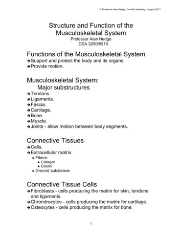
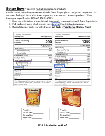
Scheele et al. Journal of Orthopaedic Surgery and Research(2019) 14:331BackgroundIn view of excellent functional results, good long-termsurvivorship, and advantages in terms of cost efficiency,unicompartmental knee arthroplasty (UKA) can make asignificant contribution to meet the rapidly growing demand for knee arthroplasty [1–5]. Since the aseptic loosening of the tibial component, misinterpretation ofradiolucent lines and cementation errors remain majorreasons for revision in UKA [6–9]; improvements of theinterface between prosthesis, cement, and trabecularbone, as well as the optimization of minimally invasivesurgical techniques, are of outstanding importance [10,11].While former studies focused on levers to improvecemented fixation that can be controlled by the surgeon,such as bone bed preparation, cementing technique, orsurgical access [11–17], little is known about the impactof bone density, which can hardly be altered, on cemented tibial fixation in UKA.The goal of this study was to quantify the impact ofregional bone density on different morphological aspectsof cement penetration in minimally invasive UKA. Inaddition, the reliable applicability of Hounsfield unit(HU) measurement, which is a simple but precise toolfor local quantification of bone density, has been validated against the established method of bone mineraldensity (BMD).Page 2 of 9MethodsEighteen fresh-frozen human cadaver knees (age 72.2,SD 14.9 years, 4 females, 14 males) were used for thisin vitro study. CT scans (Sensation 64 Somatom, Siemens AG Munich, Germany) of all tibiae were acquiredprior to the implantation in order to exclude specimenswith osseous abnormalities and to quantify BMD. Cortical and trabecular BMD [milligrams (Ca2 HA)/milliliter] were determined using Syngo Osteo CT software(Siemens AG Munich, Germany) on the proximal tibiain seven layers each in 3 mm, using a relative calibrationto water (0 HU) and calcium (200 HU). Correspondingly, local bone density was determined by measuringthe mean HU of six regions of interest (ROI) per slice,1.6 cm2 each, in the anterior, central, and posterior areaof the medial and lateral tibial plateau (Fig. 1). To capture the relevant area of implantation, both evaluationsstarted in the most cranial slice without visible corticalbone or subchondral sclerosis and assessed 2 cm distally.UKAs were implanted on the medial side of each kneevia a minimally invasive approach without eversion ofthe patella. Tibial resection was performed with an anatomical posterior slope and the removal of the cartilagein all areas of the tibial plateau was ensured intraoperatively. Bone preparation was performed using pulsatilejet lavage (Pulsavac Plus, Zimmer Biomet, USA) and ahigh viscosity bone cement (Palacos R 20 g powder/10Fig. 1 The mean Hounsefield units of six ROIs of 1.6 cm2 each, which were strictly located within the trabecular bone, were measured on eachCT slice for correlation with mean BMD. The measurement of local bone density in the anterior (red), central (blue), and posterior (yellow) area ofthe medial tibial plateau-based mean Hounsfield units was used for correlation analysis between bone density and cement penetration pattern
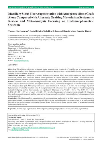
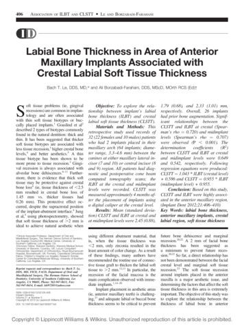
Scheele et al. Journal of Orthopaedic Surgery and Research(2019) 14:331ml monomer, Heraeus Medical Wehrheim, Germany)was mixed manually for cemented fixation. The implantation of the components was performed according tothe manufacturer’s instructions (Univation X, AesculapTuttlingen, Germany) and all operations were performedby one experienced orthopedic surgeon. The knees werethen positioned in 45 flexion and a spacer was insertedto ensure adequate pressurization during polymerizationof the cement.Afterwards, the surrounding soft tissue was removedand the medial tibial plateaus with the cemented tibialtrays were dissected (sagittal plane, at the eminentiaintercondylaris; transversal plane, 20 mm below the tibialplateau). The 18 specimens were then imbedded inTechnovit 4004 (Technovit 4004, Kulzer GmbH, Hanau,Germany) and cut into 10 frontal slices of identicalthickness. Scans with a resolution of 100 pixel/mm madethe implant–cement–bone interface accessible for morphologic evaluation using Adobe (Photoshop CS6,Adobe, San Jose, USA). Morphologic indicators of cementation were evaluated on both sides of nine cutsthrough the specimen. The anterior three serial cutsthrough the implant–cement–bone interface representedthe anterior area, the central three cuts represented thecentral area and the posterior three cuts represented theposterior area of the medial tibial plateau.The average total cement thickness was calculatedby dividing the total area of bone cement by thelength of the prosthesis (white and purple area, blackarrow in Fig. 2). Cement mantle thickness was calculated by dividing the cement area above thePage 3 of 9resection line (white in Fig. 2) by the length of thecement-prosthesis interface (defined as contact between the white area and tibial component). Cementpenetration depth was calculated by dividing the cement area below the resection line (purple in Fig. 2)by the length of the prosthesis. Furthermore, thelength proportion of the horizontal underside of thetibial component (zone 1) without visible cement intrusion into the trabecular bone was determined(Fig. 3). Finally, the proportion of interfaces with visible cement interdigitation in the area adjacent tothe anchoring peg (zone 2) of the prosthesis wasdocumented.For correlation with morphological cement penetration, bone density is quantified using HU, as this allowsselective density measurements in the individually investigated subareas of the tibial head.Results in the text are presented as an arithmeticmean, standard deviation, and minimum and maximumof the evaluated values. To calculate Pearson’s correlation coefficient between BMD and HU, all six ROIs perCT slice were taken into account. Differences in bonedensity in the different ROIs were analyzed using repeated measures ANOVA and Bonferroni’s multiplecomparison test. The effect of bone density on cementpenetration was examined by calculating regression linesand Pearson’s correlation coefficients based on all 54ROIs. All tests were two-sided and a P value of 0.05 wasconsidered significant. Statistical analysis was performedwith GraphPad Prism 5 (GraphPad Software, Inc., LaJolla, USA).Fig. 2 Image of the prosthesis-cement-bone interface: white, cement mantle (above resection line); purple, cement penetration (below resectionline). The area adjacent to the anchoring peg was defined additionally as “zone 2”
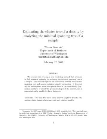
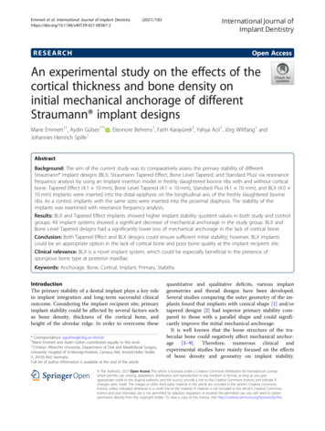
Scheele et al. Journal of Orthopaedic Surgery and Research(2019) 14:331Page 4 of 9Fig. 3 Area without visible cement penetration of bone cement into the trabecular bone in zone 1 (arrow)ResultsThe mean cortical BMD was 327.7 (SD 99.4 mg/ml)[185.8–535.6], mean trabecular BMD was 103.6 (SD29.5 mg/ml) [67.4–183.0], and mean HU was 154.0 (SD47.8) [92.0–252.8]. Between the trabecular BMD andHU, a highly significant correlation was detected with acorrelation coefficient of r 0.93 (P 0.0001; Fig. 4). Thecorrelation between the cortical BMD and HU was r 0.65 (P 0.0037). The correlation between age and HUwas not significant (r 0.36; P 0.1414).Fig. 4 Correlation between BMD and HUThe mean HU was 168.0 (SD 65.6) [83.1–294.1] in theanterior, 124.1 (SD 41.8) [55.2–215.7] in the central and175.0 (SD 56.2) [94.3–295.5] in the posterior area of themedial tibial plateau, demonstrating significant differences (P 0.0003). Differences between the anterior andcentral and between the central and posterior area weresignificant (P 0.05; Fig. 5).Based on all 54 ROIs, the mean total cement thicknesswas 2.1 (SD 0.6) [0.9–3.8], mean cement mantle thicknesswas 0.8 (SD 0.6) [0.0–2.8], and mean cement penetration
Scheele et al. Journal of Orthopaedic Surgery and Research(2019) 14:331Page 5 of 9Fig. 5 Anterior-posterior development of HU. Whiskers, 10–90 percentiledepth was 1.5 (SD 0.5) [0.6–2.7]. Only the penetrationdepth was significantly negative correlated with HU (r 0.31; P 0.023; Fig. 6). On the contrary, there was no significant correlation between the total cement thickness(r 0.24; P 0.081) or cement mantle thickness (r 0.05; P 0.70) and bone density (HU).Fig. 6 Correlation between bone density and penetration depth in area 1The average interface proportion without cement penetration in zone 1 was 31.8% SD 23.7% [0.0–80.0%]. Thecorrelation between HU and the proportion without cement penetration in zone 1 was r 0.33 (P 0.014;Fig. 7). Figure 8 shows an SEM image of an implant–cement–bone interface, that provides a morphological
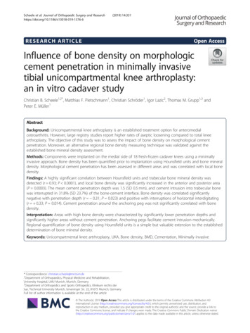
Scheele et al. Journal of Orthopaedic Surgery and Research(2019) 14:331Page 6 of 9Fig. 7 Correlation of bone density with the interruptions of penetration in area 1example of an extended area without cement intrusion inzone 1 (specimen no. 9, third cut post; approx. 164 HE).The manifestation of visible cement penetration in thearea directly adjacent to the anchoring pegs (zone 2)does not significantly correlate with bone density (r 0.24; P 0.3288). The absence of penetration in zones 1and 2 does also not correlate significantly with eachother (r 0.26; P 0.3044).DiscussionIn knee arthroplasty, achieving primary stability is crucial to prevent micromotions, which can lead to a vicious circle of membrane formation, micro fractures,radiolucent lines (RLL), and aseptic loosening [18–21].Both RLLs and aseptic loosening occur earlier on thetibial side than on the femoral side [22, 23], which emphasizes the importance of solid cementation in tibialUKA.Our results show relevant areas without cement penetration at the bone-cement interface whose occurrenceis positively correlated with bone density. In addition,the average cement penetration depth appeared reducedby high bone density. Interestingly, this relationship between bone density and penetration was not found inthe surrounding area of the anchoring pegs.As bone cement has no adhesive properties, fixation israther achieved by its intrusion into the microstructureof the trabecular bone and its area-wide mechanicalinterlocking than by a superficial conformity with irregularities of the bone bed [19, 24–26]. In other words, theprimary stability of the cemented fixation, measured asresistance to shear and tensile forces, depends oncement penetration into the adjacent trabecular bone,which is therefore is often used as an indicator of biomechanical stability [13, 24, 26–30]. Nagel et al. demonstrated that a mean tibial penetration depth of at least1.1 mm is required to maximize the fixation strength[31]. The mean cement penetration depth registered inthis study was 1.5 (SD 0.5) mm, which is well above thislower limit of penetration depth and in line with formerstudies [13, 32].The extent of cement penetration depth results fromthe interaction between the cementing technique, cement properties, and bone density. Concerning thecementing technique, it is generally accepted that cleaning of the trabecular resection surfaces is a prerequisitefor adequate cement penetration [13, 15, 32–34]. Theapplication of cement to a dry trabecular surface that isfree of blood, fat, bone marrow, and debris improves theinterlocking of the cement and thus the fixing strength[35–37]. With regard to bone cement, penetration tendsto increase with the viscosity of the cement [25, 38–40].In contrast to the previously mentioned aspects, bonedensity is a factor that cannot be easily controlled by thesurgeon. Penetration is expected to decrease with increasing bone density or decreasing porosity of the bone[26, 28, 41]. However, the specific effect of bone densityin minimally invasive implanted UKA with limited intraoperative access, thus restricted the potential of bonepreparation and component impaction, remains unclear.The presented assessment of cement penetration on serialcuts and the quantification of bone density via BMD, whichhas been adapted for extremities in other studies, can beregarded as well-established methods [15, 19, 42–45]. In
Scheele et al. Journal of Orthopaedic Surgery and Research(2019) 14:331addition, bone quality was measured via HU in the anterior,central, and posterior part of the tibial plateau.The highly significant correlation between HU and trabecular BMD underlines the applicability of HU forquantification of bone density (r 0.93; P 0.0001). Theuse of HU appears attractive as it enables a precise regional quantification of trabecular bone density. In thisanalysis, it was fundamental for the identification of thesignificantly increased average bone density in the anterior and posterior area. In combination with the pooreraccessibility for jet lavage and the more difficult pressureapplication, it uncovers the posterior tibial area, analogous to the posterior femoral condyle, as a potentialweak point of minimally invasive UKA implantation [11,15]. As regional HU measurements focused on trabecular areas, the less positive correlation between HU andcortical BMD seems plausible. The negative correlationbetween age and bone density did not reach the level ofsignificant, which might be attributable to the rather lowcase number.Based on three ROIs per specimen and the analysis of18 UKAs, the morphologic correlation analysis comprises 54 data points. Our results demonstrate a significantly negative correlation between bone density andpenetration depth (r 0.31). These values correspondto the correlation coefficient reported by Askew et al.(r 0.24) [28]. To about the same extent, bone densityrestricts area-wide cement intrusion into the trabecularbone (r 0.33). Figure 7 shows how the high bonedensity prevents the cement from penetrating into theinterspaces of the bone trabecula and thus prevents aplanar interlocking.The question to what extent the minimally invasivesurgical approach, despite the use of jet lavage, impedesthe opening of the bone substance, or if available instruments do not allow optimal pressurization of the prosthesis against the bone surface, especially in theposterior area, cannot be answered with the availabledate. In any case, we recommend the use of drill holesor special devices for microfracturing to prepare anchorPage 7 of 9wholes during the implantation of UKA in a minimallyinvasive approach.Anchoring pegs on the undersurface of the prosthesesimprove cement penetration largely independent frombone density. Our results demonstrate that in contrastto zone 1 (Fig. 8), there is no significant correlation between bone density and cement penetration around theanchoring peg in zone 2. Thus, a mechanically promotedpenetration in zone 2 may be assumed. This observationand the low correlation between penetration around thepeg and underneath the horizontal part of the component prove the sense of a differentiated assessment ofthe two zones and might influence future prosthetic design, e.g., by adding further anchoring pegs.Concerning cement application and pressurization,Dorr et al. suggested manual cement application aftermixing cement for less than 3 min and preparing thebone bed with jet lavage [46]. However, Lutz et al. statedin an in vivo study on TKA that the widespread use ofmanual cement application, despite jet lavage, is not areliable method to achieve sufficient cement penetration[14]. Experimental in vitro studies also showed thatmanual cement application leads to lower penetrationdepths than the use of a cement gun [24]. Due to theminimally invasive approach and the limited surgicalfield, manual application of bone cement was used inthis study and might add to relevant areas without cement penetration in zone 1. In addition, the choice of ahighly viscous Palacos R bone cement might also havehad a negative influence on penetration. Noble andSwarts doubted that penetration depths of more than 3mm are achievable using Palacos and a pressure of 35kPa, even after excluding all bone specimen with closedintertrabecular spaces [38]. These results are consistentwith the highest penetration of 2.7 mm observed in thisstudy.ConclusionsSpecimens with high bone density are characterized by asignificantly higher interface proportion without visibleFig. 8 SEM image, an extended area without cement penetration in area 1. Arrows, examples of how high bone density prevents bone cementfrom penetrating into the interspaces of the trabecular bone and thus prevents a mechanical interlocking
Scheele et al. Journal of Orthopaedic Surgery and Research(2019) 14:331penetration and by significantly lower penetration depthsin zone 1. In contrast, bone density does not reduce penetration around the anchoring peg, probably due to mechanical forced pressurization. Going forward, the reportedresults foster considerations of additional drill holes toopen sclerotic bone or the feasibility of pressurized cement application, additional anchoring pegs, or optimizedimpaction instruments for minimally invasive procedures.Further research is required to assess the balance of bonedensity and cement penetration with regard to biomechanical stability.AbbreviationsBMD: Bone mineral density; CT: Computed tomography; Fig.: Figure;HU: Hounsfield units; ROI: Region of interest; SEM: Scanning electronmicroscope; TKA : Total knee arthroplasty; UKA: Unicompartmental kneearthroplastyAcknowledgementsNot applicable.Authors’ contributionsHereby, I confirm that all authors have made substantial contributions to allof the following: (1) the conception and design of the study (CBS, MFP, CS,IL, TMG, PEM) acquisition of the data (CBS, MFP, CS, IL, TMG, PEM), or analysisand interpretation of the data (CBS, MFP, CS, IL, TMG, PEM); (2) drafting thearticle (CBS, MFP) or revising it critically for important intellectual content (CS,IL, TMG, PEM); and (3) final approval of the version to be submitted (CBS,MFP, CS, IL, TMG, PEM). Furthermore, each of the authors has read andconcurs with the content in the final manuscript (CBS, MFP, CS, IL, TMG,PEM).FundingAll funding and the execution of the preparation, implantation, and testingwere conducted by the Laboratory for Biomechanics and ExperimentalOrthopaedics of the Ludwig Maximilians University. There was no influenceof the manufacturer concerning the design of the study and collection,analysis, and interpretation of data and/or writing this manuscript.Availability of data and materialsThe datasets used and/or analyzed during the current study are availablefrom the corresponding author on reasonable request.Ethics approval and consent to participateThe study concept was presented to the local ethics committee of theLudwig Maximilians University of Munich (Chairman: Prof. Dr. W.Eisenmenger) and received approval. This approval included consent to theuse of human tissue (cadaver legs). The human tissue involved in this studywas obtained from the Department of Pathology of the Ludwig MaximiliansUniversity of Munich. No tissue was obtained from prisoners.Consent for publicationNot applicable.The presented manuscript does not include any details, images, or videosrelating to an individual person. The specimens pictured are entirelyunidentifiable and there are no details on individuals reported within themanuscript.Competing interestsOne author (PEM) is an advising surgeon of Aesculap R&D projects. Two ofthe authors (MFP; PEM) are getting research funding in correlation withAesculap R&D projects. The other authors declared no conflict of interest.Author details1Department of Orthopaedics, Physical Medicine and Rehabilitation,University Hospital, LMU Munich, Munich, Germany. 2Department ofOrthopedics and Sports Orthopedics, Klinikum rechts der Isar, TechnicalPage 8 of 9University Munich, Ismaninger Str. 22, 81675 Munich, Germany. 3Aesculap AGResearch & Development, Am Aesculap-Platz, 78532 Tuttlingen, Germany.Received: 21 July 2019 Accepted: 13 September 2019References1. Robertsson O, Borgquist L, Knutson K, Lewold S, Lidgren L. Use ofunicompartmental instead of tricompartmental prostheses for unicompartmentalarthrosis in the knee is a cost-effective alternative. 15,437 primarytricompartmental prostheses were compared with 10,624 primary medial orlateral unicompartmental prostheses. Acta Orthop Scand. 1999;70(2):170–5.2. Mohammad HR, Strickland L, Hamilton TW, Murray DW. Long-termoutcomes of over 8,000 medial Oxford Phase 3 Unicompartmental Knees-asystematic review. Acta Orthop. 2018;89(1):101–7.3. Inacio MCS, Paxton EW, Graves SE, Namba RS, Nemes S. Projected increasein total knee arthroplasty in the United States - an alternative projectionmodel. Osteoarthr Cartil. 2017;25(11):1797–803.4. Brown NM, Sheth NP, Davis K, Berend ME, Lombardi AV, Berend KR, DellaValle CJ. Total knee arthroplasty has higher postoperative morbidity thanunicompartmental knee arthroplasty: a multicenter analysis. J Arthroplast.2012;27(8 Suppl):86–90.5. Ghomrawi HM, Eggman AA, Pearle AD. Effect of age on cost-effectivenessof unicompartmental knee arthroplasty compared with total kneearthroplasty in the U.S. J Bone Joint Surg Am. 2015;97(5):396–402.6. Kim KT, Lee S, Lee JI, Kim JW. Analysis and treatment of complications afterunicompartmental knee arthroplasty. Knee Surg Relat Res. 2016;28(1):46–54.7. Epinette JA, Brunschweiler B, Mertl P, Mole D, Cazenave A, French Societyfor H, Knee. Unicompartmental knee arthroplasty modes of failure: wear isnot the main reason for failure: a multicentre study of 418 failed knees.Orthop Traumatol Surg Res. 2012;98(6 Suppl):S124–30.8. Saenz CL, McGrath MS, Marker DR, Seyler TM, Mont MA, Bonutti PM. Earlyfailure of a unicompartmental knee arthroplasty design with an allpolyethylene tibial component. Knee. 2010;17(1):53–6.9. van der List JP, Zuiderbaan HA, Pearle AD. Why do medial unicompartmentalknee arthroplasties fail today? J Arthroplast. 2016;31(5):1016–21.10. Liddle AD, Judge A, Pandit H, Murray DW. Adverse outcomes after total andunicompartmental knee replacement in 101,330 matched patients: a studyof data from the National Joint Registry for England and Wales. Lancet.2014;384(9952):1437–45.11. Scheele C, Pietschmann MF, Schroder C, Grupp T, Holderied M, Jansson V,Muller PE. Effect of minimally-invasive implantation of unicompartmental kneearthroplasty on cement penetration and biomechanical stability. Anexperimental study in human tibiae. Clin Biomech (Bristol, Avon). 2018;51:34–9.12. Vanlommel J, Luyckx JP, Labey L, Innocenti B, De Corte R, Bellemans J.Cementing the tibial component in total knee arthroplasty: whichtechnique is the best? J Arthroplast. 2011;26(3):492–6.13. Schlegel UJ, Siewe J, Delank KS, Eysel P, Puschel K, Morlock MM, deUhlenbrock AG. Pulsed lavage improves fixation strength of cemented tibialcomponents. Int Orthop. 2011;35(8):1165–9.14. Lutz MJ, Pincus PF, Whitehouse SL, Halliday BR. The effect of cement gunand cement syringe use on the tibial cement mantle in total kneearthroplasty. J Arthroplast. 2009;24(3):461–7.15. Clarius M, Mohr G, Jaeger S, Seeger JB, Bitsch RG. Femoral fixation pattern incemented Oxford unicompartmental knee arthroplasty--an experimentalcadaver study. Knee. 2010;17(6):398–402.16. Grupp TM, Pietschmann MF, Holderied M, Scheele C, Schroder C, Jansson V,Muller PE. Primary stability of unicompartmental knee arthroplasty underdynamic compression-shear loading in human tibiae. Clin Biomech (Bristol,Avon). 2013;28(9–10):1006–13.17. Scheele C, Pietschmann MF, Schroder C, Grupp T, Holderied M, Jansson V,Muller PE. Effect of lavage and brush preparation on cement penetrationand primary stability in tibial unicompartmental total knee arthroplasty: anexperimental cadaver study. Knee. 2017;24(2):402–8.18. Hori RY, Lewis JL. Mechanical properties of the fibrous tissue found at thebone-cement interface following total joint replacement. J Biomed MaterRes. 1982;16(6):911–27.19. Miller MA, Terbush MJ, Goodheart JR, Izant TH, Mann KA. Increased initialcement-bone interlock correlates with reduced total knee arthroplastymicro-motion following in vivo service. J Biomech. 2014;47(10):2460–6.
Scheele et al. Journal of Orthopaedic Surgery and Research(2019) 14:33120. Anthony PP, Gie GA, Howie CR, Ling RS. Localised endosteal bone lysis inrelation to the femoral components of cemented total hip arthroplasties. JBone Joint Surg Br. 1990;72(6):971–9.21. Star MJ, Colwell CW Jr, Kelman GJ, Ballock RT, Walker RH. Suboptimal (thin)distal cement mantle thickness as a contributory factor in total hiparthroplasty femoral component failure. A retrospective radiographicanalysis favoring distal stem centralization. J Arthroplast. 1994;9(2):143–9.22. Bini SA, Cafri G, Khatod M. Midterm-adjusted survival comparing the bestperforming unicompartmental and total knee arthroplasties in a registry. JArthroplast. 2017;32(11):3352–5.23. Kleeblad LJ, van der List JP, Zuiderbaan HA, Pearle AD. Regionalfemoral and tibial radiolucency in cemented unicompartmental kneearthroplasty and the relationship to functional outcomes. J Arthroplast.2017;32(11):3345–51.24. Krause WR, Krug W, Miller J. Strength of the cement-bone interface. ClinOrthop Relat Res. 1982;(163);163:290–9.25. Markolf KL, Amstutz HC. Penetration and flow of acrylic bone cement. ClinOrthop Relat Res. 1976;121:99–102.26. Walker PS, Soudry M, Ewald FC, McVickar H. Control of cement penetrationin total knee arthroplasty. Clin Orthop Relat Res. 1984;185:155–64.27. Panjabi MM, Cimino WR, Drinker H. Effect of pressure on bone cementstiffness. An in vitro study. Acta Orthop Scand. 1986;57(2):106–10.28. Askew MJ, Steege JW, Lewis JL, Ranieri JR, Wixson RL. Effect of cementpressure and bone strength on polymethylmethacrylate fixation. J OrthopRes. 1984;1(4):412–20.29. MacDonald W, Swarts E, Beaver R. Penetration and shear strength ofcement-bone interfaces in vivo. Clin Orthop Relat Res. 1993;286:283–8.30. Mann KA, Ayers DC, Werner FW, Nicoletta RJ, Fortino MD. Tensile strengthof the cement-bone interface depends on the amount of boneinterdigitated with PMMA cement. J Biomech. 1997;30(4):339–46.31. Nagel K, Bishop NE, Schlegel UJ, Puschel K, Morlock MM. The influence ofcement morphology parameters on the strength of the cement-boneInterface in tibial tray fixation. J Arthroplast. 2017;32(2):563–569 e561.32. Maistrelli GL, Antonelli L, Fornasier V, Mahomed N. Cement penetration withpulsed lavage versus syringe irrigation in total knee arthroplasty. ClinOrthop Relat Res. 1995;312:261–5.33. Miskovsky C, Whiteside LA, White SE. The cemented unicondylar kneearthroplasty. An in vitro comparison of three cement techniques. ClinOrthop Relat Res. 1992;284:215–20.34. See
Background: Unicompartmental knee arthroplasty is an established treatment option for anteromedial osteoarthritis. However, large registry studies report higher rates of aseptic loosening compared to total knee arthroplasty. The objective of this study was to assess the impact of bone density on morphological cement penetration. Moreover, an .