Transcription
HTML Version: yeasts.htmlOFD-500-P03 Antifungal Susceptibility Testing Yeasts Using Gradient Diffusion Strips1.0 Purpose/PrincipleThis document describes antifungal susceptibility testing of yeasts using gradient diffusionstrips. Currently there are at least three commercial manufacturers of gradient diffusion stripsthat measure antifungal susceptibility: Biomerieux, Liofilchem and Himed. This method ofantimicrobial testing is based upon a continuous concentration gradient of drug infused on aplastic non-porous strip. The antifungal agent diffuses into an agar plate and allows an accuratedetermination of MIC values based on elliptical growth around the antifungal gradient.2.0 ScopeThis procedure covers growth and dilution of the yeast, inoculation of the plates, and reading ofthe results.3.0 Related DocumentsTitleDocumentReference Method for Broth Dilution AntifungalSusceptibility Testing for YeastsClinical and Laboratory StandardsInstitute standard M27, 4th Ed.Performance Standards for Antifungal SusceptibilityTesting of YeastsClinical and Laboratory StandardsInstitute document M60, 2nd Ed.Gradient Diffusion strip package insertBiomerieux amphotericin B Etestpackage insert (or similar)4.0 plement and carry out the procedureTechnical SupervisorInsure training and proficiency testing5.0 Equipment5.1. Vortex5.2. Spectrophotometer or nephelometer5.3. Incubator set to 35 COFD-500-P03 Rev 2Document not ControlledEffective date: 11/12//2020
6.0 Reagents, media, and supplies6.1. Sabouraud’s Dextrose (SAB) agar plates (example: ThermoFisher Scientific, Product#R112562) or equivalent6.2. RPMI-1640 media (with glutamic acid and phenol red, without bicarbonate; example:Sigma-Aldrich, Product #R1383) or equivalent6.3. MOPS (3-[N-morpholino] propanesulfonic acid) buffer (example: Sigma-Aldrich, Product#M3183) or equivalent6.4. D-( )-glucose (example: Sigma Aldrich, Item# G8270)6.5. Sterile water (example: Fisher Scientific, Catalog #15-230-162)6.6. Sodium hydroxide (NAOH) solution, 1mol/L (example: Millipore Sigma, Product#1370311002) or equivalent6.7. 0.2 micron filter (example: Fisher Scientific, Catalog #03-377-137) or equivalent6.8. BD Bacto Dehydrated Agar (example: VWR, Catalog #214010) or equivalent6.9. Petri dish (100 x 15mm) (example: Fisher Scientific, Catalog #FB0875712)6.10. Sterile glass tubes with screw top lid or cuvettes for use with your spectrometer ornephelometer – (make sure they fit your instrument)6.11. Micropipet, size 10-100µL6.12. Micropipet tips, size 10-100µL6.13. Plastic box with lid for incubator to keep up humidity6.14. Sterile cotton swabs (example: BD Falcon Polyester Fiber-tipped Applicator Swab, Ref220690) or equivalent6.15. Plastic transfer pipets (example: Fisherbrand, Cat # 13-711-20) or equivalent6.16. Gradient Diffusion strips ( store at -20 C) (example: Amphotericin B Etest strips;BioMerieux, SKU 526348) or equivalent6.17. Quality control isolates C. parapsilosis ATCC 22019 and C. krusei ATCC 6258 which areavailable from the American Type Culture Collection (www.atcc.org)6.18. Plastic 1 µL inoculating loop (example: Thermo Scientific 253287)6.19. 0.5 McFarland Standard if using a nephelometer (example: Thermo Scientific R20410)6.20. Forceps6.21. RPMI agar and Sabouraud Dextrose agar plates are available from commercial sources.When necessary, RPMI agar may be prepared using the formulation in Section 7.0.OFD-500-P03 Rev 2Document not ControlledEffective date: 11/12//2020
7.0 Preparation of RPMI plates with 20g/L D-glucose:7.1. Dissolve 10.4 g RPMI 1640 powder (with glutamic acid and phenol red, withoutbicarbonate), 34.5 g MOPS (3-[N-morpholino] propanesulfonic acid) buffer, and 20g Dglucose into 450 ml of distilled H2O.7.2. Adjust to pH 7.0 at 25 C using 1mol/L NaOH.7.3. Filter sterilize using a 0.2 micron filter and place in hot water bath to increase temperatureto 50 C.7.4. Add 15 g of agar to 400 mL of distilled H2O.7.5. Autoclave for 20 min to sterilize.7.6. Add the sterile agar to the RPMI broth, adjust the final volume to 1 L with sterile distilledH2O and stir to mix.7.7. In a BSC, aseptically dispense 25 mL per petri dish and allow the agar to solidify, with lidajar. Once the dishes have come to room temperature, store at 4 C for up to 6 months.8.0 Safety PrecautionsNote: All institutional safety procedures must be followed when performing this standard operatingprocedure.8.1. Standard personal protective equipment should follow institutional guidelines but shouldconsist of at least labcoat, gloves, and safety glasses which should be worn at all times.8.2. Surfaces and supplies should be decontaminated using 1:128 Lysol solution or otherdecontaminant for yeasts approved by your institution.8.3. Most Candida species can be processed at the bench using standard BSL2 precautions (labcoat, gloves, and safety glasses), if consistent with your institution’s safety procedures. Oneexception: Candida auris cultures must be handled in a BSC. Gloves should be worn at alltimes and BSC decontaminated with OxivirTB or freshly made 10% bleach.9.0 Quality Control9.1. Quality control isolates C. parapsilosis ATCC 22019 and C. krusei ATCC 6258 shouldboth be run each time susceptibility testing is performed. QC isolates are aliquoted andstored frozen. A new frozen aliquot is plated every two weeks. Repeated passage of the QCisolates will result in susceptibility results that are outside of the QC range.9.2. The QC range for these isolates can be found in CLSI document M60. (See Appendix Bbelow). If the QC isolates are out of range, all of the other results should be discarded.OFD-500-P03 Rev 2Document not ControlledEffective date: 11/12//2020
10.0Protocol for isolate preparation10.1. Starting from a frozen yeast stock10.1.1. Streak the test isolate and two control isolates (see note below) on separate SABagar plates and allow them to grow for 24 hours at 35-37 C.10.1.2. Re-streak from this plate onto a new SAB agar plate, streaking for isolation ofcolonies, and allow this second plate to grow for 24 hours at 35-37 C.10.1.3. The original SAB agar plate containing growth directly from the freezer (step 10.1.1)may be kept at 4 C and used for up to 2 weeks. Discard this plate after 2 weeks.10.2. Starting from water stock or an original plate10.2.1. Streak the test isolate and two control isolates (see note below) for isolation onseparate SAB agar plates and allow them to grow for 24 hours at 35-37 C. It is onlynecessary to subculture once.NOTE: Be sure to also use a fresh culture of the two quality control isolates ATCC 22019 (C.parapsilosis) and ATCC 6258 (C. krusei).11.0Protocol for susceptibility testing using gradient diffusion strips11.1. Remove RPMI agar plates from 4 C storage and allow them to reach room temperature.11.2. Take the appropriate number of gradient diffusion strips out of -20 C storage and allowthem to come to room temperature. NOTE: Do not touch the face of the strips, and do notlay down on a non-sterile surface. Always handle the strips by the end, and always useforceps.11.3. If using a spectrophotometer, follow the instructions in the owners manual for use. Brieflyturn on and allow to warm up for approximately 5-10 minutes. Once the machine iswarmed up, choose percent (%) transmittance option. Set the wavelength to 530 nm.Note: If using a nephelometer, it does not need to be warmed up.11.4. Label one 13x100 glass tube (or whatever tube fits the instrument) per isolate with theisolate identifying number. Aliquot 2mL of sterile water into the glass tube. Place tube intothe spectrophotometer and press the button labelled “0 ABS 100% T” to blank themachine to 0% transmittance.11.5. Using the wooden end of a sterile wooden cotton swab, making sure not to touch orcontaminate the wooden end (or you can use a sterile plastic 1 µL inoculating loop), pick2-3 small yeast colonies (1 mm or smaller) from the fresh culture plate (prepared in step10) and suspend them in the 2 ml of sterile water in the glass tube by tapping against theinside of the tube.11.6. Carefully vortex the resulting suspension for 15 seconds to remove clumps.OFD-500-P03 Rev 2Document not ControlledEffective date: 11/12//2020
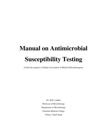
11.7. Density reading of the yeast suspension using a spectrophotometer.11.7.1. Place the tube in the spectrophotometer. The percent transmittance of light of thesuspension should fall within 80% to 82%.11.7.1.1.If percent transmittance of light is below 80%, add additional sterile water11.7.1.2.If percent transmittance is of light above 82%, add additional yeast cells11.7.2. When percent transmittance of light falls within 80% to 82% the yeast stocksuspension will yield 1-5 x 10⁶ cells per ml.11.8. Density reading of the yeast suspension using the nephelometer.11.8.1. Place the tube in the nephelometer. The reading should fall within the 0.5McFarland range.11.8.1.1.If the density is too high, add additional sterile water11.8.1.2.If the density is too low, add additional yeast cells.11.8.1.3. When the reading falls within the 0.5 McFarland range the yeast stocksuspension will yield 1-5 x 10⁶ cells per ml.11.9. Dip a sterile cotton swab in the glass tube with the adjusted yeast cell suspension, andremove excess fluid by pressing it against the inside wall of the tube.11.10. Streak an RPMI agar plate completely in three directions as shown below, making surethe entire plate is covered. Do not re-dip into the adjusted yeast cell suspension atany time (this will result in an inoculum that is too dense and can lead to falseresistance). This is different from what the package insert suggests to do.11.11. Allow the plate to air dry in the BSC with the lid cracked open for up to 15 minutes if itis excessively moist.11.12. Pick up the gradient diffusion strip using forceps, being careful to only touch the stripupward of the dark black line. NOTE: The area below this dark black line containsantifungal drug and should not be touched by hand or forceps.11.13. Apply the gradient diffusion strip to plates with the MIC numbers facing upward, beingcareful to not allow bubbles under the strip. Note: If bubbles occur, remove them byOFD-500-P03 Rev 2Document not ControlledEffective date: 11/12//2020
gently pressing down with the forceps, being careful not to slide the strip. DO NOT liftthe strip and replace it.11.14. Replace plate lid and place plate in a container that will maintain humidity (such as aplastic shoe box) and incubate at 35 C for 24 hours.12.0Protocol for reading gradient diffusion strips (Etests) and interpretation of the results12.1. After 24 hours of growth at 35 C the gradient diffusion strips are ready to read.12.2. Gradient diffusion strips are read visually by opening the plate and observing how thegrowth intersects with the testing strip.12.3. First read the gradient strips for the two QC isolates. Using Appendix A, make sure theQC isolates are in range. If either of the isolates are not in range, the values for theantifungal in question should not be used.12.4. If a sufficient lawn of growth is not obtained after 24 hours, the plate can be incubated forup to another 24 hours. Note: Only in rare occasions and with certain species would thisoccur if the steps in Section 11 are performed correctly.12.5. An ellipse where growth is not present may be seen in the lawn of growth, with the growthconcentrated near the MIC endpoint. Isolates which are entirely resistant to that antifungaldrug will show no ellipse.12.6. Always read the value on the side of the strip with the higher of the MIC values. If growthis inhibited between two MIC values, use the greater of the two values as the MIC.12.7. The interpretation of the MIC value is dependent upon the antifungal drug class. Forpolyenes (amphotericin B), the MIC is interpreted as the value where there is 100%growth inhibition. For azole and echinocandin antifungals, the MIC is interpreted as thevalue where there is 80% growth inhibition.12.8. See Appendix B (Etest manufacturer’s instructions for interpretive guidelines)13.0Reference Values, Alert Values13.1. Reference and alert values for resistant isolates can be found in CLSI document M6014.0References14.1. Clinical and Laboratory Standards Institute. 2017. Reference Method for Broth DilutionAntifungal Susceptibility Testing of Yeasts; Approved Standard – Fourth Edition, CLSIDocument M27-A4. Clinical and Laboratory Standards Institute, Wayne, PA.14.2. Clinical and Laboratory Standards Institute. 2017. Performance Standards for AntifungalSusceptibility Testing of Yeasts; Approved Standard – First Edition, CLSI DocumentM60. Clinical and Laboratory Standards Institute, Wayne, PA.OFD-500-P03 Rev 2Document not ControlledEffective date: 11/12//2020
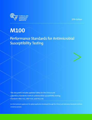
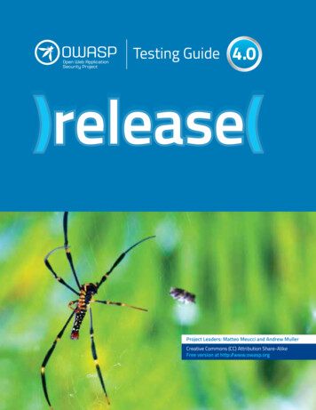
Appendix AQuality Control RangesRecommended 24-hour Minimal Inhibitory Concentration Limits for Two Quality ControlStrains for Broth Microdilution ProceduresReference: Clinical and Laboratory Standards Institute (CLSI). Performance Standards forAntifungal Susceptibility Testing of Yeasts. 1st ed. CLSI supplement M60 (ISBN 1-56238828-2 [Print]; ISBN 1-56238-829-0 [Electronic]). Clinical and Laboratory StandardsInstitute, 950 West Valley Road, Suite 2500, Wayne, Pennsylvania 19087 USA, 2017.OFD-500-P03 Rev 2Document not ControlledEffective date: 11/12//2020
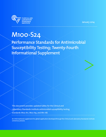
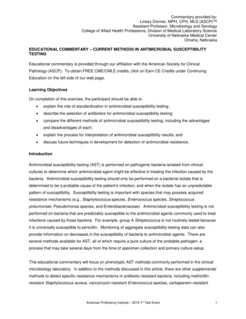
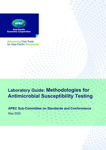
Appendix BEtest Antifungal Reading Guide from the package insert for Etests from hlib/techlib/documents/docLink/Package Insert/3590400135905000/Package Insert - 9305056 - D - en - Etest - AFST WW.pdf) Edition 2013/02.Accessed 9/1/2020.OFD-500-P03 Rev 2Document not ControlledEffective date: 11/12//2020
OFD-500-P03 Rev 2Document not ControlledEffective date: 11/12//2020
OFD-500-P03 Rev 2Document not ControlledEffective date: 11/12//2020
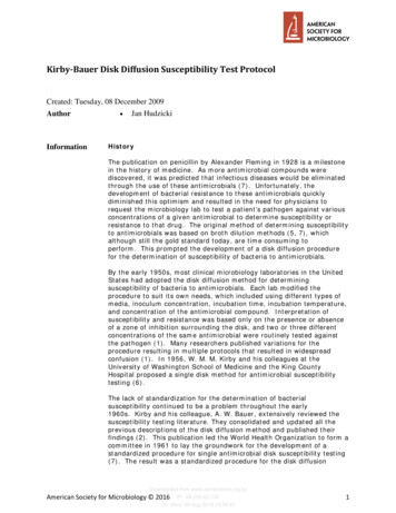
OFD-500-P03 Rev 2Document not ControlledEffective date: 11/12//2020
DISCLAIMER:The Mycotic Diseases Branch laboratory developed this document as an example test procedure forAntifungal Susceptibility Testing by Gradient Diffusion. It is the responsibility of the testinglaboratory to ensure content and format are modified as necessary to meet applicable regulatoryrequirements, quality management system standards, and chemical, radiological and biologicalsafety requirements. This is not a controlled document, and the described test methods are subject tochange without notice. It is the responsibility of the testing laboratory to ensure the informationwithin this document remains applicable. Contact the test developer at gyi2@cdc.gov to find outwhether any changes have been made.Use of trade names and commercial sources is for identification only and does not constituteendorsement by the Public Health Service or by the United States Department of Health and HumanServices.OFD-500-P03 Rev 2Document not ControlledEffective date: 11/12//2020
This document describes antifungal susceptibility testing of yeasts using gradient diffusion . This method of antimicrobial testing is based upon a continuous concentration gradient of drug infused on a plastic non-porous strip. The antifungal agent diffuses into an agar plate and allows an accurate . Performance Standards for Antifungal .