Transcription
Kirby-Bauer Disk Diffusion Susceptibility Test Protocol Created: Tuesday, 08 December 2009AuthorInformation Jan HudzickiHistoryThe publication on penicillin by Alexander Fleming in 1928 is a milestonein the history of medicine. As more antimicrobial compounds werediscovered, it was predicted that infectious diseases would be eliminatedthrough the use of these antimicrobials (7). Unfortunately, thedevelopment of bacterial resistance to these antimicrobials quicklydiminished this optimism and resulted in the need for physicians torequest the microbiology lab to test a patient’s pathogen against variousconcentrations of a given antimicrobial to determine susceptibility orresistance to that drug. The original method of determining susceptibilityto antimicrobials was based on broth dilution methods (5, 7), whichalthough still the gold standard today, are time consuming toperform. This prompted the development of a disk diffusion procedurefor the determination of susceptibility of bacteria to antimicrobials.By the early 1950s, most clinical microbiology laboratories in the UnitedStates had adopted the disk diffusion method for determiningsusceptibility of bacteria to antimicrobials. Each lab modified theprocedure to suit its own needs, which included using different types ofmedia, inoculum concentration, incubation time, incubation temperature,and concentration of the antimicrobial compound. Interpretation ofsusceptibility and resistance was based only on the presence or absenceof a zone of inhibition surrounding the disk, and two or three differentconcentrations of the same antimicrobial were routinely tested againstthe pathogen (1). Many researchers published variations for theprocedure resulting in multiple protocols that resulted in widespreadconfusion (1). In 1956, W. M. M. Kirby and his colleagues at theUniversity of Washington School of Medicine and the King CountyHospital proposed a single disk method for antimicrobial susceptibilitytesting (6).The lack of standardization for the determination of bacterialsusceptibility continued to be a problem throughout the early1960s. Kirby and his colleague, A. W. Bauer, extensively reviewed thesusceptibility testing literature. They consolidated and updated all theprevious descriptions of the disk diffusion method and published theirfindings (2). This publication led the World Health Organization to form acommittee in 1961 to lay the groundwork for the development of astandardized procedure for single antimicrobial disk susceptibility testing(7). The result was a standardized procedure for the disk diffusionDownloaded from www.asmscience.org byIP: 66.208.62.130On: Wed, 29 Aug 2018 12:55:47American Society for Microbiology 20161
susceptibility test, henceforth called the Kirby-Bauer disk diffusion test(2).Currently, the Clinical Laboratory Standards Institute (CLSI) isresponsible for updating and modifying the original procedure of Kirbyand Bauer through a global consensus process. This ensures uniformityof technique and reproducibility of results as pathogens develop newmechanisms of resistance and new antimicrobials are developed to fightthese organisms. Interpretative guidelines for zone sizes are included intheir publications (3). The CLSI publication, Performance Standards forAntimicrobial Disk Susceptibility Tests; Approved Standard 9th Edition,represents the standard for clinical laboratories performing susceptibilitytesting today.PurposeThe purpose of the Kirby-Bauer disk diffusion susceptibility test is todetermine the sensitivity or resistance of pathogenic aerobic andfacultative anaerobic bacteria to various antimicrobial compounds inorder to assist a physician in selecting treatment options for his or herpatients. The pathogenic organism is grown on Mueller-Hinton agar inthe presence of various antimicrobial impregnated filter paper disks. Thepresence or absence of growth around the disks is an indirect measure ofthe ability of that compound to inhibit that organism.TheoryDetermination of bacterial resistance to antimicrobials is an importantpart of the management of infections in patients. The disk diffusionmethod of Kirby and Bauer has been standardized and is a viablealternative to broth dilution methods for laboratories without theresources to utilize the newer automated methods for broth microdilutiontesting.When a 6-mm filter paper disk impregnated with a known concentrationof an antimicrobial compound is placed on a Mueller-Hinton (MH) agarplate, immediately water is absorbed into the disk from the agar. Theantimicrobial begins to diffuse into the surrounding agar. The rate ofdiffusion through the agar is not as rapid as the rate of extraction of theantimicrobial out of the disk, therefore the concentration of antimicrobialis highest closest to the disk and a logarithmic reduction in concentrationoccurs as the distance from the disk increases (7). The rate of diffusionof the antimicrobial through the agar is dependent on the diffusion andsolubility properties of the drug in MH agar (2) and the molecular weightof the antimicrobial compound. Larger molecules will diffuse at a slowerrate than lower molecular weight compounds. These factors, incombination, result in each antimicrobial having a unique breakpointzone size indicating susceptibility to that antimicrobial compound.If the agar plate has been inoculated with a suspension of the pathogento be tested prior to the placing of disks on the agar surface,simultaneous growth of the bacteria and diffusion of the antimicrobialcompounds occurs. Growth occurs in the presence of an antimicrobialDownloaded from www.asmscience.org byIP: 66.208.62.130On: Wed, 29 Aug 2018 12:55:47American Society for Microbiology 20162
compound when the bacteria reach a critical mass and can overpower theinhibitory effects of the antimicrobial compound. The estimated time of abacterial suspension to reach critical mass is 4 to 10 hours for mostcommonly recovered pathogens, but is characteristic of each species, andinfluenced by the media and incubation temperature (7). The size of thezone of inhibition of growth is influenced by the depth of the agar, sincethe antimicrobial diffuses in three dimensions, thus a shallow layer ofagar will produce a larger zone of inhibition than a deeper layer.The point at which critical mass is reached is demonstrated by a sharplymarginated circle of bacterial growth around the disk. The concentrationof antimicrobial compound at this margin is called the criticalconcentration and is approximately equal to the minimum inhibitoryconcentration obtained in broth dilution susceptibility tests.Zone size observed in a disk diffusion test has no meaning in and ofitself (7). The interpretation of resistance and susceptibility toantimicrobials is determined through in vivo testing of blood and urine tocalculate the obtainable level of a given antimicrobial that results inresolution of an infection. This information is correlated with zone sizesresulting in the interpretive standards. The current interpretationstandards can be found in the Clinical Laboratory StandardsInstitute Performance Standards for Antimicrobial Disk SusceptibilityTests: Approved Standards 9th Edition (3).RECIPESterile saline in 2-ml tubes0.5 McFarland standardWickerham cardMueller-Hinton agar plates, 100 mm or 150 mmCaliper or rulerAntibiotic disksbForcepsAntibiotic disk dispenser (optional)18- to 24-hoVortexSterile swabsInoculating loBact-cineratoAlcohol pads35 C to 37 CaRecommended organisms for quality assurance purposesare Staphylococcus aureus ATCC 25923 (Biosafety level (BSL)2), Escherichia coli ATCC 25922 (BSL 1), and Pseudomonasaeruginosa ATCC 27853 (BSL 2) (www.atcc.org), as the zone of inhibitionfor these organisms is known. Because the zone sizes are known forthese organisms, they are recommended for use in the educationalsetting, although the use of unknowns should also be incorporated intothe educational experience. For quality control testing, the zone sizes forthese three organisms can be found on the package insert from anyantimicrobial disk you purchase.bSelection of antimicrobial is based on the type of organism being testedand source of the isolate (blood, urine, wound, etc.). See theInterpretative Standards Tables for suggested antimicrobials to use inthis exercise.Additional NotesDownloaded from www.asmscience.org byIP: 66.208.62.130On: Wed, 29 Aug 2018 12:55:47American Society for Microbiology 20163
Mueller-Hinton agarMH agar is considered the best medium to use for routine susceptibilitytesting of nonfastidious bacteria for the following reasons:·It shows acceptable batch-to-batch reproducibility for susceptibilitytesting·It is low in sulfonamide, trimethoprim, and tetracycline inhibitors·It supports satisfactory growth of most nonfastidious pathogens·A large body of data and experience has been collected concerningsusceptibility tests performed with this medium (6).Please note that the use of media other than Mueller-Hinton agar mayresult in erroneous results. Also note that only the aerobic or facultativebacteria that grow well on unsupplemented MH agar should be testedusing this protocol. Fastidious organisms require MH agar supplementedwith additional nutrients and require that modification to this protocol bemade. Neither the supplements nor the procedural modification arediscussed in this basic protocol.MH agar may be purchased as prepared agar platesfrom Remel (Lenexa, KS), BD BBL (Franklin Lakes, NJ ), or anyother supplier of prepared agar plates. Follow the manufacturer’srecommendation for storage of prepared plates. MH agar can also beprepared from dehydrated media available from companies such asRemel, BD BBL, or any other supplier of dehydrated media. Be sure toprepare the media according to the manufacturer’s directions.Formula for Mueller-Hinton agar per liter of purified water (4)Beef, Infusion fromCasamino acid, technicalStarchAgar300.0 g17.5 g1.5 g17.0 gSuspend the components listed above in 1 liter of purified water. Mixthoroughly. Heat with frequent agitation and boil for 1 minute tocompletely dissolve the components. Autoclave at 121 C for 15minutes. Dispense as desired. Allow to solidify at room temperature,then store at 4 to 8 C. Mueller-Hinton agar is stable for approximately70 days (per Remel Technical Services, 1 September 2009) from the dateof preparation. Each lab should verify the quality and functionality ofeach batch of prepared media by testing known strains of organismsagainst each antimicrobial compound being used as the 70-day expirationdate approaches.·If you prepare the MH agar plates from dehydrated media, theplates must be poured to a depth of 4 mm (approximately 25 ml of liquidagar for 100-mm plates and 60 ml of liquid agar for 150-mm plates, butin any case to a measured depth of 4 mm). Plates that are too shallowwill produce false susceptible results as the antimicrobial compound willdiffuse further than it should, creating larger zones ofDownloaded from www.asmscience.org byIP: 66.208.62.130On: Wed, 29 Aug 2018 12:55:47American Society for Microbiology 20164
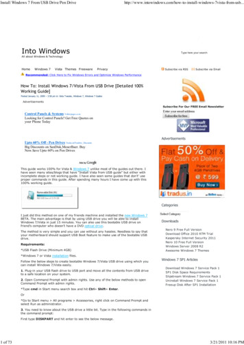
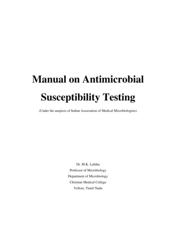
inhibition. Conversely, plates poured to a depth 4 mm will result infalse resistant results.·pH of the MH agar should fall between 7.2 and 7.4 at roomtemperature after solidification and should be tested when the media isfirst prepared. If the pH is 7.2 certain drugs will appear to lose potency(aminoglycosides, quinolones, macrolides), while other agents mayappear to have excessive activity (tetracycline). If the pH is 7.4, theopposite results may occur.·Excessive thymidine or thymine can reverse the inhibitory effectsof sulfonamides and trimethoprim resulting in smaller and less distinctzones of inhibition, or no zones at all.·The incorrect concentration of divalent cations (calcium andmagnesium) will affect the results of aminoglycoside and tetracyclinetests against Pseudomonas aeruginosa. Excess cation concentration willresult in reduced zone sizes and low concentration will increase zonesizes. Excess calcium will increase the zone size of P. aeruginosa againstdaptomycin. Excess zinc ions may reduce the zone size of carbapenemsagainst P. aeruginosa.·MH agar should be tested with known strains of organism at leastweekly in order to verify that the media and disks are working asexpected.Antibiotic susceptibility disksAntimicrobial disks can be purchased from any reputable suppliers, suchas Remel or BD BBL. They are packaged in spring-loaded cartridgescontaining 25 or 50 disks and can be ordered as individual cartridges orin packages of 10 cartridges. Proper storage of these disks is essentialfor reproducible results.Sealed cartridges containing commercially prepared paper disks shouldbe stored at either 8 C or frozen at -14 C in a non-self-defrostingfreezer. Allow disks to come to room temperature prior to removing theprotective plastic packaging. Once opened, store the cartridges in astorage container containing desiccant for no more than 1 week.Semiautomatic disk dispensers are available from companies such asRemel and BD BBL. Be aware that disk cartridges from one companymay not fit the dispenser of another company.McFarland standardMcFarland standards are suspensions of either barium sulfate or latexparticles that allow visual comparison of bacterial density (Fig.1). Commercially prepared standards are available for purchase fromcompanies such as Remel or BD BBL. These often include a Wickerhamcard, which is a small card containing parallel black lines. A 0.5McFarland standard is equivalent to a bacterial suspension containingbetween 1 x 10⁸ and 2 x 10⁸ CFU/ml of E. coli.Downloaded from www.asmscience.org byIP: 66.208.62.130On: Wed, 29 Aug 2018 12:55:47American Society for Microbiology 20165
A 0.5 McFarland standard may be prepared in-house as describe below.1.Add a 0.5-ml aliquot of a 0.048 mol/liter BaCl2 (1.175% wt/volBaCl2 2H20) to 99.5 ml of 0.18 mol/liter H2SO4 (1% vol/vol) withconstant stirring to maintain a suspension.2.Verify the correct density of the turbidity standard by measuringabsorbance using a spectrophotometer with a 1-cm light path andmatched cuvette. The absorbance at 625 nm should be 0.08 to 0.13 forthe 0.5 McFarland standard.3.Transfer the barium sulfate suspension in 4- to 6-ml aliquots intoscrew-cap tubes of the same size as those used in standardizing thebacterial inoculums.4.Tightly seal the tubes and store in the dark at room temperature.Use of the McFarland standard in the Kirby-Bauer procedure.1.Prior to use, vigorously agitate the barium sulfate standard on amechanical vortex mixer and inspect for a uniformly turbidappearance. Replace the standard if large particles appear. If using astandard composed of latex particles, mix by inverting gently, not on avortex mixer.2.As the student adds bacterial colonies to the saline in the“preparation of the inoculum” step of the procedure, he or she shouldcompare the resulting suspension to the McFarland standard. This isdone by holding both the standard and the inoculum tube side by sideand no more than 1 inch from the face of the Wickerham card (withadequate light present) and comparing the appearance of the linesthrough both suspensions. Do not hold the tubes flush against thecard. If the bacterial suspension appears lighter than the 0.5 McFarlandstandard, more organisms should be added to the tube from the cultureplate. If the suspension appears more dense than the 0.5 McFarlandstandard, additional saline should be added to the inoculum tube in orderto dilute the suspension to the appropriate density. In some cases itmay be easier to start over rather than to continue to dilute a bacterialsuspension that is too dense for use.Downloaded from www.asmscience.org byIP: 66.208.62.130On: Wed, 29 Aug 2018 12:55:47American Society for Microbiology 20166
FIG. 1. McFarland standards (left to right) 0.5, 1.0, 2.0, 3.0, positionedin front of a Wickerham card. McFarland standards are used to preparebacterial suspensions to a specified turbidity. In the Kirby-Bauer diskdiffusion susceptibility test protocol, the bacterial suspension of theorganism to be tested should be equivalent to the 0.5 McFarlandstandard.PROTOCOLPreparation of Mueller-Hinton plate1.Allow a MH agar plate (one for each organism to be tested) tocome to room temperature. It is preferable to allow the plates to remainin the plastic sleeve while they warm to minimize condensation.2.If the surface of the agar has visible liquid present, set the plateinverted, ajar on its lid to allow the excess liquid to drain from the agarsurface and evaporate. Plates may be placed in a 35 C incubator or in alaminar flow hood at room temperature until dry (usually 10 to 30minutes).3.Appropriately label each MH agar plate for each organism to betested.Preparation of inoculum1.Using a sterile inoculating loop or needle, touch four or five isolatedcolonies of the organism to be tested.2.Suspend the organism in 2 ml of sterile saline.3.Vortex the saline tube to create a smooth suspension.Downloaded from www.asmscience.org byIP: 66.208.62.130On: Wed, 29 Aug 2018 12:55:47American Society for Microbiology 20167
4.Adjust the turbidity of this suspension to a 0.5 McFarland standardby adding more organism if the suspension is too light or diluting withsterile saline if the suspension is too heavy.5.Use this suspension within 15 minutes of preparation.Additional NotesInoculum preparationOrganisms to be tested must be in the log phase of growth in order forresults to be valid. It is recommended that subcultures of the organismsto be tested be made the previous day.Never use extremes in inoculum density. Never use undiluted overnightbroth cultures or other unstandardized inocula for inoculating plates.If the organism is difficult to suspend directly into a smooth suspension,the growth method of preparing the inoculums should be used. However,the recommended organisms listed in this procedure all produce smoothsuspensions with little difficulty. See the Clincial Laboratory StandardsInstitute document (3) for the growth procedure method for preparingthe inoculums, if needed.Inoculation of the MH plate1.Dip a sterile swab into the inoculum tube.2.Rotate the swab against the side of the tube (above the fluid level)using firm pressure, to remove excess fluid. The swab should not bedripping wet (Fig. 2).3.Inoculate the dried surface of a MH agar plate by streaking theswab three times over the entire agar surface; rotate the plateapproximately 60 degrees each time to ensure an even distribution of theinoculum (Fig. 3).4.Rim the plate with the swab to pick up any excess liquid (Fig. 4).5.Discard the swab into an appropriate container.6.Leaving the lid slightly ajar, allow the plate to sit at roomtemperature at least 3 to 5 minutes, but no more than 15 minutes, forthe surface of the agar plate to dry before proceeding to the next step.Downloaded from www.asmscience.org byIP: 66.208.62.130On: Wed, 29 Aug 2018 12:55:47American Society for Microbiology 20168
FIG. 2. Kirby-Bauer disk diffusion susceptibility test protocol, inoculationof the test plate. Step 2. Rotate the swab against the side of the tubewhile applying pressure to remove excess liquid from the swab prior toinoculating the plate.ADownloaded from www.asmscience.org byIP: 66.208.62.130On: Wed, 29 Aug 2018 12:55:47American Society for Microbiology 20169
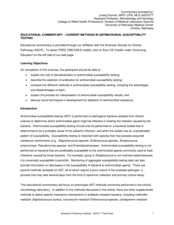
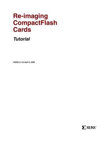
BFIG. 3. Kirby-Bauer disk diffusion susceptibility test protocol, inoculationof the Mueller-Hinton agar plate. Step 3. (A) Inoculate the plate withthe test organism by streaking the swab in a back-and-forth motion veryclose together as you move across and down the plate. Rotate the plate60 and repeat this action. Rotate the plate once more and repeat thestreaking action. This ensures an even distribution of inoculum that willresult in a confluent lawn of growth. (B) Diagram illustrating the patternthe swab should follow as it is drawn across the plate.FIG. 4. Kirby-Bauer disk diffusion susceptibility test protocol, inoculationof the Mueller-Hinton agar plate. Step 4. After streaking the MuellerHinton agar plate as described in Step 3, rim the plate with the swab byrunning the swab around the edge of the entire the plate to pick up anyexcessive inoculum that may have been splashed near the edge. TheDownloaded from www.asmscience.org byIP: 66.208.62.130On: Wed, 29 Aug 2018 12:55:47American Society for Microbiology 201610
arrow indicates the path of the swab.Placement of the antibiotic disks1.Place the appropriate antimicrobial-impregnated disks on thesurface of the agar, using either forceps to dispense each antimicrobialdisk one at a time, or a multidisk dispenser to dispense multiple disks atone time. (See steps a. through d. for the use of the multi-diskdispenser or steps e. through g. for individual disk placement withforceps.a.To use a multidisk dispenser, place the inoculated MH agar plate ona flat surface and remove the lid (Fig. 5).b.Place the dispenser over the agar plate and firmly press theplunger once to dispense the disks onto the surface of the plate.c.Lift the dispenser off the plate and using forceps sterilized by eithercleaning them with an alcohol pad or flaming them with isopropyl alcohol,touch each disk on the plate to ensure complete contact with the agarsurface. This should be done before replacing the petri dish lid as staticelectricity may cause the disks to relocate themselves on the agarsurface or adhere to the lid (Fig. 6d).d.Do not move a disk once it has contacted the agar surface even ifthe disk is not in the proper location, because some of the drug begins todiffuse immediately upon contact with the agar.e.To add disks one at a time to the agar plate using forceps, placethe MH plate on the template (Fig. 7) provided in this procedure (Fig.6a). Sterilize the forceps by cleaning them with a sterile alcohol pad andallowing them to air dry or immersing the forceps in alcohol then igniting.f.Using the forceps carefully remove one disk from the cartridge(Fig. 6b).g.Partially remove the lid of the petri dish. Place the disk on theplate over one of the dark spots on the template and gently press thedisk with the forceps to ensure complete contact with the agar surface.Replace the lid to minimize exposure of the agar surface to room air (Fig.6c, d).h.Continue to place one disk at a time onto the agar surface until alldisks have been placed as directed in steps f. and g. above.2.Once all disks are in place, replace the lid, invert the plates, andplace them in a 35 C air incubator for 16 to 18 hours. Whentesting Staphylococcus against oxacillin or vancomycin,or Enterococcus against vancomycin, incubate for a full 24 hours beforereading.Downloaded from www.asmscience.org byIP: 66.208.62.130On: Wed, 29 Aug 2018 12:55:47American Society for Microbiology 201611
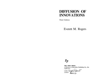
ABCFIG. 5. Kirby-Bauer disk diffusion susceptibility test protocol, placementof antibiotic disks using an automated disk dispenser. Step 1, a. throughd. An automatic disk dispenser can be used to place multiple diskssimultaneously on a MH agar plate. () Set the dispenser over the plate.(B) Place the palm of your hand on the top of the handle. (C) Press downfirmly and completely to dispense the disks. The spring loaded handlewill return to the original position when pressure is removed.Downloaded from www.asmscience.org byIP: 66.208.62.130On: Wed, 29 Aug 2018 12:55:47American Society for Microbiology 201612
ABCDownloaded from www.asmscience.org byIP: 66.208.62.130On: Wed, 29 Aug 2018 12:55:47American Society for Microbiology 201613
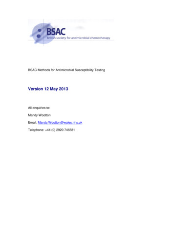
DFIG. 6. Kirby-Bauer disk diffusion susceptibility test protocol, placementof antibiotic disks using forceps to manually place the disks. Step 1, e.through h. Antibiotic disks can be manually placed on the MH agar plateif desired. (A) Place the Mueller-Hinton agar plate over the disktemplate. (B) Remove one disk from the cartridge using forceps thathave been sterilized. (C) Lift the lid of the plate and place the disk overone of the positioning marks. (D) Press the disk with the forceps toensure complete contact with the agar surface. Replace the lid of theplate between disks to minimize exposure to air-borne contaminants.Additional NotesDisk placementDisks should not be placed closer than 24 mm (center to center) on theMH agar plate. Ordinarily, no more than 12 disks should be placed on a150-mm plate or more than 5 disks on a 100-mm plate. However, thesemiautomatic disk dispensers hold 16 and 8 disks respectively and maynot maintain the recommended 24 mm center to center spacing. Thetemplate provided in this protocol (Fig. 7) maintains the recommended24 mm center to center spacing and allows the placement of up to 8disks on the plate.You should avoid placing disks close to the edge of the plate as the zoneswill not be fully round and can be difficult to measure.Each disk must be pressed down with forceps to ensure complete contactwith the agar surface or irregular zone shapes may occur.If the surface of the agar is disrupted in any way (a disk penetrating thesurface, visible lines present due to excessive pressure of the swabagainst the plate during inoculation, etc.) the shape of the zone may beaffected.When printing the template for use in your microbiology lab, be sure thatthe diameter of the circle on the template is the same size as theMueller-Hinton agar plates that you use in lab (100 mm). The "reduce" orDownloaded from www.asmscience.org byIP: 66.208.62.130On: Wed, 29 Aug 2018 12:55:47American Society for Microbiology 201614
"enlarge" function on a photocopier can be used to change the size of thetemplate if needed. You may also make your own template by drawing acircle around a MH agar plate on a sheet of paper. Add the placementmarks based on the number of disks you plan to use in your lab session,maintaining the recommended spacing as indicated above.Incubation of the platesA temperature range of 35 C 2 C is required.Note that temperatures above 35 C may not allow the detection ofmethicillin-resistant Staphylococcus.Do not incubate plates in CO2 as this will decrease the pH of the agarand result in errors due to incorrect pH of the media.Results can be read after 18 hours of incubation unless you aretesting Staphylococcus against oxacillin or vancomycin,or Enterococcus against vancomycin. Read the results for the otherantimicrobial disks then reincubate the plate for a total of 24 hoursbefore reporting vancomycin or oxacillin.FIG. 7. Manual disk placement template for eight disks on a 100-mmplate. Place the MH agar plate on the figure above so that the edge ofthe plate lines up with the outer circle. Remove the lid from the plateand place one antibiotic disk on each dark gray circle. If fewer than eightantibiotics are used, adjustments can be made to the spacing of thedisks. See "Additional Notes" disk placement for suggestions for printingthis template.Measuring zone sizesDownloaded from www.asmscience.org byIP: 66.208.62.130On: Wed, 29 Aug 2018 12:55:47American Society for Microbiology 201615
1.Following incubation, measure the zone sizes to the nearestmillimeter using a ruler or caliper; include the diameter of the disk in themeasurement (Fig. 8, 9b).2.When measuring zone diameters, always round up to the nextmillimeter.3.All measurements are made with the unaided eye while viewing theback of the petri dish. Hold the plate a few inches above a black,nonreflecting surface illuminated with reflected light (Fig. 9a).4.View the plate using a direct, vertical line of sight to avoid anyparallax that may result in misreading.5.Record the zone size on the recording sheet.6.If the placement of the disk or the size of the zone does not allowyou to read the diameter of the zone, measure from the center of thedisk to a point on the circumference of the zone where a distinct edge ispresent (the radius) and multiply the measurement by 2 to determinethe diameter (Fig. 10).7.Growth up to the edge of the disk can be reported as a zone of 0mm.8.Organisms such as Proteus mirabilis, which swarm, must bemeasured differently than nonswarming organisms. Ignore the thin veilof swarming and measure the outer margin in an otherwise obvious zoneof inhibition.9.Distinct, discrete colonies within an obvious zone of inhibitionshould not be considered swarming. These colonies are either mutantorganisms that are more resistant to the drug being tested, or theculture was not pure and they are a different organism. If it isdetermined by repeat testing that the phenomenon repeats itself, theorganism must be considered resistant to that drug.Downloaded from www.asmscience.org byIP: 66.208.62.130On: Wed, 29 Aug 2018 12:55:47American Society for Microbiology 201616
FIG. 8. Measuring zones of inhibition. Gray shading represents aconfluent lawn of bacterial growth.The white circle represents no growth of the test organism.ADownloaded from www.asmscience.org byIP: 66.208.62.130On: Wed, 29 Aug 2018 12:55:47American Society for Microbiology 201617
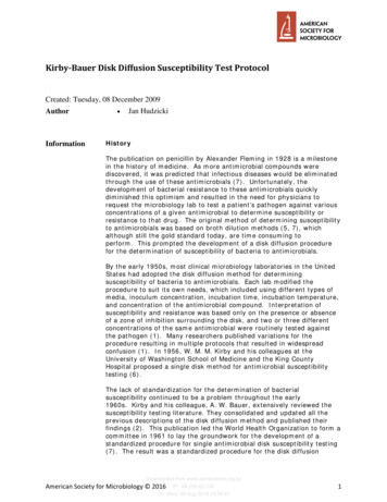
BFIG. 9. Kirby-Bauer disk diffusion susceptibility test protocol, measuringzone sizes. (A) Using a ruler or caliper measure each zone with theunaided eye while viewing the back of the petri dish. Hold the plate afew inches above a black, nonreflecting surface illuminated with reflectedlight. (B) The size of the zone for this organism-antibiotic combination is26 mm.FIG. 10. Kirby-Bauer disk diffusion susceptibility test protocol, measuringzone sizes; an alternate method for measuring zones. If the zones ofadjacent antibiotic disks overlap, the zone diameter can be determinedby measuring the radius of the zone. Measure from the center of theantibiotic disk to a point on the circumference of the zone where adistinct edge is present. Multiply this measurement by 2 to determinethe diameter of the zone of inhibition. In this example, the radius of thezone is 16 mm. Multiply this measurement by 2
their publications (3). The CLSI publication, Performance Standards for Antimicrobial Disk Susceptibility Tests; Approved Standard 9th Edition, represents the standard for clinical laboratories performing susceptibility testing today. Purpose The purpose of the Kirb