Transcription
HindawiComputational and Mathematical Methods in MedicineVolume 2022, Article ID 8192832, 8 pageshttps://doi.org/10.1155/2022/8192832Research ArticleEfficacy and Safety Analysis of Multislice Spiral CT-GuidedTransthoracic Lung Biopsy in the Diagnosis of PulmonaryNodules of Different SizesHuitong Liu , Xiao Yao, Bingqiang Xu, Wei Zhang, Yu Lei, and Xiaolong ChenShaanxi Provincial People’s Hospital, Xi’an, Shaanxi Province 710068, ChinaCorrespondence should be addressed to Xiaolong Chen; chenxl1976@126.comReceived 9 June 2022; Revised 14 July 2022; Accepted 26 July 2022; Published 25 August 2022Academic Editor: Pan ZhengCopyright 2022 Huitong Liu et al. This is an open access article distributed under the Creative Commons Attribution License,which permits unrestricted use, distribution, and reproduction in any medium, provided the original work is properly cited.Objective. This study is aimed at investigating the efficacy and safety of multislice spiral CT-guided transthoracic lung biopsy inthe diagnosis of pulmonary nodules of different sizes. Methods. Data of 78 patients with pulmonary nodules who underwent CTguided transthoracic lung biopsy in our hospital from January 2020 to December 2021 were retrospectively analyzed, and theywere divided into the small nodules group (n 12), medium nodules group (n 35), and large nodules group (n 31)according to the diameter of pulmonary nodules. The results of puncture biopsy and final diagnosis of pulmonary nodules ofdifferent sizes were compared. The incidence of complications in patients with pulmonary nodules of different sizes wascompared. Univariate analysis was used to compare the incidence of complications in 78 patients. Logistic multiple regressionanalysis was used to analyze the independent risk factors of pneumothorax in patients with pulmonary nodule puncture.Logistic multiple regression analysis was used to analyze the independent risk factors of pulmonary hemorrhage in patientswith pulmonary nodule puncture. Results. The diagnostic accuracy, sensitivity, and specificity were 83.33%, 100.00%, and77.78% in small nodules group. The diagnostic accuracy, sensitivity, and specificity of medium nodules group were 85.71%,100.00%, and 73.68%, respectively. The diagnostic accuracy, sensitivity, and specificity of large nodules group were 93.55%,100.00%, and 33.33%, respectively. There was no significant difference in the incidence of pneumothorax among the threegroups (P 0:05). The incidence of pulmonary hemorrhage in small nodule group was higher than that in the medium nodulegroup and large nodule group, and the difference was statistically significant (P 0:05). There was no significant difference inthe incidence of total complications among the three groups (P 0:05). There were statistically significant differences inclinical data such as the needle tract length, the puncture position, and the distance of the puncture needle passing through thelung tissue in patients with or without pneumothorax (P 0:05). There were statistically significant differences in needle tractlength, distance of puncture needle passing through lung tissue, and size of pulmonary nodules in patients with or withoutpulmonary hemorrhage (P 0:05). Logistic multivariate analysis showed that needle tract length 50 mm, lateral decubitusposition, and the distance of puncture needle passing through lung tissue 14 mm were independent risk factors forpneumothorax after puncture in patients with pulmonary nodules (P 0:05). The needle tract length 50 mm, the distance ofpuncture needle passing through lung tissue 14 mm, and small nodules (pulmonary nodules diameter 10 mm) wereindependent risk factors for pulmonary hemorrhage after puncture in patients with pulmonary nodules (P 0:05). Conclusion.Multislice spiral CT-guided transthoracic lung biopsy is effective in diagnosing pulmonary nodules of different sizes.1. IntroductionPulmonary nodules refer to lesions with a diameter of 30 mm in the lungs. The lung nodules are round or quasiround in shape and surrounded by air-filled lung tissue.According to CT imaging morphology, pulmonary nodulescan be divided into solid nodules, partially solid nodules,and ground glass nodules [1]. The incidence of pulmonarynodules is 35.5%, of which pulmonary nodules diagnosedas lung cancer account for 0.54%, respectively; the incidenceof pulmonary nodules among people who smoke more than30 packs per year is 25.9%, of which pulmonary nodules
2diagnosed as lung cancer accounted for 1.1% [2]. The etiology of pulmonary nodules is complex and diverse, includinginfectious factors, tumor factors, and pulmonary infectiousfactors [3, 4]. It is reported that pulmonary nodules withdiameter 5 mm have a 0.4% probability of developing intocarcinoma in situ, pulmonary nodules with diameter of 510 mm have a 1.3% probability of developing into malignancy, pulmonary nodules with diameter of 10-20 mm havea 50% probability of developing into malignancy, and pulmonary nodules with diameter 20 mm have an 87.5%probability of developing into malignancy. Pulmonary nodules of different sizes have different probability of developinginto malignancy, so early diagnosis of pulmonary nodules isvery important [5]. Lung biopsy has high diagnostic accuracy and can also judge the differentiation degree of malignant tumor, which can effectively guide clinical treatment.Transthoracic lung biopsy was first applied to the microbiological diagnosis of patients with lung infection in 1882, andthe first lung cancer biopsy was performed in 1886. Later, itwas gradually developed to use ultrasound, CT, etc. for lungbiopsy. CT is the most widely used guidance method in clinical practice, with high soft tissue contrast ability and spatialresolution. CT-guided puncture biopsy of pulmonary nodules also has high diagnostic accuracy and safety [6–9]. Multislice spiral CT refers to an imaging system with a multirowdetector structure and a single exposure of the tube, whichcan simultaneously obtain data from multiple slices ofimages [10]. Compared with traditional CT-guided lungbiopsy, multislice CT-guided lung biopsy has the advantagesof fast scanning speed and shorter scanning time. In addition, the repeated scanning procedure of multislice spiralCT-guided lung biopsy can make the guidance easier tooperate [11]. The purpose of this study was to investigatethe diagnostic efficacy of multislice spiral CT-guided transthoracic lung biopsy for pulmonary nodules of differentsizes. The report is as follows.2. Materials and Methods2.1. General Information. Data of 78 patients with pulmonary nodules who underwent multislice spiral CT-guidedtransthoracic lung biopsy in our hospital from January2020 to December 2021 were retrospectively analyzed. Therewere 55 males and 23 females, aged from 20 to 78 years old,with an average age of 49:55 5:63 years old; 12 cases withpulmonary nodule diameter 10 mm were divided intosmall nodule group; 35 cases with pulmonary nodulediameter 20 mm were divided into middle nodule group;31 cases with pulmonary nodule diameter 20 mm and 30 mm were divided into large nodule group. This studywas approved by the hospital ethics committee.2.2. Inclusion Criteria. Inclusion criteria are as follows: (1)0 mm pulmonary nodule diameter 30 mm; (2) age 18years old; (3) no contraindications for CT-guided transthoracic lung biopsy; (4) complete clinical and imaging data;(5) the chest CT examination showed pulmonary nodules;and (6) patients volunteered to participate in this study.Computational and Mathematical Methods in Medicine2.3. Exclusion Criteria. Exclusion criteria are as follows: (1)patients with intrapulmonary vascular disease; (2) patientswith pulmonary fibrosis, pulmonary hypertension, andsevere emphysema; (3) patients with severe bleeding tendency or coagulation dysfunction; (4) patients who cannotcooperate with breathing; (5) the heart and blood vesselscannot be avoided during the puncture of the lesion; (6)patients with lung bullae lesions in puncture approach; (7)patients with severe pulmonary infection; and (8) there isskin infection at the site to be punctured.2.4. Methods2.4.1. Operation Instrument. Canon Aquilion ONE 640CTwas used, and the puncture needle was a shotgun-cut biopsyneedle. The imaging data of the patients were carefullyobserved before surgery, and the lateral, supine, and pronepositions were adopted according to the size and locationof the lung nodules. First, local scan of the lesion was carriedout with a layer thickness of 2.7 mm. The best puncturepoint was selected by CT positioning ruler, and the distancebetween the puncture point and the edge and center of thelesion as well as the puncture direction and angle were measured. Routine disinfection and draping were performed,local anesthesia was performed, anesthesia needle wasinjected 1 2 cm in accordance with the expected puncturedirection and angle, the puncture biopsy scanning procedurewas used to scan to verify whether there was any error inpuncture point, puncture direction, and angle, and the scancould be performed again after appropriate adjustment. Thepatient was told to keep calm breathing and hold his breath,and after confirmation, the shotgun-cut biopsy needle wasused to rapidly puncture the lung lesions of the patient. Afterreaching the expected depth, the scan was carried out to confirm that the needle tip was in the patient’s lesion. Then thepatient was told to hold his breath, the lesion tissue wascut, the needle was removed quickly, and the tissue specimen was fixed with formalin and then sent for examination. If the pathologist is not satisfied, the needle biopsyshould be performed again and the liquefaction necroticarea should be avoided. After surgery, the puncture sitewas pressed for 2 3 minutes, external dressing wasapplied, and then the lungs were scanned to observewhether the patient had bleeding, pneumothorax, andother complications.2.5. Judgment Criteria for Puncture Results. (1) If the biopsytissue is normal lung tissue and diaphragmatic muscleaccording to pathological results, it is regarded as failure ofsampling. (2) If the pathological examination result ofbiopsy tissue is malignant tumor, the pathological diagnosisis definite. (3) If the pathological examination result ofbiopsy tissue is benign and confirmed by surgery, or thelesions disappear, shrink, and stabilize for more than half ayear after treatment, it is regarded as correct pathologicaldiagnosis. The accuracy of puncture biopsy can be obtainedby comparing the final clinical examination results with theresults of needle biopsy.
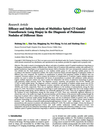
Computational and Mathematical Methods in Medicine2.6. Observation Indicators. Observation indicators are asfollows: (1) general information of all patients; (2) comparison of puncture biopsy and final diagnosis results ofpulmonary nodules of different sizes; (3) comparison ofthe incidence of complications among patients with pulmonary nodules of different sizes; (4) univariate analysisof the incidence of complications in all patients; (5) logistic multiple regression analysis was used to analyze theindependent risk factors of pneumothorax in patients withpulmonary nodule puncture; and (6) logistic multipleregression analysis was used to analyze the independentrisk factors of pulmonary hemorrhage in patients withpulmonary nodule puncture.2.7. Statistical Methods. All data in this study were input intoExcel by two persons without communication and analyzedand processed by the SPSS24.0 statistical software. The measurement data were expressed as mean SD (x̅ s). Whenthe data were in line with normal distribution and the variance was uniform, the t test was adopted, and one-wayANOVA was used comparison among multiple groups.Counting data were described by n and %, and disorderedclassification data were compared by the χ2 test or Fisher’sexact probability method. Multivariate analysis was performed by the logistic regression model. All were two-sidedtests, and P 0:05 was considered statistically significant.3. Results3.1. General Information of Patients. The general information of all patients is shown in Table 1.3.2. Comparison of Puncture Biopsy and Final DiagnosisResults of Pulmonary Nodules of Different Sizes. In the smallnodules group, the diagnostic accuracy, sensitivity, and specificity were 83.33%, 100.00%, and 77.78%, respectively, thepositive prediction rate was 60.00%, the negative predictionrate was 81.82%. In the medium nodules group, the diagnostic accuracy, sensitivity, and specificity were 85.71%,100.00%, and 73.68%, respectively, the positive predictionrate was 76.19%, and the negative prediction rate was79.17%. In the large nodules group, the diagnostic accuracy,sensitivity, and specificity were 93.55%, 100.00%, and33.33%, respectively, the positive prediction rate was93.55%, and the negative prediction rate was 60.00% (seeTables 2 and 3 and Figure 1).3.3. Comparison of Complications among the Three Groups.There was no significant difference in the incidence of pneumothorax among the three groups (P 0:05); the incidenceof pulmonary hemorrhage in the small nodules group washigher than that in the middle nodules group and the largenodules group, and the difference was statistically significant(P 0:05). There was no significant difference in the totalincidence of complications among the three group(P 0:05) (see Table 4).3.4. Univariate Analysis of Complications in 78 Patients.There were statistically significant differences in clinical datasuch as the needle tract length, the puncture position, and3Table 1: General information of patients (cases (%)).ItemLung nodule locationRight upper lobeRight middle lobeRight lower lobeLeft upper lobeLeft lower lobePrevious tumor historyYesNoSmoking historyYesNoPuncture positionSupine positionProne positionLateral positionComposition ratio29 (37.18)9 (11.54)17 (21.79)12 (15.38)11 (14.10)3 (3.85)75 (96.15)41 (52.56)37 (47.44)25 (32.05)44 (56.41)9 (11.54)the distance of the puncture needle passing through the lungtissue in patients with or without pneumothorax (P 0:05).There were statistically significant differences in needle tractlength, distance of puncture needle passing through lung tissue, and size of pulmonary nodules in patients with or without pulmonary hemorrhage (P 0:05), as shown in Table 5.3.5. Logistic Multivariate Regression Analysis of the Incidence ofPneumothorax in 78 Patients. Factors with statistically significant differences in the above univariate analysis of pneumothorax, including needle track length, puncture position, anddistance of puncture needle passing through lung tissue, wereincluded in the logistic regression analysis and assigned (needle track length 50 mm 1, 50 mm 0; puncture position:supine position 2, prone position 1, and lateral position 0; the distance of the puncture needle passing through the lungtissue 14 mm 1, 14 mm 0). Logistic multivariate analysis showed that needle length 50 mm, lateral position, andthe distance of puncture needle passing through lung tissue 14 mm were independent risk factors for pneumothorax afterpuncture in patients with pulmonary nodules (P 0:05), asshown in Table 6.3.6. Logistic Multivariate Regression Analysis of the Incidence ofPulmonary Hemorrhage in 78 Patients. Factors with statistically significant differences in the above univariate analysis ofpulmonary hemorrhage, including needle track length, distance of puncture needle passing through lung tissue, and thesize of the pulmonary nodule, were included in Logistic regression analysis and assigned (needle track length 50 mm 1, 50 mm 0; distance of puncture needle passing through lungtissue 14 mm 1, 14 mm 0; large nodule ð20 mm pulmonary nodule diameter 30 mmÞ 2, medium nodule ð10 mm pulmonary nodule diameter 20 mmÞ 1,andsmall nodules ðpulmonary nodule diameter 10 mmÞ 0).
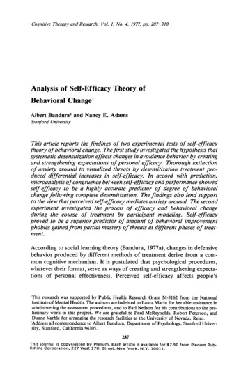
4Computational and Mathematical Methods in MedicineTable 2: Comparison of puncture biopsy and final diagnosis results of pulmonary nodules of different sizes (cases (%)).GroupBiopsy diagnosis resultBenignMalignantSmall nodules group (n 12)Medium nodules group (n 35)Large nodules group (n 31)Total7 (58.33)14 (40.00)1 (3.23)22Final diagnosis resultBenignMalignant5 (41.67)21 (60.00)30 (96.77)569 (75.00)19 (54.29)3 (9.68)313 (25.00)16 (45.71)28 (90.32)47Table 3: Comparison of diagnostic accuracy, sensitivity, specificity, positive predictive rate, and negative predictive rate of puncture biopsyamong the three groups (%).GroupSmall nodules group (n 12)Medium nodules group (n 35)Large nodules group (n 31)AccuracySensitivitySpecificityPositive predictive rateNegative predictive 3360.0076.1993.3381.8279.1760.00(a)(b)(c)(d)Figure 1: A 54-year-old male patient, with a nodule in the left lower lobe found by physical examination before 2 weeks. (a) A solid nodulein the lateral basal segment of the left lower lobe, with a diameter of about 1.3 cm (indicated by the arrow). (b) The needle was obliquelyinserted from the adjacent intercostal space, avoiding the scapula. (c) After needle extraction, there was a small amount of bleeding andpneumothorax (indicated by the arrow), and the pneumothorax were absorbed after observation. (d) The surgical pathological resultswere the same after puncture, and invasive adenocarcinoma was considered ( 100, HE staining).Table 4: Comparison of the incidence of complications among the three groups (cases (%)).GroupSmall nodules group (n 12)Medium nodules group (n 35)Large nodules group (n 31)PneumothoraxPulmonary hemorrhageTotal complication rate2 (16.67)6 (17.14)7 (22.58)7 (58.33)8 (22.86)5 (16.13)9 (75.00)14 (40.00)12 (38.71)Logistic multivariate analysis results showed that the needletract length 50 mm, the distance of the puncture needlepassing through the lung tissue 14 mm, and the small nodule(pulmonary nodule diameter 10 mm) were independent riskfactor for pulmonary hemorrhage after puncture in patientswith pulmonary nodules (P 0:05) (see Table 7).4. DiscussionLung biopsy, open lung biopsy, thoracoscopy biopsy, andfiberoptic bronchoscopy biopsy can all obtain histopathological specimens of lung lesions, but open lung biopsy andthoracoscopic biopsy are more traumatic to the body, and
Computational and Mathematical Methods in Medicine5Table 5: Univariate analysis of complications in 78 patients.PneumothoraxP/Fisher’s exactt/χ2Yes (n 15) No (n 63)value probability valueItemAge 60 years old (n 29)6 (20.69)23 (79.31) 60 years old (n 49)9 (18.37)40 (81.63)GenderMale (n 55)11 (20.00) 44 (80.00)Female (n 23)4 (17.39)19 (82.61)Nodule density (cases)Solid (n 67)14 (20.90) 53 (79.10)Nonsolid (n 11)1 (9.09)10 (90.91)Needle track length 50 mm (n 17)7 (41.18)10 (58.82) 50 mm (n 61)8 (13.11)53 (86.89)Puncture lung lobeRight upper lobe (n 29)4 (13.79)25 (86.21)Right middle lobe (n 9)2 (22.22)7 (77.78)Right lower lobe (n 17)4 (23.53)13 (76.47)Left upper lobe (n 12)2 (16.67)10 (83.33)Left lower lobe (n 11)3 (27.27)8 (72.73)Smoking historyYes (n 41)8 (19.51)33 (80.49)No (n 37)7 (18.92)30 (81.08)Puncture positionSupine position (n 25)2 (8.00)23 (92.00)Prone position (n 44)7 (15.91)37 (84.09)Lateral position (n 9)6 (66.67)3 (33.33)Number of punctures1 3 times (n 35)7 (20.00)28 (80.00) 4 times (n 43)8 (18.60)35 (81.40)Distance of puncture needle passing22:15 2:55 13:56 2:35through lung tissue (mm)Pulmonary hemorrhaget/χ2Yes (n 20) No (n 58)valuePvalue0.0630.8015 (17.24)15 (30.61)24 (82.76)34 (69.39)1.7080.1910.0710.79013 (23.64)7 (30.43)42 (76.36)16 (69.57)0.3930.5310.8480.35716 (23.88)4 (36.36)51 (76.12)7 (63.64)0.7720.3806.7400.0091 (5.88)19 (31.15)16 (94.12)42 (68.85)4.4510.0351.3150.8597 (24.14)1 (11.11)4 (23.53)3 (25.00)5 (45.45)22 (75.86)8 (88.89)13 (76.47)9 (75.00)6 (54.55)3.3380.5030.0040.94711 (26.83)9 (24.32)30 (73.17)28 (75.68)0.0640.800—0.0017 (28.00)12 (27.27)1 (11.11)18 (72.00)32 (72.73)8 (88.89)1.1310.5680.0240.87610 (28.57)10 (23.26)25 (71.43)33 (76.74)0.2860.59312.520 0.00124:19 5:68 12:68 3:25 11.101 0.001Table 6: Logistic multivariate regression analysis of the incidence of pneumothorax in 78 patients.βFactorSEWald χ2OR95% CIPNeedle track length 50 mm0.5210.2255.3621.6841.083 2.6170.021Lateral positionDistance of the puncture needle passing through the lung tissue 14 mm0.3550.1347.0191.4261.097 1.8550.0081.5260.5547.5874.6001.553 13.6240.006Table 7: Logistic multivariate regression analysis of the incidence of pulmonary hemorrhage in 78 patients.βSEWald χ2OR95% CIPNeedle track length 50 mm1.1590.4526.5753.1871.314 7.7290.011Distance of puncture needle passing through lung tissue 14 mm1.5940.6865.3994.9231.283 18.8890.021Small nodules1.4030.5456.6274.0671.398 11.8370.010Factor
6their application is limited in elderly patients and patientswith cardiopulmonary dysfunction [12–14]. Lung puncturebiopsy has little trauma, low cost, and less pain to patients.Some patients do not need to be hospitalized after punctureand can live a normal life the next day. In addition, CT scanhas good contrast and resolution, which can clearly displaythe morphology, size, and adjacent important structure oflesions and can display small lesions beside the mediastinum, behind the heart shadow, and beside the spine [15,16]. It has been reported that the accuracy of CT-guidedlung biopsy in the diagnosis of benign lesions is 80% andthat of malignant lesions is 90% [17]. In this study, thediagnostic efficiency of multislice spiral CT for pulmonarynodules of different sizes was observed. The results showedthat the diagnostic accuracy of the small nodule group was83.33%, the sensitivity was 100.00%, the specificity was77.78%, the positive predictive rate was 60.00%, and the negative predictive rate was 81.82%. In the middle nodulegroup, the diagnostic accuracy was 85.71%, the sensitivitywas 100.00%, the specificity was 73.68%, the positive predictive rate was 76.19%, and the negative predictive rate was79.17%. In the large nodule group, the diagnostic accuracywas 93.55%, sensitivity was 100.00%, specificity was33.33%, the positive predictive rate was 93.33%, and negativepredictive rate 60.00%. Multislice spiral CT-guided lungbiopsy has the highest diagnostic accuracy for diagnosinglargenodules(20 mm pulmonary nodule diameter 30 mm). The sensitivity of multislice spiral CT-guided lung biopsy in the diagnosis of lung nodules of different sizes was up to 100.00%,indicating that multislice spiral CT-guided lung biopsy isan effective method for the diagnosis of lung cancer.Common complications of transthoracic lung biopsyinclude pneumothorax, pulmonary hemorrhage, andhemoptysis, and the rare serious complications include airembolism and tumor needle passage metastasis [18, 19]. Inthis study, complications of multislice spiral CT-guided lungbiopsy were observed, and the results showed that there wasno statistical significance in the incidence of pneumothoraxin groups of different sizes of pulmonary nodules. Accordingto relevant reports, the incidence of pneumothorax of CTguided transthoracic lung biopsy is 15.4% 42.0%, only4.3% 7.3% patients need thoracic catheter drainage, andthe incidence of hemoptysis is 0.5% 14.4%, most of whichdo not need intervention [20, 21]. The common risk factorsof pneumothorax include long puncture path, multiplecrossing of pleura, and small lesion [22, 23]. In this study,univariate and multivariate analyses were conducted on therisk factors for pneumothorax and pulmonary hemorrhage,and it was found that needle tract length 50 mm, lateralposition, and the distance of puncture needle passingthrough lung tissue 14 mm were independent risk factorsfor pneumothorax after puncture in patients with pulmonary nodules. The results of this study suggest that whenthe puncture needle tract is 50 mm, the risk of pneumothorax increases, which is contrary to the results of previousstudies [24, 25]. The reason may be that the shorter thepuncture needle tract, the gas in the lung will pass throughthe needle in a short time, thus causing pneumothorax. InComputational and Mathematical Methods in Medicinethis study, the poor postural stability of patients in lateralposition also easily led to an increase in the incidence ofpneumothorax after puncture. In this study, the distance ofpuncture needle passing through the lung tissue is closelyrelated to the occurrence of pneumothorax, which is manifested as the greater the distance of puncture needle passingthrough the lung tissue, the more likely pneumothorax willoccur. This may be because when people breathe, there issliding between visceral pleura and parietal pleura, and indirect friction can be formed when the needle is fixed withchest wall muscle layer, which can lead to pneumothorax.The longer the distance of puncture needle passing throughthe lung tissue, the more obvious the friction effect is. Therefore, the needle tract length 50 mm, lateral position, andthe distance of puncture needle passing through the lungtissue 14 mm are independent risk factors for pneumothorax in patients with pulmonary nodules after puncture. It issuggested that full attention should be paid to patients withneedle tract length 50 mm, lateral position, and thedistance of puncture needle passing through lung tissue 14mm to prevent pneumothorax in time.In this study, logistic multivariate analysis showed thatthe needle tract length 50 mm, the distance of thepuncture needle passing through the lung tissue 14 mm,and small nodules (pulmonary nodule diameter 10 mm)were independent risk factors for pulmonary hemorrhageafter puncture in patients with pulmonary nodules(P 0:05). It was reported that the incidence of pulmonaryhemorrhage after puncture was 16%-33%. In this study,the incidence of pulmonary hemorrhage was higher in thesmall nodules group, and the overall incidence of pulmonaryhemorrhage was consistent with the results of previous studies [26–28]. This study found that the longer the needletrack, the easier it is to cause pulmonary hemorrhage, whichis consistent with previous research results [29, 30]. The longer the needle track is, the more it passes through the lungtissue, which can lead to injury and bleeding. Therefore,the needle track length 50 mm and the distance of theneedle passing through the lung tissue 14 mm are closelyrelated to pulmonary hemorrhage. In this study, the pulmonary nodule diameter 10 mm was an independent risk factor for pulmonary hemorrhage. This may be because whenthe lesion is small, the puncture process is greatly affectedby breathing, and the needle needs to be adjusted severaltimes during the puncture process, which is likely to causepulmonary hemorrhage. In addition, if the lesion is small,normal tissue may be removed to obtain sufficient tissuespecimens, resulting in increased bleeding rate. Therefore,prevention of pulmonary hemorrhage should be done wellfor patients with needle tract length 50 mm, the distanceof puncture needle passing through lung tissue 14 mm, andsmall nodules (lung nodules diameter 10 mm).In conclusion, multislice spiral CT-guided lung biopsyhas high accuracy in diagnosing pulmonary nodules, andthe complication rates of pneumothorax and pulmonaryhemorrhage are consistent with previous research results[31], which has clinical application value. However, in thisstudy, the sample size is small and the classification methodof nodule size is different from that of most literatures. In
Computational and Mathematical Methods in Medicinefuture research, the sample size of the study will be furtherexpanded and the grouping of nodule size will be improved,to provide more scientifically rigorous clinical researchresults in the diagnosis of pulmonary nodules by multislicespiral CT-guided lung biopsy.Data AvailabilityThe labeled dataset used to support the findings of this studyare available from the corresponding author upon request.Conflicts of InterestThe authors declare no competing interests.References[1] S. Y. Liu, “Pay attention to the problem of over-diagnosis inthe clinical management of pulmonary nodules,” Chinese Journal of Radiology, vol. 55, no. 11, p. 4, 2021.[2] Lung cancer group of respiratory disease branch of ChineseMedical Association and expert group of China Lung CancerPrevention and Control Alliance, “Chinese expert consensuson diagnosis and treatment of pulmonary nodules (2018 edition),” Chinese Journal of Tuberculosis and Respiratory Medicine, vol. 41, no. 10, p. 9, 2018.[3] Q. Wu, B. Cao, Y. Zheng et al., “Feasibility and safety offine positioning needle-mediated breathing control in CTguided percutaneous puncture of small lung/liver nodulesadjacent to diaphragm,” Scientific Reports, vol. 11, no. 1,p. 3411, 2021.[4] K. M. Bhatt, Y. K. Tandon, R. Graham et al., “Electromagnetic navigational bronchoscopy versus CT-guided percutaneous sampling of peripheral indeterminate pulmonarynodules: a cohort study,” Radiology, vol. 286, no. 3,pp. 1052–1061, 2018.[5] Y. Xu, L. Lu, L. N. E et al., “Application of radiomics in predicting the malignancy of pulmonary nodules in different sizes,”AJR. American Journal of Roentgenology, vol. 213, no. 6,pp. 1213–1220, 2019.[6] J. R. Andrade, R. D. Rocha, P. M. Falsarella et al., “CT-guidedpercutaneous core needle biopsy of pulmonary nodulessmaller than 2 cm: technical aspects and factors influencingaccuracy,” Jornal Brasileiro de Pneumologia, vol. 44, no. 4,pp. 307–314, 2018.[7] D. Lendeckel, M. L. Kromrey, T. Ittermann, S. Schäfer,B. Mensel, and J. P. Kühn, “Pulmonary emphysema is a predictor of pneumothorax after CT-guided transthoracic pulmonary biopsies of pulmonary nodules,” PLoS One, vol. 12,no. 6, article e0178078, 2017.[8] Y. Y. Chang, C. K. Chen, Y. C. Yeh, and M. H. Wu, “Diagnosticfeasibility and safety of CT-guided core biopsy for lung nodules less than or equal to 8 mm: a single-institution experience,” European Radiology, vol. 28, no
The diagnostic accuracy, sensitivity, and specificity were 83.33%, 100.00%, and 77.78% in small nodules group. The diagnostic accuracy, sensitivity, and specificity of medium nodules group were 85.71%, . Excel by two persons without communication and analyzed . J. Hou and Z. K. Ou, "A report of 14 cases of limited thoracot-omy lung .