Transcription
Qi et al. BMC Musculoskeletal Disorders(2020) SEARCH ARTICLEOpen AccessClinical efficacy of short-term pre-operativehalo-pelvic traction in the treatment ofsevere spinal deformities complicated withrespiratory dysfunctionLongtao Qi, Beiyu Xu, Chunde Li and Yu Wang*AbstractBackground: Halo traction has been used as an adjunctive method in the treatment of severe spinal deformities.But there are few reports on the clinical efficacy of halo-pelvic traction (HPT) in the treatment of severe spinaldeformities complicated with respiratory dysfunction. This study was to evaluate the clinical efficacy andcomplications associated with pre-operative HPT in the treatment of severe spinal deformities with respiratorydysfunction.Methods: Thirty patients with severe spinal deformities complicated with respiratory dysfunction treated withshort-term pre-operative HPT were retrospectively reviewed. Inclusion criteria were: (1) patients with severekyphoscoliosis (coronal Cobb angle or kyphosis angle 100 ) and respiratory failure, (2) patients undergoing HPTuntil posterior fusion surgery. All patients underwent general anesthesia for HPT application, which the pelvic ringused in this study was a half-ring, and the rods were all placed on the anterolateral side of the truck.Results: The major coronal curve scoliosis averaged 116.00 16.70 and was reduced to 63.23 14.00 after HPT,46.33 10.70 after surgery. The major kyphosis was 102.40 27.67 and was reduced to 52.23 14.16 after HPT,42.0 11.92 after surgery. A significantly increased FVC was observed after HPT (p 0.001), with a significantlyimproved FVC% (p 0.001). Similarly, a significantly increased FEV1 was also observed (p 0.001), with a significantlyimproved FEV1% (p 0.001).Conclusion: This study indicated that the modified HPT could be used to help patients with severe spinaldeformities complicated with respiratory dysfunction achieve significant correction in both the coronal and sagittaldeformities during the pre-operative treatment period along with improved respiratory function and in the absenceof severe complications.Keywords: Severe spinal deformity, Halo-pelvic traction, Short-term, Pulmonary function* Correspondence: qlt6219@126.comDepartment of Orthopaedics, Peking University First Hospital, XichengDistrict, Beijing 100034, China The Author(s). 2020 Open Access This article is licensed under a Creative Commons Attribution 4.0 International License,which permits use, sharing, adaptation, distribution and reproduction in any medium or format, as long as you giveappropriate credit to the original author(s) and the source, provide a link to the Creative Commons licence, and indicate ifchanges were made. The images or other third party material in this article are included in the article's Creative Commonslicence, unless indicated otherwise in a credit line to the material. If material is not included in the article's Creative Commonslicence and your intended use is not permitted by statutory regulation or exceeds the permitted use, you will need to obtainpermission directly from the copyright holder. To view a copy of this licence, visit http://creativecommons.org/licenses/by/4.0/.The Creative Commons Public Domain Dedication waiver ) applies to thedata made available in this article, unless otherwise stated in a credit line to the data.
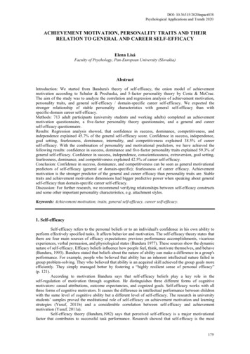
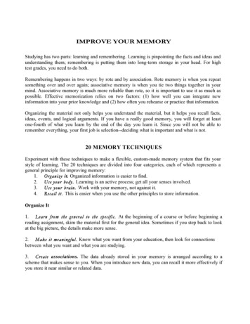
Qi et al. BMC Musculoskeletal Disorders(2020) 21:665BackgroundSevere spinal deformities are usually accompanied bycardiopulmonary impairment, and this often results insignificantly increased morbidity and mortality. Despitesubstantial improvements in surgical techniques for thetreatment of severe spinal deformities, the managementof these patients remains a major challenge due to theassociated poor pulmonary function, high potential forbleeding, and related neurological complications [1–3].To minimize the risks and complications associated withthese patients, especially in regard to neurological complications, numerous authors have advocated that onestage surgery should be avoided, and adjunctivemethods, such as traction (halo-femoral, halo-pelvic,halo-gravity), should be used for the reduction of stiffcurves prior to surgery as well as to reduce the useof acute corrective maneuvers [3–5]. Currently, numerous surgeons advocate for halo-gravity traction(HGT) as a safe adjunctive method, as prior studieshave demonstrated that pre-operative HGT can obtain15 to 38% correction in scoliosis and 17 to 35% correction in kyphosis; in addition, some reports indicatesignificant improvements in pulmonary function withthis procedure [3–5].In the 1970s, O’Brien et al. introduced the applicationhalo-pelvic traction (HPT) for patients with scoliosis in aseries of 118 patients [6]. The authors reported an impressive correction of the deformities. Although thepowerful traction forces generated by HPT can effectively correct various spinal deformities, the traditionalHPT procedure has some drawbacks, including a number of various complications and poor tolerance due tothe rods that are circularly distributed around the trunk,which tend to affect the patient’s supine sleeping position. With the development of internal fixation techniques, which do not present the drawbacks of HPT, theuse of HPT has gradually declined. To the best of ourknowledge, there are few reports on the clinical efficacyof HPT in the treatment of severe spinal deformitiescomplicated with respiratory dysfunction over the last10 years. However, our experience has led to an improved HPT technique, and the purpose of this studywas to evaluate the clinical efficacy and complicationsassociated with pre-operative HPT used for the treatment of severe spinal deformities with respiratorydysfunction.MethodsA total of 30 patients with severe spinal deformitiescomplicated with respiratory dysfunction treated withpre-operative HPT were retrospectively reviewed. All ofthe surgeries were performed by the same senior authorbetween 2017 and 2019. Clinical and radiographic datawere collected and evaluated by an independent spinalPage 2 of 8surgeon who was not involved in the surgical treatment.Patients who met the following inclusion criteria wererecruited in the study: (1) patients with severe kyphoscoliosis (coronal Cobb angle or kyphosis angle 100 ) andrespiratory dysfunction (defining as pressure of oxygen(PaO2) 60 mmHg), and (2) patients undergoing HPTuntil posterior fusion surgery. Exclusion criteria were:(1) patients who had prior spinal surgery and (2) patientswith revision surgery or anterior release. Informed consent was obtained from each patient prior to recruitment. The demographic data collected included age, sex,standing height, diagnosis, and duration of traction.Halo-Pelvic Traction (HPT) protocolThe HPT device consisted of a head ring, a pelvic ring,and retractable connecting rods. The number of pinswas based according to the patient’s bone condition andanatomical variation. All patients underwent localanesthesia for HPT application. Briefly, with the patientin the supine position, three pins (4.5 mm in diameter)were inserted into the area between the inner and outertable of the ilium on each side. The skull halo devicewas then placed using standard techniques, as previouslyreported [7]. Different from traditional HPT, the pelvicring used in this study was a half-ring, and the rods wereall placed on the anterolateral side of the truck (Fig. 1).The frame was constructed 3–5 days after HPT application so that the patient had time to become accustomedto the pins. Traction was initiated after the constructionof the frame. For the first week, the frame was elongatedat a rate of 0.5 cm per day. Beginning in the secondweek, the elongation rate was decreased to 0.3–0.5 cmevery 2–3 days. Additionally, daily pin site care was performed. Cranial nerve and upper/lower extremity neurological examinations were also performed every day,especially after each increase in traction length. If anycomplication occurred, such as upper-extremity numbness, the traction length was reduced to the previouslength.Radiographic analysisThe major scoliosis and kyphosis (Cobb angle) weremeasured from biplanar, full-standing, standard radiographs of the whole spine at pre-traction, post-traction,and post-operative. Radiographs consisted of standingposteroanterior (PA) and lateral. In each case, radiographs were repeated every 2 weeks while the patientswere in traction. Radiographic analysis of the coronalplane included the major curve Cobb angle on thestanding PA radiograph. For the sagittal plane, we included the major curve Cobb angle and sagittal verticalaxis (SVA), which was measured as the horizontal distance between the center of the C7 vertebral body andthe posterior superior aspect of the S1 vertebral body.
Qi et al. BMC Musculoskeletal Disorders(2020) 21:665Page 3 of 8Fig. 1 The halo-pelvic traction. The posterior (a) and lateral (b/c) views photographs, and the standing anteroposterior (d) and lateral digitalradiographs (e)All measurements were performed using Surgimap software (version 2.3.2.1; New York, USA).Pulmonary Function Test (PFT)The PFT was performed at both pre-traction and posttraction. The pulmonary function value included forcedvital capacity (FVC), FVC %, forced expiratory volume atthe end of the first second (FEV1), and FEV1%. To ensureaccurate and reliable data, the PFT was measured in triplicate, and the highest values were selected. According to theAmerican Thoracic Society’s guidelines for the severity ofpulmonary impairment, ‘no’ pulmonary impairment wasconsidered when the FVC% was 80% of the predictedvalue, ‘mild’ when the FVC% was 80% but 65%, ‘moderate’ when the FVC% was 65% but 50%, and ‘severe’when the FVC% was 50% [8]. When the pulmonary function (in terms of the FVC %) at the completion of HPT hadimproved by at least 5%, the patients were defined as ‘pulmonary responders’ to HPT. If there was an improvementor decrease of 0–5%, patients were defined as ‘non-responders’ to HPT. If the FVC % dropped more than 5%during HPT, patients were defined as ‘decline’ [9].Statistical analysisAll of the data were analyzed using SPSS version 20.0(SPSS, Chicago, IL, USA). Statistical data are presentedas the mean standard deviation. Statistical comparisonof the radiographic measurements and PFT results wereperformed using a parametric paired t-test; p 0.05 wasconsidered statistically significant.ResultsDemographicsThe study included a total of 30 patients undergoingHPT treatment, of which 22 were female (73%) and 8were male (27%). The mean age of the patients was30.00 9.33 (12–46) years at the time of surgery. Etiological diagnoses were neuromuscular (n 5), idiopathic(n 15), and congenital (n 10) scoliosis. All 30 patientswere treated with posterior instrumentation and fusionwithout anterior release. Typical cases are shown inFigs. 2 and 3. The average pre-traction height was139.63 8.67 cm (124-160 cm), and the height after traction was 150.37 9.08 cm (132-170 cm). The average increase was 10.73 3.70 cm (6-15 cm; p 0.001), and theaverage traction duration was 5.37 0.93 weeks (4–6weeks).Radiological parametersDuring traction, the major correction of kyphosis andscoliosis occurred in the first 2 weeks (Fig. 4). Over thefirst 2 weeks, correction of 30.24% for scoliosis and33.54% for kyphosis was obtained. At 4 weeks, the correction of scoliosis and kyphosis was 44.10 and 44.57%,respectively. At 6 weeks, the correction of scoliosis andkyphosis was 49.81 and 56.22%, respectively. After HPTtraction, the degree of scoliosis on the final-HPT radiographs demonstrated a significant improvement, asshown in Table 1. The major scoliosis correction averaged 52.77 13.68 (4 to 75 ; p 0.0001) or 45.15 11.41% (6–65%; p 0001). After the surgery, the correction of scoliosis was 69.67 13.11 (17 -94 ; p 0001) or59.81 8.82% (27–73%; p 0001) compared with thecurves prior to traction. Additionally, the major kyphosispresented a significant correction of 50.17 22.81 (11–102 ; p 0.001) or 47.59 13.33% (16–68%; p 0.001) onthe final-HPT radiographs and 60.33 20.87 (24–105 ;p 0.001) or 53.67 25.95% (23–69%; p 0.001) aftersurgery compared with the kyphosis before traction. Forthe total correction, the HPT traction demonstrated an
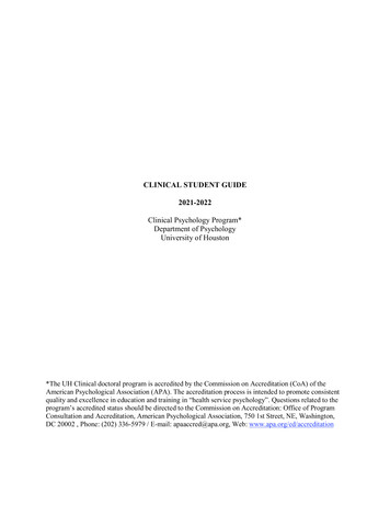
Qi et al. BMC Musculoskeletal Disorders(2020) 21:665Page 4 of 8Fig. 2 A 21-year-old female with congenital scoliosis and respiratory dysfunction. The major scoliosis was 120 and the major kyphosis angle was74 before traction. After 5 weeks of HPT, the scoliosis and kyphosis had decreased to 63 and 46 respectively, and the FVC% had significantlyincreased from 36.00 to 57.57%. Posterior correction with instrumentation was performed. After surgery, the scoliosis had decreased to 45 , thekyphosis had decreased to 40 . At 2 years follow-up, no loss of correction was observedaverage correction of 74.46% for scoliosis and 81.23% forkyphosis.The pre-traction SVA was 18.8 36.3 mm ( 35 mm –97 mm). After HPT, the SVA was 61.2 35.6 mm ( 141 mm – 2 mm), and this demonstrated a significantchange of 80.0 50.0 mm ( 7 mm – -178 mm; p 0.001). The post-surgery SVA was significantly differentrelative to the post-traction SVA ( 75.3 43.2 mm; p 0.001) but similar to the pre-traction SVA ( 4.7 35.4mm; p 0.475).Pulmonary functionThe changes in FVC, FVC%, FEV1, and FEV1%, asshown in Table 2, were used to analyze the effect ofHPT on pulmonary function. Regarding the results ofthe PFT, a significantly increased FVC was observedafter HPT (0.91 L vs. 1.18 L; p 0.001), with a significantly improved FVC% (34.15% vs. 44.18%; p 0.001).Similarly, a significantly increased FEV1 was also observed (0.79 L vs. 1.02 L; p 0.001), with a significantlyimproved FEV1% (34.10% vs. 44.25%; p 0.001). At thetime of pre-traction, 3 patients (10%) demonstratedmoderate pulmonary impairment and 27 (90%) demonstrated severe pulmonary impairment. A total of 6 patients were identified as non-responders, presenting onlya slight improvement in PFT, and the remaining 24 patients were classified as responders to HPT. The statistical analysis of the relationship between HPT andpulmonary function showed that there was no significantcorrelation between the improvements in the PFT results with the correction of the radiographic parameters(p 0.05; Table 3).ComplicationsTwo patients developed complications after HPT traction. One patient experienced a decrease in musclestrength in the right lower extremity after 3 weeks oftraction, and this patient recovered their muscle strengthafter the traction length was reduced to that of the previous length. Another patient experienced nausea andvomiting that resolved 1 week after halo placement, andthis returned to normal after reducing the traction.There was no pin loosening, pin site infection, nystagmus, paraplegia, cranial nerve injury, hypoglossal nerveinjury, or any other kind of neurological complicationsobserved. Because of the continuity of the traction, thepatients often had cervical discomfort and trapezial soreness, and all these symptoms disappeared after undergoing surgery.
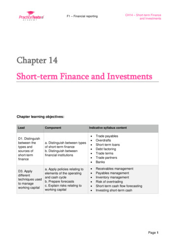
Qi et al. BMC Musculoskeletal Disorders(2020) 21:665Page 5 of 8Fig. 3 A 24-year-old female with severe kyphosis (165 ) and respiratory dysfunction. After 6 weeks of HPT, the kyphosis had decreased to 55 , thescoliosis had decreased from 64 to 54 , and the FVC% had significantly increased from 34.10 to 44.69%. Posterior correction with Ponteosteotomy and instrumentation was performed. After the surgery, the kyphosis had decreased to 48 , the scoliosis had decreased to 42 DiscussionThe rapid correction of severe scoliosis complicated withrespiratory impairment can increase the risk of neurologic injury, morbidity, and mortality [10]. Halo tractionhas been used as an adjunctive method in the treatmentof severe spinal deformities, which can reduce the risk ofspinal cord injury while effectively obtaining correctionof severe spinal deformities in a controlled and safemanner. As a result, less correction is needed duringsurgery, and aggressive procedures, such as vertebralFig. 4 Improvement in coronal and sagittal plane deformities during the traction period (0–6 weeks, n 30)
Qi et al. BMC Musculoskeletal Disorders(2020) 21:665Page 6 of 8Table 1 Radiographic parameters of the patients with severescoliosis (n 30)Pre-tractionPost-traction①Table 3 Pearson’s coefficients between radiographic parametersand PFT results after traction (n 30) Major CobbPost-surgery①② Kyphosis CobbCoefficientp valueCoefficientp value FVC 0.2530.0520.1580.224 FVC % 0.2400.0630.1410.276Note: ①Compared with pre-traction data, p 0.05. ②Compared with posttraction data, p 0.05 FEV1 0.2340.0760.2110.104 FEV1% 0.2450.1930.1600.218column resection, might be avoided. Finally, this approach can also help improve pre-operative pulmonaryfunction, allowing a better tolerance for more aggressiveprocedures [5, 6, 11]. Unlike HGT, HPT can providepowerful traction forces throughout the day to effectively correct spinal deformities. However, traditionalHPT has a number of various complications and poortolerance. As such, the use of HPT has gradually declined [12]. In this study, the pelvic ring of HPT devicewas a half-ring, and the rods were all placed on the anterolateral side of the truck (Fig. 1). This modifiedmethod allowed the patients to sleep in a supine position, wear clothes, and move by themselves while stillachieving 24 h of continuous traction. The gradual traction applied during the pre-operative period might alsohelp in evaluating the neurologic function and estimating the amount of correction that can safely be obtained.O’Brien et al. first used HPT in the treatment of scoliosis in the 1970s [6], but there are few subsequent literature reports on the use of HPT for the treatment ofsevere spinal deformities, especially over the past 10years. In this current study, with the help of HPT, thecorrection rate after HPT was 45.15% for scoliosis and47.59% for kyphosis. This outcome was superior to thepre-operative HGT documented in prior reports, whichdemonstrated 15 to 38% correction in scoliosis and 17to 35% correction in kyphosis [3–5, 11]. In the study byJanus et al., the correction of pre-operative HGT wassimilar to the final correction after surgery when compared with the initial curve (32% vs. 36% for scoliosis,24% vs. 27% for kyphosis), but the total correction wassignificantly lower than that observed in this currentstudy (36% vs. 59.81% for scoliosis, 27% vs. 53.67% forkyphosis) [13]. The correction of pre-operative HGTwas similar to Janus in the studies by Koller et al. [9, 14].Xia et al. reported a significant correction of spinal deformity conferred by pre-operative HGT in conjunction : The improvements in the PFT results. For example, FVC means theimprovement of FVC in post-traction compared with that in pre-tractionScoliosis angle ( )116.00 16.7063.23 14.0046.33 10.70Kyphosis angle ( )102.40 27.6752.23 14.16①42.0 11.92①②SVA (mm)18.8 36.3① 61.2 35.6②14.1 23.7Table 2 Comparison of PFT results before and after HPT (n 30)Parameters of PFTPre-tractionPost-tractionp valueFVC (L)0.91 0.421.18 0.53p 0.001FVC %34.15 13.98%44.18 16.26%p 0.001FEV1 (L)0.79 0.411.02 0.44p 0.001FEV1%34.10 15.63%44.25 17.43%p 0.001with posterior fusion; however, the correction conferredby pre-operative HGT in this prior study was lower(18.4 to 23%) [15], which confirms the effectiveness ofour modified pre-operative HPT technique for improving severe spinal deformities prior to surgery due to thestrong traction force and the subsequently reduced difficulty of the second-stage orthopedic corrective surgery.Moreover, the sagittal correction conferred by preoperative HPT was superior to the coronal correction(45.15% for scoliosis, 47.59% for kyphosis), which wasdissimilar to that reported for HGT [9, 13–15]. This difference might result because the force of HPT was primarily in front of the body in this study, or it might bedue to the selection of patients with severe kyphosis. Inthis study, the sagittal imbalance of the patients becameworse, with the center of the C7 vertebral body positioned further behind the sacrum after HPT traction.However, after surgery, the patients obtained sagittalbalance. Thus, according to these results, the use of preoperative HPT of severe spinal deformities had no measurable negative impact on sagittal balance.Severe spine deformities can reduce the compliance ofthe respiratory system by affecting the thoracic cage aswell as muscular and diaphragmatic function [16, 17].When Cobb 100 , respiratory system compliance is decreased to levels comparable to adult respiratory distresssyndrome [8]. Therefore, halo traction is recommendedto improve the poor respiratory function and to decreasecomplications in patients with severe spine deformities[9, 11, 18]. However, there are few reports regarding theeffect of pre-operative HPT on the respiratory functionin patients with severe spine deformities. In our sample,all of the patients had moderate or severe pulmonaryimpairment prior to traction, and significant improvements in the PFT results were achieved after HPT.These results indicated an increase of 10.03 9.60% inthe FVC%, which was similar to previously reported dataon pediatric and adolescent populations. The meanchange in FVC% after pre-operative HGT ranged from10 to 14% [5, 9, 19]. This also confirmed that our preoperative HPT could significantly improve pulmonaryfunction in patients with severe spine deformities.
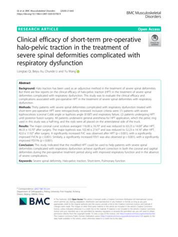
Qi et al. BMC Musculoskeletal Disorders(2020) 21:665However, it would still be necessary to use some pulmonary exercises to help improve muscle endurance andstrength in these patients [11, 20]. We used feedbackbreathing exercises in our patients to help improve endurance and muscle strength.The length of traction depended on the response ofthe various curves to the traction and the patient’s systemic, respiratory, and nutritional conditions. Currently,the duration of HGT varies from 2 to 12 weeks, as basedon reports in previous studies [5]. However, there is noconsensus on the optimal duration of traction for HPT,and there are few studies reporting the relationship between the duration of traction and the correction ofHPT. In this current study, the major correction of HPTon scoliosis and kyphosis was obtained in the first 2weeks, and the correction plateaued at nearly 50% afterapproximately 4–6 weeks after a rapid period of initialcorrection. In regard to pulmonary function, Koller et al.reported that prolonged traction might not help furtherimprove the pulmonary function [9]. For optimal correction and reduction in surgical risk, we recommend HPTtraction for at least 4 weeks, and appropriately prolongedperiod of traction according to the patient’s condition.However, the optimal length of traction remains to bedetermined and requires additional studies.O’Brien et al. previously described the complicationsassociated with HPT in detail. These included perforation of the intestine during insertion of the pelvic pins,infection of the pelvic pins, injury to the cervical spine,both acute and degenerative nerve injuries, and paraplegia [6]. Different from the HPT method used in thestudy by O’Brien et al., the pelvic pins used in the ourHPT did not need to be drill through the iliac crestPage 7 of 8(Fig. 5), and there was no perforation of the intestine orinjuries of other pelvic organs observed in this study.Strict protocols were used, including daily pin checksand prudent hygiene, and there were no other complications related to the pins, including infection of the pinsor pin loosening. As for injury to the cervical spine,O’Brien et al. reported that more than 50% of patientsshowed some degeneration of the cervical spine whenthe traction was prolonged over 3 months [6]. Tredwelland O’Brien noted that the incidence of apophyseal jointdegeneration was 47.4% in patients with HPT occurringover 9.47 months [21]. In this current study, the averagetime of traction was only 5.37 weeks, which was substantially shorter than that presented in O’Brien et al. orTredwel and O’Brien. Additionally, significant degeneration of the cervical spine was not observed in our study;however, our patients often complained of cervical discomfort and trapezial soreness, but these symptoms disappeared after surgery. Neurologic complications, suchas cranial nerve injuries and paraplegia, are known tooccur during traction, and warning signs must be monitored daily. In our study, two patients (6.7%) developedneurologic complications after HPT traction, thoughtthese occurrences returned to normal after reducing thetraction. Moreover, due to the lack of immobilization ofthe patients receiving the modified HPT, any occurrenceof osteoporosis could be avoided [22, 23].To our knowledge, this was the first study to report onthe use of this modified HPT, in which the pelvic ringwas changed to a half-ring and the rods were all placedon the anterolateral side of the truck. All of thesechanges were made to improve patient tolerance andcomfort while ensuring traction strength andFig. 5 Photographs of bony pelvis with the pelvic pins. The anteroposterior digital radiographs (a), and the CT scan (b-d)
Qi et al. BMC Musculoskeletal Disorders(2020) 21:665effectiveness. However, this study has several limitations.The study was a retrospective review of a single cohortwithout comparison to a control group to confirm theefficacy of the HPT since the standard protocol in ourcenter is surgical treatment after HPT in severe scoliosiscases. Secondly, this study is missing the post-operativedata regarding PFT and the results of 1 year and 2 yearfollow-ups, as the acquisition of post-operative PFT wasnot a regular protocol and some patients did not achievea 2 year follow-up. In the future, the results of 1 year and2 years follow-ups will be assessed to complete the postoperative data set.Page 8 of 83.4.5.6.7.8.ConclusionsThis study indicated that the modified HPT could beused to help patients with severe spinal deformities complicated with respiratory dysfunction achieve significantcorrection in both the coronal and sagittal deformitiesduring the pre-operative treatment period along withimproved respiratory function and in the absence of severe complications.AbbreviationsHPT: Halo-pelvic traction; HGT: Halo-gravity traction; SVA: Sagittal vertical axis;PFT: Pulmonary function test; FVC: Forced vital capacity; FEV1: Forcedexpiratory volume at the end of the first second9.10.11.12.AcknowledgmentsInterdisciplinary Clinical Research Project of Peking University First Hospital,No. 2019CR41.13.Authors’ contributionsLTQ and BYX analyzed and interpreted the data, and drafted the manuscript.YW and CDL designed the concept of this study. All authors have read andapproved the final manuscript.14.FundingThis research didn’t receive grants from any funding agency in the public,commercial or not-for-profit sectors.Availability of data and materialsThe datasets used or analysed during the current study are available fromthe corresponding author on reasonable request.Ethics approval and consent to participateNot applicable (radiographic follow-up). The study was a retrospective study, wedid not collect the patient’s privacy Information and had none follow-up for patients. We had an exemption from requiring ethics approval from the local ethicscommittee. (No. 2020–458, Peking university first hospital ethics committee).Consent for publicationThe patient’s written consent to publish the study and anonymized imagingwas obtained prior to submission.Competing interestsAll authors declare they have no conflict of interest.Received: 27 July 2020 Accepted: 1 October 2020References1. Sucato DJ. Management of severe spinal deformity: scoliosis and kyphosis.Spine (Phila Pa 1976). 2010;35(25):2186–92.2. Gollogly S, Smith JT, Campbell RM. Determining lung volume with threedimensional reconstructions of CT scan data: a pilot study to evaluate the15.16.17.18.19.20.21.22.23.effects of expansion thoracoplasty on children with severe spinaldeformities. J Pediatr Orthop. 2004;24(3):323–8.Bouchoucha S, Khelifi A, Saied W, Ammar C, Nessib MN, Ben Ghachem M.Progressive correction of severe spinal deformities with halo-gravity traction.Acta Orthop Belg. 2011;77(4):529–34.Garabekyan T, Hosseinzadeh P, Iwinski HJ, Muchow RD, Talwalkar VR, WalkerJ, Milbrandt TA. The results of preoperative halo-gravity traction in childrenwith severe spinal deformity. J Pediatr Orthop B. 2014;23(1):1–5.Yang C, Wang H, Zheng Z, Zhang Z, Wang J, Liu H, Kim YJ, Cho S. Halogravity traction in the treatment of severe spinal deformity: a systematicreview and meta-analysis. Eur Spine J. 2017;26(7):1810–6.O'Brien JP, Yau AC, Hodgson AR. Halo pelvic traction: a technic for severespinal deformities. Clin Orthop Relat Res. 1973;93:179–90.O'Brien JP, Yau AC, Smith TK, Hodgson AR. Halo pelvic traction. A preliminaryreport on a method of external skeletal fixation for correcting deformities andmaintaining fixation of the spine. J Bone Joint Surg Br. 1971;53(2):217–29.Quanjer PH, Tammeling GJ, Cotes JE, Pedersen OF, Peslin R, Yernault JC. Lungvolumes and forced ventilatory flows. Report working party standardization oflung function tests, European Community for steel and coal. Official statementof the European Respiratory Society. Eur Respir J Suppl. 1993;16:5–40.Koller H, Zenner J, Gajic V, Meier O, Ferraris L, Hitzl W. The impact of halogravity traction on curve rigidity and pulmonary function in the treatmentof severe and rigid scoliosis and kyphoscoliosis: a clinical study andnarrative review of the literature. Eur Spine J. 2012;21(3):514–29.Schroeder M, Viezens L, Kunkel P, Ridderbu
Background: Halo traction has been used as an adjunctive method in the treatment of severe spinal deformities. But there are few reports on the clinical efficacy of halo-pelvic traction (HPT) in the treatment of severe spinal deformities complicated with respiratory dysfunction. This study was to evaluate the clinical efficacy and