Transcription
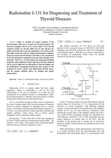
Radioiodine I-131 for Diagnosing and Treatment ofThyroid DiseasesWal‘a Al-jubeh, Ansam Shaheen, and Othman ZalloumApplied Physics Department, College of Applied Sciences,Palestine Polytechnic UniversityHebron, PalestineAbstract—Iodine is essential for proper function of thethyroid gland, which uses it to make the thyroid hormones. Thethyroid is equipped with an active system called “Na /I- thyroidsymporter pump” for moving iodine into its cells. Because theability of thyroid cells to take up radioiodine I-131 or RAI exactlylike iodine, it has been used by medical professionals to diagnoseand treat various thyroid diseases. RAI that is not concentratedin the thyroid gland is eliminated from the body through sweatand urine. However, I -131 has unique beta and gamma emittingproperties with a half-life of about eight days; the beta radiationcan be used in high dose for destruction of thyroid nodules andfor elimination of remaining thyroid tissue after surgery for thetreatment of Grave's disease and differentiate thyroid cancer,and the gamma radiation allows for imaging and uptakemeasurement.13154Xe * 13154 Xe rays 364 keV(2)The primary emissions of I-131 decay are thus betaparticles with a maximal energy of 606 KeV with 89.6%abundance and 364 KeV gamma rays with 81.5% abundanceas illustrated in figure 1. Beta decay, as always in this process,also produces an antineutrino, which carries off variableamounts of the beta decay energy.Keywords : Iodine 131, Radioiodine therapy, thyroid scan, RAIU.I.INTRODUCTIONRadioiodine I-131 as sodium iodide NaI, also calledradioactive iodine or radioiodide is one of the firstradioisotopes to be used in medicine. It is taken up andincorporated in an identical manner as nonradioactive iodineby the thyroid gland [1]. Therefore it has been used to evaluateand treat thyroid diseases for more than 60 years.I-131 is an artificial isotope of iodine, and it is separated inthe form of sodium iodide (NaI) from the products of uraniumfission or neutron irradiation of tellurium-130 in a nuclearreactor. According to the Environmental Protection Agency, I131 has a very short half-life of about eight days with beta andgamma radiation, which means that it decays almostcompletely in the environment within months.On decaying, 131I most often (89% of the time) expends its971 KeV of decay energy by transforming into the stable 131Xe(Xenon) in two steps, with gamma decay following rapidlyafter beta decay as shown in equations 1 and 2 :13153* I 13154 Xe v e 606 KeV(1)Figure 1. I-131 Decay Scheme.In the I-131 decay scheme, the color blue indicates that thenucleus decays by β-emission. I-131 is characterized by theemission of several β-particles with different endpointenergies. A direct transition to the ground state of the daughternuclide Xe-131 has not been observed. Transitions occurthrough the excited states of the daughter nuclide and with avery small (0.39%) probability through the metastable Xe131m that has a half-life of 11.9 d. This metastable state hasenergy difference to ground state of 164 KeV, and thetransition to the ground state is mostly due to conversionelectrons. Further weak transitions are indicated by dots in theI-131 scheme.When radioiodine is orally administered as the iodide ion,it is readily absorbed from the gastrointestinal tract anddistributed in the extracellular fluid. It is concentrated in the
salivary glands, thyroid, and gastric mucosa. RAI that is notconcentrated in the thyroid gland and other organs iseliminated from the body through sweat and urine. However,iodide trapped and organified by the normal thyroid has aneffective half-life of about 7 days; it decreases to 3–5 days inhyperthyroidism and is sometimes lower than 3 days inthyroid cancer.II.DIAGNOSTIC USES OF I-131IN THYROID GLANDI-131 is the isotope used to take pictures of the thyroidgland. A normal thyroid scan shows a small butterfly-shapedthyroid gland about 2 inch (5.1 cm) long and 2 inch (5.1 cm)wide with an even spread of radioactive tracer in the gland asshown in figure 2. On another hand an abnormal thyroid scanshows a thyroid gland that is smaller or larger than normal. Itcan also show areas in the thyroid gland where the activity isless than normal (cold nodules) or more than normal (hotnodules). Cold nodules may be related to thyroid cancer.Figure 2. Normal thyroid image obtained with 131I-iodide 24 hours afteradministration of the dosage showing uniform distribution of activity in bothlobes.Today the use of 131I is limited to follow-up diagnostics inthe case of thyroid differential cancer after thyroidectomy.This is because of it is unfavorable physical properties forscintigraphic imaging (i.e. physical half-life of 8.02 days, betaand gamma emissions, with high energy of 364 KeV, and highradiation dose ―131I emits approximately 1 rad/mCi to thethyroid gland‖) [2, 3, 4].After thyroidectomy for thyroid carcinoma, radio-ablationwith 131I can be used to eradicate any remaining thyroid tissuewithin the thyroid bed or metastases to lymph nodes or distantsites. Whole body imaging has an important role in the postoperative management of the thyroid cancer patient.A very small dose of I-131 is given to the patient, whothen returns 24 to 72 hours (sometimes 7-10 days after acancer therapy) later so pictures of the Whole Body Scan orWBS can be taken using a gamma camera that picks up thegamma radiation emitted by the RAI in the thyroid gland or intumor cells. The photons emitted from the body enter thegamma camera and interact with the crystal to producescintillations that are detected, amplified, and converted intolight. Light converted to electrical signal and sent to acomputer. The computer builds a picture by converting thediffering intensities of radioactivity emitted into differentshades of gray. Thyroid cancer cells such as papillary andfollicular type retain the ability to accumulate iodine althoughto a much lesser extent than normal thyroid tissue. Thisproperty is used in the detection of local tumor recurrence aswell as distant metastatic spread of thyroid cancer aftersurgery by administration of iodine and imaging the wholebody. This is the reason that cancer nodules appear cold onroutine thyroid scans.In addition to getting the scan or picture, the amount ofradiation being given off can also be counted to determinehow active the thyroid gland is Radioactive Iodine Uptake,RAIU. Thyroid uptake test is based on the principle that thethyroid gland absorbs iodine from the blood and uses it tomake certain hormones in the body. The average thyroid glandweighs around 20 grams, with each gram taking up a round 1percent of the iodine available from diet [5]. Therefore, whenthe radioactive iodine is given, it is quickly taken up by thetissues of the thyroid gland. Cells which are most active in thethyroid tissue will take up more of the radioiodine. So, activeparts of the tissue or hyperthyroidism cells will emit moregamma rays than less active or inactive parts. Likewise,thyroid cells that are not working well such as Thyroiditisusually take up less iodine, and so it will emit less gamma raysthan other thyroid cells. However, the gamma rays which areemitted from inside the thyroid gland are detected by thegamma probe, and then they are converted into an electricalsignal, and sent to a computer to count it, where the number ofcounts detected is proportional to the amount of radioactiveiodine in the low neck.To measure this, the patient swallows a small, oral dose,approximately 10 to 15 μCi (0.37–0.56 MBq) of sodiumiodide in the form of capsule or fluid. At given time afteradministration (usually at 4 to 6 hours and 24 hours) ascintillation crystal of gamma probe is placed 25 cm from thepatient neck‘s and the radioactivity at this point is counted forone minute as shown in figure 3. In order to eliminate thosecounts which did not come from the thyroid, the patient isrecounted for one minute with a 1/2-inch-thick lead shieldover the thyroid area. The difference of the two measurementsis called the ―net patient count.‖ A standard dose, identicalwith that given to the patient, is counted in a neck phantomunder the same conditions. A one-minute background count isalso taken of the phantom, with the standard dose removed.The difference of these two counts is the ―net standard count‖,and the method of counting the 131I standard is the same for allpatients [6,7]. The ratio of the net patient count to the netstandard count times one hundred equals the percent iodineuptake as in Formula [5]:% Thyroid Uptake Net counts per min in Thyroid 100 %Net counts of Standard Dose per min decay correct(3)
Decay correct in equation 3 is a method of adjusting themeasurements of radioactive decay obtained at two differenttime points (i.e. the time elapsed between swallowing ofiodine capsule and the uptake measure of theradiopharmaceutical) .However, the Decay correction factoris exp(-λt ) , where "λ" is the decay constant of I-131, and "t"is the elapsed time ( t time of measurement of the net thyroidcount per min - the net standard count per min time ).The percentage of radioiodine cumulated at specific times,such as 6 and 24 h, provides quantitation. This can becompared to the normal range in the community; for example,the normal 24-h uptake is and 7% to 20% for 6 hr and 7% to20% for 6 hours 10% to 35% for 24 hours. However, twovariables that determine the uptake are the function of thegland and the quantity of iodine in the diet [5,8,9].thyroid cancer (FTC), and mixed tumors will concentrateradioiodine.Although 131I is a beta emitter, there is a predominantenergetic gamma emission (364 KeV), which can be used toimage the biodistribution. This gamma photon also can resultin measurable absorbed radiation doses to persons near thepatient. Because excretion is via the urinary tract, and, to alesser extent, via saliva and sweat, special radiation protectionprecautions need to be taken for days after these patients aretreated.A. Heperthyrodisim DiseasesPhysicians usually prefer to treat the hyperthyroidismpatients with a dose of I-131 to render the patientshypothyroid, because calculation of a dose that would result ina euthyroid state is difficult, and it is easier to treathypothyroidism than hyperthyroidism. Patients can go homeafter the RIT, although they are asked to follow someprecautions. It is common for patients to experience some painin the thyroid after RIT for hyperthyroidism. Aspirin,ibuprofen or acetaminophen can treat this pain, and theysubsequently require thyroid hormone replacement for life.The RIT may take up to several months to have its effect.B. Well-Differentiated ThyroidFigure 3. RAIU. The detector of gamma probe is positioned 25cm from thepatient‘s thyroid.People who eat foods that are high iodine (iodized salt,dairy product, kelp, seafood, and so on), will have a lowerRAIU. This is because, although their thyroid‘s ability to suckup iodine has not been altered, the nonradioactive dietaryiodine dilutes out of portion of the radioactive iodine that goesinto the thyroid gland, resulting in falsely in a lower RAIU. Sothese foods should be stopped at least 4 to 5 days before anRAIU test.III.Therapy for Well-differentiated Thyroid Cancer (DTC)starts with near-total thyroidectomy because the thyroid cancerdoes not concentrate iodine as well as normal thyroid tissue. AWDS is done to visualize the remaining functional thyroidtissue, and depending on the amount of residual thyroid tissueor tumor and the location and extent of disease, an appropriatedose of I-131 is administered. Scans are often obtained a fewdays after treatment to further evaluate the extent of disease.Follow-up another WBS are usually done 1 year after RIT toassess for residual or recurrent thyroid cancer after the patienthas been off thyroid hormone replacement for 6 weeks (Figure4).RADIOIODINE I-131 THERAPYI-131 emitted beta particles with an average energy of 192KeV. When beta particles of I-131 are released inside thyroidcells, they travel an average of 0.8 millimeters [6] before theirenergy is completely absorbed by the thyroid deposit. Theymay collide with atoms of critical parts of the cells, impartingtheir energy to these atoms and killing the cells. This permitsthe I-131 treatment to specifically target thyroid abnormalcells without bothering other nearby body cells [5,9].Therefore, radioiodine therapy (RIT) is an important option inthe treatment of hyperthyroidism associated with Graves'disease, toxic thyroid adenoma, and toxic multinodular goiteror Plummer‘s disease. RIT is also used as adjuvant treatmentafter surgery for Well-differentiated thyroid carcinomas(DTC) such as papillary thyroid cancer (PTC), follicularFigure 4. Whole-body scan of a 15-year old girl with metastatic DTC before(left) and after (right) effective radioiodine therapy.If the patients are given a higher dose, they may be askedto stay isolated in a special room in the hospital for about 24hours to avoid exposing other people to radiation, especially ifthere are small children living in the same home with them.The regulations that determine whether a patient needs to beisolated or can go home after the treatment are different indifferent states. Since the salivary glands weakly concentrate
iodine, there may be pain and swelling of the salivary glandsafter high doses of RIT for WDC. This can be prevented orreduced by sucking on lemon drops after the therapy.Different doses of I-131 are given for different thyroiddisorders. Controversies still exist regarding the administereddosage of 13lI-sodium iodide in each of these conditions, e.g.there are three approaches dosimetric method to choosing adose of I-131 for WDC therapy: a fixed dose method thatadministers empiric fixed amounts of I-131 for different tumorstages, quantitative tumor dosimetry that calculates theradiation delivered by I-131 to a tumor site, and a dosimetryguided technique that calculates the upper limits of a safeblood and whole body level of irradiation from I-131. Therehas been no prospective randomized trial to determine whichis better. Several analyses and studies have suggested thatthese approaches yield similar results [10-14].IV.THE ABSORBED DOSE FROM RADIOACTIVE IODINE I131 WITHIN THE BODYBecause thyroid gland contains the radioiodine I-131, itbecomes an emitting source ‗S‘, which can also irradiate theneighboring tissues (defined as target regions ‗T‘) such as lungand liver. Consequently, each body organ or tissue couldreceive a radiation dose after administration of radioiodineeven if no activity is present in it. Therefore, RIT requirepatient-specific planning of the absorbed dose to the targetvolumes, in order to achieve an expected biological effect andbenefits of their use, taking into account that the absorbeddoses to normal organs and tissues should be kept as low asreasonably achievable (ALRA principle) [15,16]. So, thecalculation of absorbed doses to the lungs and the dose toother organs has to be as accurate as possible.V.Despite being more than 70 years old, 131I remains a majoragent of choice for the treatment of thyroid diseases. It is stillused to image thyroid cancer and quantify thyroid uptakemeasurements. Iodine I-131 has unique beta and gammaemitting properties; the beta radiation is used for therapy, andthe gamma radiation allows for imaging and uptakemeasurement. But it is high radiation dose and unfavorableimaging characteristics. The introduction of newer radiotracerswith lower radiation doses, such as radioiodine 123I andpertechnetate 99mTc, has resulted in a decreasing role of 131I inthe imaging and uptake evaluation of thyroid disease.However, 131I continues to play a major role in the imaging ofpatients with thyroid cancer after thyroidectomy and thetreatment of hyperthyroidism and thyroid cancer.When a radioactive iodine I-131 is given to a patient foreither diagnosis or therapy, the nuclei end up in differentorgans; such as lung and liver, in varying amounts. Therefore,the calculation of absorbed doses to the lungs and the dose toother organs has to be as accurate as possible.ACKNOWLEDGMENTThe authors are indebted to the program committee of theStudent Innovation Conference (SIC) which is endowed by theDeanship of Graduate Studies and Scientific Research at thePalestine Polytechnic University (PPU) for their excellentwork that recognizes student innovation, research excellenceand presentation skills. The first two authors would like tothank their teachers in Department of Applied Physics at thePPU for their support and encouragement.For this purpose a group of physicists and biologists wereformed a committee called the Medical Internal RadiationDose (MIRD) Committee of the Society of Nuclear Medicine.REFERENCESThe total energy emitted by the thyroid organ is the[1]fraction ( i) of this energy is deposited in the target organ,which is the location of interest in the dose calculation. Withthese quantities and knowledge of the mass of the target organ[2] product of and the cumulated activity, A . However, only a[3](m), the mean absorbed dose D delivered to an organ in gray(Gy) is [17,18]:[4](4)[5]where i Фi (T S) is called the S value. Also known as thedose per cumulated activity, S has SI units of dose per decayor dose rate per activity (e.g., grays per Becquerel second). Avalue of S for radioiodine is published in MIRD Pamphlet no.11.[6][7] D A i i (T S )iDISCUSSION AND CONCLUSION[8][9]Peter F. Sharp, Howard G. Gemmell, and Alison D. Murray.Practical nuclear medicine, third edition. Spring, New York,USA.2005.Fred A. Mettler, Jr., and Milton J. Guiberteau. Essential Physicsof Nuclear Imaging, sixth edition. Elsevier Science Health ScienceDivision, Philadelphia, USA. 2012.Richard Zimmermann. Nuclear Medicine: Radioactivity forDiagnosis and Therapy. L'Editeur : EDP Sciences, 2007Judith M. Joyce, and Andrew Swihart. ―Thyroid: NuclearMedicine Update‖. Radiologic. North America. Vol 49, pp. 425434, May 2011.John R. Cameron , and Douglas B. Bell, ―Thyroid Uptake Studieswith I131 in Very Small Doses,‖ Radiology. North America. vol.79, pp. 452-456, September edhealth/brt/radscape/case-theneck-phantom.html.I. Ross Mcdougall, Benjamin Franc and Jason Cohen. ―ThyroidImaging‖. Encyclopedia of Endocrine Diseases, Elsevier Inc. Vol4, pp.496-504, June 2004.Kenneth Ain, and M. Sara Rosenthal. The Complete Thyroidbook, second edition. McGraw-Hill Professional, 2010.
[10][11][12][13][14][15]Max H. Lombardi. Radiation Safety for Nuclear medicine, secondedition. Taylor & Francis, New York, USA. 2006.Abdel hamid H. Elgazzar. A Concise Guide to Nuclear Medicine.Spring, New York, USA. aceuticals in Nuclear Pharmacy and NuclearMedicine, second edition. The American Pharmacists Association,Washington, USA. 2004.Robert J. Amdur, and Ernest L. Mazzaferri. Essentials of ThyroidCancer Management. Birkhäuser, 2005.Ernest L. Mazzaferri. Practical Management of Thyroid Cancer.Springer, New York, USA. 2006.R E Toohey, M G Stabin, E Watson. ―Internal RadiationDosimetry: Principles and Applications‖. Vol 20, no 2, pp. 533-[16][17][18]564. March-April 2000.Douglas J. Simpkin . ―Radiation Interactions and InternalDosimetry in Nuclear Medicine‖. RadioGraphics, Volume 19,Pages 155-167. January 1999.Russell K. Hobbie, and Bradley J. Roth . Intermediate Physics forMedicine And Biology, fourth edition. Springer, New York, USA.2007.Michael G. Stabin.Fundamentals of Nuclear MedicineDosimetry. Springer, New York, USA. 2008.
A direct transition to the ground state of the daughter . energy is completely absorbed by the thyroid deposit. They may collide with atoms of critical parts of the cells, imparting . Therefore, radioiodine therapy (RIT) is an important option in the treatment of hyperthyroidism associated with Graves' disease, toxic thyroid adenoma, and .

