Transcription
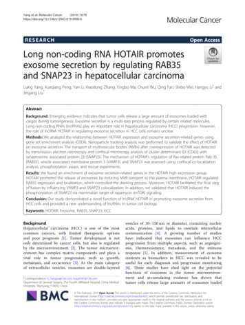
Yang et al. Molecular Cancer(2019) ARCHOpen AccessLong non-coding RNA HOTAIR promotesexosome secretion by regulating RAB35and SNAP23 in hepatocellular carcinomaLiang Yang, Xueqiang Peng, Yan Li, Xiaodong Zhang, Yingbo Ma, Chunli Wu, Qing Fan, Shibo Wei, Hangyu Li* andJingang Liu*AbstractBackground: Emerging evidence indicates that tumor cells release a large amount of exosomes loaded withcargos during tumorigenesis. Exosome secretion is a multi-step process regulated by certain related molecules.Long non-coding RNAs (lncRNAs) play an important role in hepatocellular carcinoma (HCC) progression. However,the role of lncRNA HOTAIR in regulating exosome secretion in HCC cells remains unclear.Methods: We analyzed the relationship between HOTAIR expression and exosome secretion-related genes usinggene set enrichment analysis (GSEA). Nanoparticle tracking analysis was performed to validate the effect of HOTAIRon exosome secretion. The transport of multivesicular bodies (MVBs) after overexpression of HOTAIR was detectedby transmission electron microscopy and confocal microscopy analysis of cluster determinant 63 (CD63) withsynaptosome associated protein 23 (SNAP23). The mechanism of HOTAIR’s regulation of Ras-related protein Rab-35(RAB35), vesicle associated membrane protein 3 (VAMP3), and SNAP23 was assessed using confocal co-localizationanalysis, phosphorylation assays, and rescue experiments.Results: We found an enrichment of exosome secretion-related genes in the HOTAIR high expression group.HOTAIR promoted the release of exosomes by inducing MVB transport to the plasma membrane. HOTAIR regulatedRAB35 expression and localization, which controlled the docking process. Moreover, HOTAIR facilitated the final stepof fusion by influencing VAMP3 and SNAP23 colocalization. In addition, we validated that HOTAIR induced thephosphorylation of SNAP23 via mammalian target of rapamycin (mTOR) signaling.Conclusion: Our study demonstrated a novel function of lncRNA HOTAIR in promoting exosome secretion fromHCC cells and provided a new understanding of lncRNAs in tumor cell biology.Keywords: HOTAIR, Exosome, RAB35, SNAP23, HCCBackgroundHepatocellular carcinoma (HCC) is one of the mostcommon cancers, with limited therapeutic optionsand poor prognosis [1]. Tumor development is notonly determined by cancer cells, but also is regulatedby the microenvironment [2]. The tumor microenvironment has complex matrix components and plays avital role in tumor progression, such as growth,metastasis, and occurrence [3]. As the main categoryof extracellular vesicles, exosomes are double-layered* Correspondence: li hangyu@126.com; liujg0347@126.comDepartment of General Surgery, The Fourth Affiliated Hospital, China MedicalUniversity, Shenyang 110032, Chinavesicles of 30–150 nm in diameter, containing nucleicacids, proteins, and lipids to mediate intercellularcommunication [4]. A growing number of studieshave indicated that exosomes can influence HCCprogression from multiple aspects, such as angiogenesis, chemoresistance, metastasis, and the immuneresponse [5]. In addition, measurement of exosomecontents as biomarkers in HCC was revealed to beuseful for early diagnosis and progression monitoring[6]. These studies have shed light on the potentialfunctions of exosomes in the tumor microenvironment and accumulating evidence has shown thattumor cells release large amounts of exosomes loaded The Author(s). 2019 Open Access This article is distributed under the terms of the Creative Commons Attribution 4.0International License (http://creativecommons.org/licenses/by/4.0/), which permits unrestricted use, distribution, andreproduction in any medium, provided you give appropriate credit to the original author(s) and the source, provide a link tothe Creative Commons license, and indicate if changes were made. The Creative Commons Public Domain Dedication o/1.0/) applies to the data made available in this article, unless otherwise stated.
Yang et al. Molecular Cancer(2019) 18:78with cargos during tumorigenesis [7, 8]; however, themolecular mechanisms of exosome secretion in tumorcells remain unclear.The biogenesis of exosomes is generated from theinward budding of the membranes of multivesicularbodies (MVBs) to form intraluminal vesicles (ILVs),which finally mature and are contained within MVBs[9]. This process involves various sorting machineries,including endosomal sorting complex required for transport (ESCRT)-dependent and ESCRT-independent processes [10]. The MVBs containing ILVs are transportedalong microtubules and fuse with the plasma membrane,causing the ILVs to be released outside the cell as exosomes. Before exosome release, multiple intracellulartrafficking steps are required to regulate MVB motility,docking, and fusion with the plasma membrane, whichinvolve different effectors and molecular mechanisms[11]. Rab GTPases are required for the motility of MVBsand docking at the plasma membrane [12]. Rab GTPasescontrol MVBs transport by acting as molecular switchesthat convert between the GTP-bound active form andthe GDP-bound inactive form [13]. The Rab family comprises almost 70 subtypes that show varied subcellulardistributions. Several Rab GTPases play different roles inthe exosomal pathway and mediate exosome secretion[14, 15]. Different Rab GTPases localize in distinct subcellular locations and are specific to cell types; however,the molecular mechanisms of Rab GTPases’ effects onexosome secretion in tumor cells require further study.MVB fusion with the plasma membrane results inthe release of ILVs as exosomes, which is the finaland key step of exosome secretion. SolubleN-ethylmaleimide-sensitive fusion factor attachmentprotein receptor (SNARE) proteins have been shownto mediate this final step [16]. The SNARE complexinvolved in exocytic release comprises v-SNAREs onmembranes of the MVBs and t-SNAREs on the cell membrane, forming a stable ternary complex that mediatesexosome secretion. As v-SNAREs, vesicle-associatedmembrane protein (VAMP) 2, VAMP3, VAMP7, andVAMP8 are known to regulate exocytosis in tumor cells[17, 18]. In addition, synaptosome associated protein 23(SNAP) 23 is an important t-SNARE that is mainly locatedat the plasma membrane [19]. Phosphorylation ofSNAP23 not only promotes the formation of the SNAREcomplex, but also increases exosome secretion [20]. However, whether long non-coding RNAs (lncRNAs) participate in the regulation of SNARE proteins and mediateexosome secretion is unclear. HOX transcript antisenseRNA (HOTAIR) is a 2158 nucleotide lncRNA transcribedfrom the HOXC locus and is frequently upregulated inmany types of human cancer, including HCC [21].Previously, we demonstrated that HOTAIR could induceautophagy by upregulating autophagy related 3 (ATG3)Page 2 of 12and ATG7 expression in HCC cells [22]. Although agrowing number of studies revealed that HOTAIR couldinfluence multiple biological functions as an oncogene[23], a relationship between lncRNA HOTAIR and exosome secretion has not been identified. To determinewhether HOTAIR contributes to exosome secretion inHCC cells, we analyzed the function of HOTAIR in exosome secretion using nanoparticle tracking analysis(NTA). We demonstrated that HOTAIR could promotethe release of exosomes from HCC cells. Given the importance of Rab GTPases and SNAREs in the mediationof exosome secretion, our mechanistic study revealed thatHOTAIR facilitates the transport of MVBs towards theplasma membrane by regulating Ras-related proteinRab-35 (RAB35), which controls the docking process.Furthermore, we identified that HOTAIR promotes thecolocalization of VAMP3 with SNAP23, which influencesSNARE complex formation, leading to MVB fusion withthe plasma membrane. Our research demonstrated therole of HOTAIR in exosome secretion and provides a newunderstanding of lncRNAs in tumor cell biology.MethodsGene set enrichment analysis (GSEA)Liver hepatocellular carcinoma (LIHC) RNA sequencing(RNA-seq) data (374 cases) and normal tissues RNA-seqdata (50 cases) were generated from The Cancer Genome Atlas (TCGA). The 374 cases were categorized intoa HOTAIR high expression group and a HOTAIR lowexpression group. We performed GSEA analysis usingGSEA v2.0 software ). Statistical significance was assessed bycomparing the enrichment score with the enrichmentresults generated from 1000 random permutations ofthe gene set to obtain p-values.Cell cultureHepG2 (an HCC cells line) was obtained from ZhongQiao Xin Zhou (Shanghai, China). The cell line wasmaintained in minimal essential medium (MEM)(HyClone, Logan, UT, USA) with 10% fetal bovine serum(HyClone) containing 100 U/mL penicillin and 100 μg/mLstreptomycin and incubated at 37 C under 5% CO2. Cellsor culture medium were collected for experimentation atthe indicated times.Cell transfectionAll the HOTAIR plasmids and small interfering RNAs(siRNAs) were purchased from Genechem (Shanghai,China). HepG2 cell transfection was performed in 6-wellplates using Invitrogen Lipofectamine 3000 (ThermoFisher Scientific, Shanghai, China) according to themanufacturer’s instructions. At 48 h after transfection,the cells or culture supernatants were harvested to
Yang et al. Molecular Cancer(2019) 18:78perform further experiments. The full length cDNAsequences of HOTAIR were cloned into pcDNA3.1(GV144) vector (GeneChem Shanghai, China) toconstruct HOTAIR overexpression plasmid (pcDNA-HOTAIR). The two siRNA sequence for RAB35 was asfollows: Rab35 siRNA#1: 5′-CTGGTCCTCCGAGCAAAGAAA-3′. Rab35 siRNA#2: 5′-GAUGAUGUGUGCCGAAUAU-3′.Exosome isolationExosomes were prepared from the culture supernatantfrom a 48-h culture of HepG2 cells. The culture supernatant was centrifuged (Beckman XPN-100) at 2000 gfor 10 min (4 C) and 1000 g for 30 min (4 C) toremove debris. Then, the supernatant was centrifuged at100,000 g for 70 min (4 C). The pellet was resuspendedand washed in phosphate-buffered saline (PBS) andthe supernatant was centrifuged at 100,000 g for 70min (4 C) again [24]. Finally, the exosomes werecollected from the pellet, washed, and resuspended inPBS as described previously [24].Nanoparticle tracking analysisThe number and size of the exosomes were directlytracked by the rate of Brownian motion of exosomesusing the NanoSight NS 300 system (NanoSight Technology, Malvern, UK), configured with a high-sensitivitysCMOS camera, fast video capture, and particle-trackingsoftware (NanoSight, Amesbury, UK). The samples werediluted 150–3000 times with Dulbecco’s PBS (DPBS)without any nanoparticles to attain a concentration of1–20 108 particles per milliliter for analysis. Eachsample was measured in triplicate at the camera, whichrecorded and tracked each visible particle. Exosomenumbers and size distribution were explored using theStokes-Einstein equation.Phos-tag SDS-PAGE and western blottingCells and exosomes were lysed in Radioimmunoprecipitation assay (RIPA) buffer containing protease inhibitorsstored at 20 C until use. The proteins were separatedby SDS-PAGE and then transferred to polyvinylidenefluoride (PVDF membranes. Trichloroacetic acids (TCA)precipitation was used to reduce the impurity content inthe Phos-tag SDS-PAGE (#193–16,711) samples. Metalions (Mn2 or Zn2 ) were removed from the gel usingethylenediaminetetraacetic acid (EDTA) before filmtransfer. After incubation with the appropriate primaryantibodies, horseradish peroxidase-conjugated secondaryantibodies were incubated with the membranes. TheLi-Cor Odyssey protein imaging system was used toanalyze protein bands. Antibodies used were: rabbitpolyclonal anti-tumor susceptibility 101 (TSG101)(#14497–1-AP), rabbit polyclonal anti-cluster determinantPage 3 of 1263 (CD63) (#25682–1-AP), rabbit polyclonal anti-RAB35(#11329–2-AP), rabbit polyclonal anti-β-Actin (#60008–1-Ig), rabbit polyclonal anti-SNAP23 (#ab4114), rabbitpolyclonal anti-mammalian target of rapamycin (mTOR)(#ABP54398), and rabbit polyclonal anti-mTOR (phosphoSer2448) (#ABP50363).Real-time PCRWe used the TRIzol reagent (Invitrogen) to extract totalRNA from the control and treated cells, as previouslydescribed [22]. The RNA was reversely transcribed tocDNA using a Reverse Transcription Kit (Takara, Dalian,China). Real-time PCR was conducted using theSYBR-Green PCR Master Mix kit. The following primerswere used: RAB5 forward 5′-AGACCCAACGGGCCAAATAC-3′, and reverse, 5′-GCCCCAATGGTACTCTCTTGAA-3′; RAB7 forward 5′-CTCATTATCGTCGGAGCCATTG-3′, and reverse, 5′-AGTGTGGTCTGGTATTCCTCATA-3′; RAB11 forward 5′-GCTCGGCCTCGACAAGTTC-3′, and reverse, 5′-ACTTATACCACTGCGTCTTCCT-3′; RAB27A forward 5′-GGAGAGGTTTCGTAGCTTAACG-3′, and reverse, 5′-CCACACAGCACTATATCTGGGT-3′; RAB27B forward 5′-TAGACTTTCGGGAAAAACGTGTG-3′, and reverse, 5′-AGAAGCTCTGTTGACTGGTGA-3′; RAB35 forward 5′-TTAAGCTTCGATGGCCCGGGACTACGACC-3′, and reverse, 5′-TTGGATCCTTAGCAGCAGCGTTTCTTTCGTTTACTG-3′; glyceraldehyde-3-phosphate dehydrogenase (GAPDH) forward 5′-AAAGATGTGCTTCGAGATGTGT-3′, and reverse, 5′-CACTTTGTCAGTTACCAACGTCA-3′. GAPDH was used as an endogenouscontrol.Immunofluorescence and confocal microscopyFor immunofluorescence assays, cells were fixed with 4%paraformaldehyde for 25 min at 25 C and then stainedwith the indicated primary antibodies (1:100) at 4 Covernight. Cells were then incubated with secondaryantibodies for 1 h at 37 C. Finally, nuclei were midine(DAPI) (Beyotime) for 3 min at room temperature.Immunofluorescence was captured using a Nikon A1rconfocal microscope. The antibodies used were asfollows: rabbit polyclonal anti-SNAP23 (#ab4114), yclonal anti-CD63 (#GTX28219), mouse polyclonalanti-VAMP3 (#66488–1-Ig), Goat anti-rabbit IgG H&L(fluorescein isothiocyanate (FITC)) (#ab6717), and Goatanti-mouse IgG H&L (Cy3) (#ab97035).Transmission electron microscopySamples of cells were quickly removed and fixed in 5%glutaraldehyde (protected from light) in 0.1 M phosphatebuffer at 4 C. The fixed cells were then subjected
Yang et al. Molecular Cancer(2019) 18:78ultracryomicrotomy to generate slices of about 70 nm inthickness. The isolated exosomes (20–40 μm) in heavysuspension droplets were placed on the special coppermesh of the electron microscope, and then subjected tonegative staining with 20 μL 2% phosphotungstic acidfor 10 min. All samples were analyzed using a H-7650electron microscope at 100KV.RNA immunoprecipitation (RIP) assayRIP assay was performed using RNA-Binding ProteinImmunoprecipitation Kit (Millipore, Bedford, MA) according to the manufacturer’s instructions. Cell lysateswere incubated with RIP buffer containing magneticbeads conjugated with RAB35 antibody or negative control IgG (Millipore). Immunoprecipitated RNA was puried, and then subjected to real-time PCR analysis todetect the relative levels of HOTAIR.Pull-down assayPull-down assay was was examined using PierceMagnetic RNA-Protein Pull-Down Kit (Thermo fisher)according to the manufacturer’s protocols. Biotin-labeledHOTAIR or antisense RNA was co-incubated withprotein extract of HepG2 cells and magnetic bead. Theretrieved protein was detected by western blot withβ-actin as control.Statistical analysisAll data are presented as the mean standard error (SD)from three independent experiments, analyzed by SPSSversion 17.0 software (IBM Corp., Armonk, NY, USA).Student’s t-test or analysis of variance were used toperform the statistical analyses. Statistically significancewas concluded at *P 0.05, **P 0.01.ResultsAbnormal expression of HOTAIR is associated withexosome secretionRecent evidence shows that exosome secretion is amulti-step process regulated by certain related molecules[11]. Our previous review and other studies have summarized a series of regulators involved in exosome secretion [5, 11]. However, very little is known about therelationship between lncRNA HOTAIR and exosome secretion. We aimed to study the link between HOTAIRand exosome secretion-related genes. To date, 31 genesencoding proteins that regulate the process of exosomesecretion have been identified, which are listed in Fig. 1a.We used the RNA-seq data from 374 cases of liver cancergenerated from TCGA and categorized these cases intotwo groups: The low HOTAIR expression group (less thanthe median) and the high HOTAIR expression group(greater than the median). Using GSEA, we identified asignificant enrichment of exosome secretion-related genesPage 4 of 12in the patients with HCC in the high HOTAIR group(normalized enrichment score (NES) 1.548, p-value 0.028, Fig. 1b). In addition, we showed that the expressionlevels of RAB35, SNAP23, and VAMP3 were significantlyhigher in HCC tissues than in normal tissues (Fig. 1c–e).Moreover, we divided the HCC tissues into relative highHOTAIR group and relative low HOTAIR group. Wefound that he expression levels of RAB35, SNAP23 wereincreased in HOTAIR high group (Fig. 1f–h). Theseresults suggested that HOTAIR might play a role inregulating exosome secretion in HCC.HOTAIR promotes exosome secretion in HCC cellsTo detect exosome secretion, we first isolated exosomesfrom HCC cell culture medium using ultracentrifugation. We then analyzed the purified exosomes usingtransmission electron microscopy. As shown in Fig. 2a,exosomes are extracellular vesicles with a double membrane, and are 50–100 nm in diameter. To analyze theeffect of lncRNA HOTAIR on exosome secretion, weconstructed HOTAIR overexpression cell line bytransfection pcDNA3.1-HOTAIR in HepG2 cells. Thetransfection efficiency was about 56-fold compared withnegative control (Additional file 1: Figure S1a). Subcellular fractionation and real-time PCR analysis showed thatHOTAIR was mainly located in the cytoplasm of HCCcells, which indicated that HOTRAIR may function incytoplasm in HCC cells (Additional file 1: Figure S1b).Then we detected the exosome markers CD63 andTSG101 in exosomes purified from the culture mediumof cells overexpressing HOTAIR compared with that inthe negative control. The results showed that overexpression of HOTAIR could increase the secretion ofexosomes containing CD63 and TSG101 (Fig. 2b). Inaddition, NTA indicated that the sizes of releasedexosomes were about 100 nm (Fig. 2c). We also foundthat when HepG2 cells overexpressed HOTAIR, they secreted more exosomes (Fig. 2d).These results suggestedan important role of HOTAIR in promoting exosomesecretion from HCC cells.HOTAIR enhances the transport of MVBs towards theplasma membraneTo determine the mechanisms by which HOTAIRinfluences exosome release, we analyzed the process ofexosome generation intracellularly. Before exosomes aresecreted into the extracellular environment, they arecontained within MVBs, which are transported alongmicrotubules to the plasma membrane [25]. Consequently, the effect of HOTAIR on the transport of MVBswas studied. Previously, CD63 has been used as a markerof MVBs. Overexpression of HOTAIR resulted in CD62being distributed further away from the nucleus (Fig. 3a).In addition, SNAP23 is part of the SNARE complexes
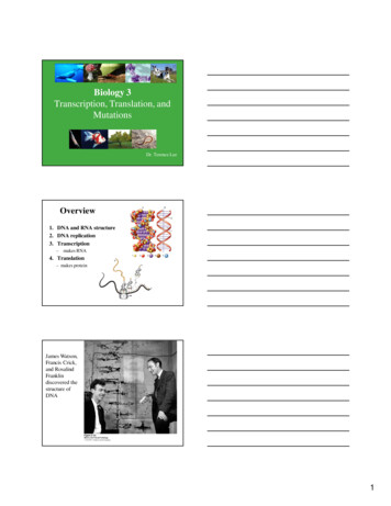
Yang et al. Molecular Cancer(2019) 18:78Page 5 of 12Fig. 1 Abnormal expression of HOTAIR is associated with exosome secretion. a Heatmap showing the relative expression values for 31 exosomesecretion-related genes in a series of 374 cases of liver cancer generated from TCGA and categorized into subgroups according to their medianHOTAIR expression. The list on the right shows 31 genes involved in exosome secretion. b Enrichment plot showing enrichment of exosomesecretion-related genes in the HOTAIR high expression group. c-e The mRNA expression of RAB35, SNAP23, and VAMP3 were analyzed in HCCtissues compared with normal tissues. f-h The mRNA expression of RAB35, SNAP23, and VAMP3 were analyzed in HOTAIR relative high groupcompared with HOTAIR relative low group.t-test *P-value 0.05that are located mainly in the plasma membrane andregulate MVB docking and fusion with the plasma membrane. The results showed that overexpression ofHOTAIR also increased the colocalization of SNAP23with CD63 (Fig. 3b). Electron microscopy results showedthat MVBs were more abundant in HOTAIR overexpressing cells that in the negative control cells (Fig. 3c).Collectively, these results demonstrated that HOTAIRinduces the transport of MVBs towards the plasmamembrane.HOTAIR regulates the expression and the localization ofRAB35To further investigate the molecular mechanisms bywhich HOTAIR affects the motility of MVBs, weanalyzed members of the Rab GTPase family. Previousstudies have found that several Rab GTPases are locatedin a subcellular position coincident with MVBs and mediate MVB transport along microtubules [14, 15]. Ourprevious review summarized the Rab GTPases involvedin the release of exosomes, which include RAB5, RAB7,RAB11, RAB27a, RAB27b, and RAB35 [5]. We screenedthe expression of these Rab GTPases using real-timePCR. The results showed that RAB35 was the mostupregulated Rab GTPase gene in response to HOTAIRoverexpression in HCC cells (Fig. 4a). Consistent withthis result, overexpression of HOTAIR increased theabundance of the of RAB35 protein, as assessed usingwestern blotting analysis (Fig. 4b). The same effectwas demonstrated in Huh7 cells (Additional file 2:Figure S2a-b). Next, we investigated whether HOTAIRinfluenced the subcellular localization of RAB35. Ourresults indicated that overexpression of HOTAIRcould induced stronger staining of RAB35 and an increased the colocalization of RAB35 with CD63,which suggested that HOTAIR regulated thelocalization of RAB35 at MVBs and upregulatedRAB35 expression (Fig. 4c). RIP assay results showedthat HOTAIR was significantly enriched by Rab35antibody compared with control IgG (Fig. 4d). Thespecific association between HOTAIR and Rab35 wasfurther validated by pull-down assay using in vitrotranscribed biotin-labeled HOTAIR (Additional file 2:Figure S2d). Our findings demonstrate a direct interaction between HOTAIR and Rab35. Thus, the mainmechanism of HOTAIR is to promote MVBs transport tothe plasma membrane. Furthermore, we demonstratedthat HOTAIR’s promotion of exosome secretion was
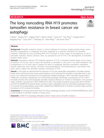
Yang et al. Molecular Cancer(2019) 18:78Page 6 of 12Fig. 2 HOTAIR promotes exosome secretion in HepG2 cells. a–c Isolated exosomes from HepG2 cells were assessed by a transmission electronmicroscopy, b western blotting and c NTA. d NTA analysis of the effect of HOTAIR on exosome release in HepG2 cells. Data are reported as themean standard error (SD) from three independent experiments, t-test *P-value 0.05eliminated by cotransfection with HOTAIR and RAB35specific siRNAs (Fig. 4e). The knock-down efficiency ofsi-RAB35 is detected by western blot (Additional file 2:Figure S2c). By cotransfection assay, we aimed to demonstrated HOTAIR influence exosome secretion via regulation Rab35. These results support the view that HOTAIRpromotes the motility of MVBs by regulating the expression and localization of RAB35.HOTAIR induces the translocation of VAMP3 and SNAP23When MVBs are transported to the cell membrane, theymust fuse with the plasma membrane to release the ILVsas exosomes. This process is mediated by SNAREtransmembrane proteins. One SNARE molecule on theMVB membrane (v-SNARE) binds to SNAREs onplasma membrane (t-SNARE), forming a stable ternarycomplex (trans-SNARE) that mediates MVB fusion withthe plasma membrane [16]. A previous study indicatedthat SNAP23, as an important t-SNARE located atplasma membrane, plays a vital role in regulatingsecretion by binding to VAMP3 as a v-SNARE [19].Overexpression of HOTAIR induced SNAP23 to becomediffusely located at the plasma membrane comparedwith the negative control (Fig. 5a). Furthermore, overexpression of HOTAIR also promoted an increasedcolocalization of VAMP3 with SNAP23, which suggestedthat HOTAIR could induce SNARE complex formationto influence the fusion process of MVBs (Fig. 5b).HOTAIR promotes the release of exosome viaphosphorylation of SNAP23Based on the result that HOTAIR regulates the locationof SNAP23, we next tested whether HOTAIR couldinfluence the activity of SNAP23. A previous studyindicated that SNAP23 phosphorylation is required forexosome secretion [26]. To test whether HOTAIR couldregulate SNPA23 phosphorylation, we performed aphosphorylation assay to detect the level of phosphorylated SNAP23 in HepG2 cells transfected withpcDNA3.1-HOTAIR. As shown in Fig. 6a, overexpression of HOTAIR significantly increased the ratio ofp-SNAP23/SNAP23 compared with that in the negativecontrol (Fig. 6a). This result suggested that HOTAIRcould induce SNAP23 phosphorylation. We next soughtto determine how HOTAIR phosphorylates SNAP23.mTOR signaling is involved in regulating SNAREcomplexes [27]. Our results showed that overexpressionof HOTAIR increased the amount of phosphorylated(p)-mTOR (Additional file 3: Figure S3a). Using rapamycin(an inhibitor of mTOR) to perform a rescue experiment,
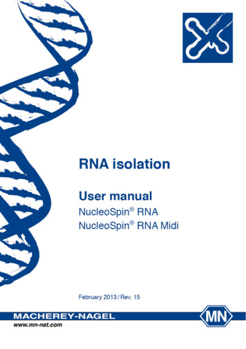
Yang et al. Molecular Cancer(2019) 18:78Page 7 of 12Fig. 3 HOTAIR enhances the transport of MVBs towards the plasma membrane. a Confocal microscopy analysis of CD63 (red) in HepG2 cellstransfected with pcDNA3.1-HOTAIR. Nuclei were stained with DAPI. b Confocal co-localization analysis of CD63 (red) and SNAP23 (green) inHepG2 cells transfected with pcDNA3.1-HOTAIR. The rectangular box indicates the small punctate structures where CD63 and SNAP-23 wereco-localized. c Electron microscopy images showing MVBs in HepG2 cells transfected with pcDNA3.1 or pcDNA3.1-HOTAIR. Red arrowsindicated MVBswe found that the effect of HOTAIR in inducing SNPA23phosphorylation was attenuated when the transfected cellswere co-treated with rapamycin (Fig. 6b). Moreover,exosome release induced by overexpression HOTAIR wassignificantly decreased after rapamycin-induced inhibitionof mTOR (Additional file 3: Figure S3b). These resultssuggested that HOTAIR could promote the release ofexosomes via phosphorylation of SNAP23.DiscussionCells can secrete many extracellular vesicles, such asmicrovesicles (shedding vesicles) and exosomes. Thebiogenesis mechanisms of these extracellular vesicles aredifferent. Microvesicles originate from evagination of theplasma membrane, while exosomes originate from theendosomal system as ILVs and are released via the fusionof MVBs with the cell membrane [11]. Alternatively,MVBs can fuse with lysosomes to degrade their content.Although the detailed mechanism of how MVBs finallyfuse with the lysosome or plasma membrane remainsunclear, there is some evidences that suggested that thefinal fate of MVBs can be influenced by certain specialconditions [28]. In tumor progression, tumor cells facethe loss of cellular homeostasis, such as via hypoxia,starvation, inflammation, and metabolic stress. Hypoxiais a common feature of solid tumors. Interestingly,previous studies found that hypoxia could not onlypromote the release of exosomes [8, 29, 30], but also
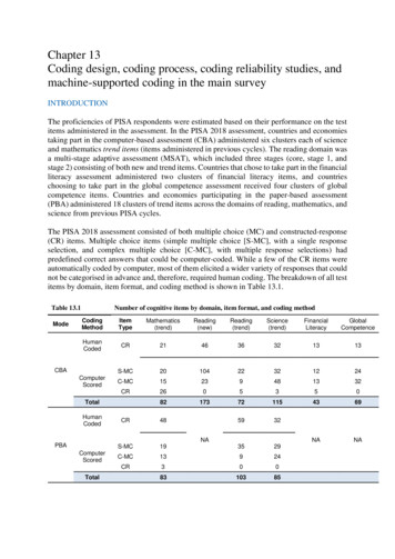
Yang et al. Molecular Cancer(2019) 18:78Page 8 of 12Fig. 4 HOTAIR regulates the expression and the localization of RAB35. a Real-time PCR analysis of the mRNA expression of RAB5, RAB7, RAB11,RAB27A, RAB27B, and RAB35, which encode GTPases involved in the release of exosomes, in HOTAIR overexpressing HepG2 cells. b Westernblotting analysis of RAB35 protein levels from the above cells. c Confocal co-localization analysis of CD63 (red) and RAB35 (green) in HepG2 cellstransfected with pcDNA3.1or pcDNA3.1-HOTAIR. The rectangular box indicates the small punctate structures where CD63 and RAB35 were colocalized. d RIP assays were performed using RAB35 antibody or control IgG antibody in HepG2 cells, and then real-time PCR analysis was usedto measure the enrichment degrees of HOTAIR coprecipitated RNA. e NTA analysis of exosome secretion in HepG2 cells co-transfected withpcDNA3.1-HOTAIR and si-Rab35. Data are reported as the mean standard error (SD) from three independent experiments, t-test *P-value 0.05altered the contents of tumor-derived exosomes, whichaffected tumor progression through communication between the tumor cells and their microenvironment [29].Moreover, aerobic glycolysis, also termed the Warburgeffect, is the characteristic of glucose metabolism intumor cells in response to metabolic stress. Wei et al.found that pyruvate kinase M1/2 (PKM2), the keyenzyme of aerobic glycolysis, plays a vital role inpromoting the release of exosomes from tumor cells[18]. Although the phenomenon of increasing exosomesecretion is observed during tumorigenesis, themolecular mechanisms controlling tumor exosomerelease remain unclear.Recently, lncRNAs have become a focus in the field ofcancer research. Many studies have demonstrated thatlncRNAs are involved in the biological functions oftumor cells, such as proliferation, invasion, metastasis,immunological function, metabolism, and drug resistance [31, 32]. However, few studies have explored the relationship between lncRNAs and the regulation ofexosome secretion from tumor cells. In the presentstudy, we analyzed the potential role of lncRNAHOTAIR in the exocytosis of exosomes. First, our GSEAanalysis demonstrated an enrichment of exosomesecretion-related genes in the group of patients showingrelatively high HOTAIR expression. Second, using
Yang et al. Molecular Cancer(2019) 18:78Page 9 of 12Fig. 5 HOTAIR induces the translocation of VAMP3 and SNAP23. a Confocal microscopy analysis of SNAP23 (green) in HepG2 cells transfectedwith pcDNA3.1-HOTAIR. Nuclei were stained with DAPI. b Confocal co-localization analysis of VAMP3 (red) and SNAP23 (green) in HepG2 cellstransfected with pcDNA3.1-HOTAIR. The rectangular box indicates the small punctate structures where VAMP3 and SNAP23 were co-localizedNanosight analysis, we demonstrated that overexpression of HOTAIR promoted exosome secretion fromHCC cells. Third, based on the intracellular process ofexosome secretion, we showed th
(SNAP) 23 is an important t-SNARE that is mainly located at the plasma membrane [19]. Phosphorylation of SNAP23 not only promotes the formation of the SNARE complex, but also increases exosome secretion [20]. How-ever, whether long non-coding RNAs (lncRNAs) partici-pate in the regulation of SNARE proteins and mediate exosome secretion is unclear.