Transcription
Miller and Rothstein Biological Procedures Online (2017) 19:7DOI 10.1186/s12575-017-0057-2METHODOLOGYOpen Access3D Printer Generated Tissue iMolds forCleared Tissue Using Single- and MultiPhoton Microscopy for Deep TissueEvaluationSean J. Miller1,2,3 and Jeffrey D. Rothstein1,2,3*AbstractBackground: Pathological analyses and methodology has recently undergone a dramatic revolution. With thecreation of tissue clearing methods such as CLARITY and CUBIC, groups can now achieve complete transparency intissue samples in nano-porous hydrogels. Cleared tissue is then imagined in a semi-aqueous medium that matchesthe refractive index of the objective being used. However, one major challenge is the ability to control tissuemovement during imaging and to relocate precise locations post sequential clearing and re-staining.Methods: Using 3D printers, we designed tissue molds that fit precisely around the specimen being imaged. First,images are taken of the specimen, followed by importing and design of a structural mold, then printed withaffordable plastics by a 3D printer.Results: With our novel design, we have innovated tissue molds called innovative molds (iMolds) that can begenerated in any laboratory and are customized for any organ, tissue, or bone matter being imaged. Furthermore,the inexpensive and reusable tissue molds are made compatible for any microscope such as single and multi-photonconfocal with varying stage dimensions. Excitingly, iMolds can also be generated to hold multiple organs in one mold,making reconstruction and imaging much easier.Conclusions: Taken together, with iMolds it is now possible to image cleared tissue in clearing medium while limitingmovement and being able to relocate precise anatomical and cellular locations on sequential imaging events in anybasic laboratory. This system provides great potential for screening widespread effects of therapeutics and diseaseacross entire organ systems.Keywords: CLARITY, Tissue molds, Microscopy, Cleared tissue, 3D printer, MedicineBackgroundHistology is a technique focused on evaluating tissuesections in many different elements. Historically, in basicscience laboratories groups would undergo tremendousefforts with high room for error to obtain histologicalsamples ready for imaging. To resolve this issue, groups* Correspondence: jrothstein@jhmi.edu1Department of Neurology, Johns Hopkins University School of Medicine,Johns Hopkins Univ., 855 N Wolfe Street, Room 250.18, Baltimore, MD 21205,USA2The Brain Science Institute at Johns Hopkins, Johns Hopkins Univ. School ofMedicine, Johns Hopkins Univ., 855 N Wolfe Street, Baltimore, MD 21205,USAFull list of author information is available at the end of the articlehave recently developed tissue clearing techniques thatrender samples transparent and intact [1–3]. This advancement in histology has allowed groups to visualizewhole anatomical specimens with 3D reconstructionsand analyses. Additionally, cleared tissue in hydrogelcomplexes are capable of antibody removal and restaining; something infeasible in prior methods [1]. However, one major limitation is the ability to limit tissuemovement and to relocate exact anatomical locations postre-staining and mounting in tissue clearing medium. Thisis especially important for the sequential evaluation ofmultiple biological markers. The Author(s). 2017 Open Access This article is distributed under the terms of the Creative Commons Attribution 4.0International License (http://creativecommons.org/licenses/by/4.0/), which permits unrestricted use, distribution, andreproduction in any medium, provided you give appropriate credit to the original author(s) and the source, provide a link tothe Creative Commons license, and indicate if changes were made. The Creative Commons Public Domain Dedication o/1.0/) applies to the data made available in this article, unless otherwise stated.
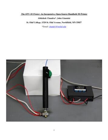
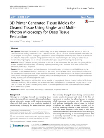
Miller and Rothstein Biological Procedures Online (2017) 19:7With current imaging setups for cleared tissue, the tissue is held by a semi-hardened material such as putty.The tissue is placed within the putty prior to and duringimaging. Post imaging, the putty is removed as well asthe tissue. For the sequential imaging, new putty has tobe used to hold the tissue. However, this leaves the tissuein unprecise locations in each imaging session making itchallenging for users to relocate exact biological targets ofinterest. Furthermore, if the user is attempting to re-imagemultiple organs at once on sequential stainings, then it isnear impossible to place the tissue in the exact locationsfrom the original imaging.The need for a device that can securely hold any tissuesample in a refined position with the tissue sample capable of being removed and replaced in the same positionis of high importance. Not only would this allow groupsto relocate areas of interest with sequential stains, itwould also allow users to easily set imaging parametersthat don’t require the user to be present during the imaging session (by pre-setting the microscope coordinates).Thus, reducing movement, improving re-evaluations ofbiological targets after re-staining, and saving time compared to the other methodologies.To develop a device that could provide these improvements we chose to generate tissue specific molds, calledinnovative molds (iMolds). iMolds are printed by inexpensive 3D printers in several different plastics that canbe sterilized and are generated specific to the tissuespecimen it is supporting. Overall the plastic materialsand 3D printers are dramatically cheaper than using excess clearing solution, iSpacers, and other materials onthe market. Lastly, iMolds allow users to evaluate andre-evaluate multiple organs in one imaging session.MethodsMaterials and ReagentsPolylactic acid (PLA) plastic was purchased from micro3D(USA); low magnification tissue images were obtained withan iPhone 6S from Apple Inc. (USA); 32% Paraformaldehyde (PFA) purchased from Electron Microscopy Sciences(USA); 10X PBS from Quality Biological Inc. (USA); Tissueclearing solution from Logos Biosystems (USA); Photoinitiator VA-044 from Wako chemicals (Germany); Histodenzfrom Sigma (USA).iMold Engineering Software.STL files were generated in Google Sketchup Software;Prototypes were printed using the Micro 3D printingsoftware.3D PrinterM3D printer was purchased from micro3D (USA); allprinting parameters were not adjusted from the manufacturer’s settings.Page 2 of 5Tissue ClearingTo achieve transparent tissue in organs and bones wefollowed the protocol previously established [4].Tissue StainingFor deep tissue staining we followed the protocol previously established for 5 days with primary antibody andthree days for secondary antibody [4].SterilizationTissue iMolds can be placed under UV light to sterilize,we recommend a duration of at least one hour [5].ResultsGeneration of the Tissue iMoldTo begin the process of generating a tissue mold that isspecific to the tissue specimen being analyzed, the usermust take a photo of the tissue being prepared for clearing and iMold generation, and place the tissue next to aruler or scale bar (Fig. 1a). Taking a photo prior to tissueclearing is preferred because once the tissue has clearedit becomes very challenging to generate iMolds due tothe tissue transparency. Next, the image is imported intoGoogle Sketchup or another 3D printer software to generate an.STL file. The Google Sketchup scale is then setto the appropriate scale bar as the photo taken with thetissue (i.e. millimeters, inches) (Fig. 1a). Next the userwill precisely trace the outside shape of the tissue specimen (this can be performed in other software packageslike Adobe Photoshop). After successfully tracing the tissue specimen, delete the original image, keeping onlythe new traced image.Next generate the slide base that will be used on themicroscope, this is generally the same size as a slide or acell culture plate. Thus, this slide base can range fromthe size of a whole plate or to a typically slide size (i.e.25 mm 75 mm); this is determined by your imagingplatform. Once the slide base is generated place thetraced tissue specimen shape into the desired location inthe slide base on the same Z-plane in the software.Now, once again remove the inside of the traced tissuethat has been just placed within the slide base. This willnow appear as a slide base with a missing tissue specimenshape (Fig. 1b). Next raise (i.e. pull) the slide specimen tothe appropriate height measured during the initial step(i.e. the height of the tissue). Now you have successfullygenerated an iMold design. Now, export the file as a.STLfor use in the 3D printer. Finally, transfer and print the 3Dmold.STL file with the 3D printer (Fig. 1c). Parametersused with the micro3D software included no raft or support material, as this would have to be removed postprinting, and a print quality of 150 μm with fill densityselected as high. Once you have imported the.STL fileand set the parameters correctly, then it is now ready
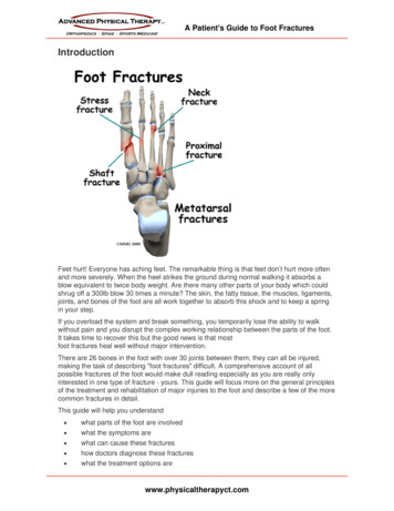
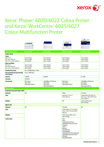
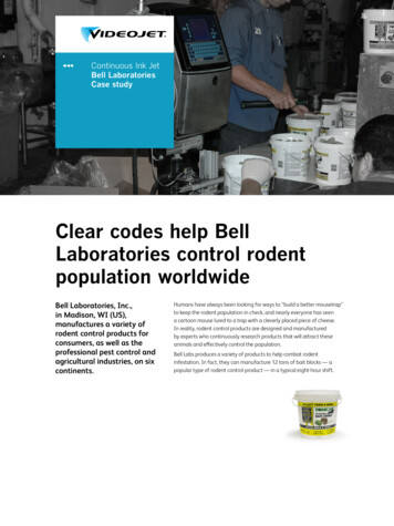
Miller and Rothstein Biological Procedures Online (2017) 19:7Page 3 of 5Fig. 1 Generation and imaging with tissue iMolds. a a photo of the tissue (tibia) to be cleared next to a ruler, b generation of a slide base withthe tissue (tibia) inserted inside, c 3D printed tissue iMold for tibiato be printed. Prior to using the finished iMold, superglue a piece of cover glass to one side of the iMold. Ifyou plan to image from both sides, then coverglass needsto be placed on both sides. After imaging the coverglasscan be removed (if double sided) with the use of forceps.However, this can result in the glass breaking, in whichthe user would have to reprint the saved.STL file. Due tothis, we highly recommend only imaging from one sidetherefore the coverglass will not have to be removed whenusing an inverted microscope.dot with a marker onto the glass and use the same Z-stacktiling parameters on the microscope. Next, we stained thetibia with marker CD31 to visualize areas of blood vessels,using the previously published protocol [4]. We found, insupport of prior literature, that the cartilage of the metaphysis and the vasculature closely interact, within a fewmicrons of each other (Fig. 2b) [8].Lastly, we generated an iMold that can fit multiple organs into the mold, such as brain and bone to emphasizePreparing for Imaging on a Single or Multi-Photon ConfocalSystemNext, it is time for imaging on single or multi-photonconfocal systems. Place the cleared organ into the iMold(it should fit appropriately to inhibit movement) andwith a pipet fill the iMold with clearing solution for imaging. If the size is too small or too large, you may goback to the original Google Sketchup file and expand orcontract the iMold. Now find the starting point of thetissue being imaged and z-stack tiled. Set the coordinatesfor tiling the entire tissue or the area of interest andbegin. Once you retrieve the image, there should be nomovement from the tissue.We chose to use the tibia for our demonstration. Boneis notoriously challenging to clear due to low lipid concentrations, undergoes immense amounts of movementwhile imaging, and therefore we wanted to show bothclearing of whole bone and the end results of the tibia inthe iMold (Fig. 2) [6]. After z-stack tiling the entire tibiain the iMold we were capable of reconstructing the tissue with internal bone and cartilage structures on the488 nM wavelength, using a Zeiss LSM 800 confocal,and Bitplane Imaris software suite (Fig. 2a, Additionalfile 1: Video 1) [7]. Simply import the image file (e.g. czi)into Imaris and use the automatic 3d rendering forvisualization. To relocate exact areas after sequential imaging sessions we recommend starting to image from aset coordinate (e.g. corner of the iMold) or by placing aFig. 2 Imaging with iMolds. a imaging of the tissue (tibia) in theiMold, b Imaging of the metaphysis vasculature and bone
Miller and Rothstein Biological Procedures Online (2017) 19:7the user flexibility and ability to customize the iMold forimaging (Additional file 2: Figure S1). This multipleorgan iMold allows groups to evaluate multiple organsand/or bones without the need for individual molds.DiscussionThe revolution of imaging cleared tissue is allowinggroups to finally image whole organs at super resolutionwith multiple targets of interest stained [7]. For therapeuticstudies that have an unknown effect on other organs, thissystem is an ideal way to rapidly scan through multiple organs. In neuroscience, it has been used to identify and follow neuronal projections throughout the central nervoussystem furthering our understanding of neuronal communications. However, there are no current devices that allowyou to place organs of interest into pre-casted moldsfor repeated imaging. Another caveat to imaging clearedtissue is the usage of semi-aqueous solutions during imaging sessions which allows the organ to move throughoutscans and challenging to relocate areas of interest duringsequential imaging events.To address the issues with the current imaging modalityof cleared tissue and to provide additional benefits wehave successfully generated tissue molds, iMolds. iMoldsfor cleared tissue limits the overall movement, can bereused and allow for exact areas to be re-analyzed post sequential staining, and are very inexpensive for any basicscience lab to produce. These provide essential improvements to prior methods [9]. iMolds are user customizedallowing for multiple organs in one mold or abnormal tissue architecture such as tumors to be imaged, in additionto usage on any stage for single and multi-photon confocals. Future studies will focus on advancing iMoldsfor use in light sheet microscopy where samples aretypically submerged in an aqueous solution during imaging [10]. In closing, we strongly believe that the openaccess to generating iMolds will allow groups to significantly improve their imaging quality and exploration ofwhole organs.ConclusionGenerating a device to repeatedly scan biological specimensin the exact locations is essential for repeated imaging modalities. With the advantage of iMolds over current methodologies, we believe that this novel design will speed upimaging and processing of cleared tissue making CLARITYand other clearing techniques more feasible for basic science labs and drug screening.Additional filesAdditional file 1: Video representation of a 3d rendering of the clearedtibia in the tissue iMold. (MOV 6251 kb)Page 4 of 5Additional file 2: Figue S1. iMolds can hold multiple organs per mold.A) iMold for brain and tibia in one slide base. (TIFF 7721 kb)AbbreviationsiMold: innovative moldAcknowledgementsWe would like to thank Haley Axelrod, Ken Valkenburg, and Ken Pienta ofthe Department of Urology at Johns Hopkins University for the tibia samplesand expertise; also Trent Button for his software expertise.FundingThis work was financially supported by the National Science Foundation (GRFP(SJM)), and National Institute of Health (NS085207 (JDR), and NS092067 (JDR)).Availability of Data and MaterialsAll datasets used and/or analyzed during the current study are available fromthe corresponding author on reasonable request.Authors’ ContributionsMSJ performed all experiments; RJD provided support throughout the durationof this project. Both authors read and approved the final manuscript.Competing InterestsThe authors declare that they have no competing interests.Consent for PublicationNot applicable.Ethics Approval and Consent to ParticipateThe care and treatment of animals is in accordance with the NIH Guide forthe Care and Use of Laboratory Animals, the Guidelines for the Use ofAnimals in Neuroscience Research, and the Johns Hopkins University IACUC.Publisher’s NoteSpringer Nature remains neutral with regard to jurisdictional claims inpublished maps and institutional affiliations.Author details1Department of Neurology, Johns Hopkins University School of Medicine,Johns Hopkins Univ., 855 N Wolfe Street, Room 250.18, Baltimore, MD 21205,USA. 2The Brain Science Institute at Johns Hopkins, Johns Hopkins Univ.School of Medicine, Johns Hopkins Univ., 855 N Wolfe Street, Baltimore, MD21205, USA. 3Cellular and Molecular Medicine Johns Hopkins School ofMedicine, Johns Hopkins Univ., 855 N Wolfe Street, Room 250.18, Baltimore,MD 21205, USA.Received: 2 May 2017 Accepted: 5 June 2017References1. Chung K, et al. Structural and molecular interrogation of intact biologicalsystems. Nature. 2013;497(7449):332–7. doi:10.1038/nature12107.2. Susaki EA, et al. Whole-brain imaging with single-cell resolution usingchemical cocktails and computational analysis. Cell. 2.3. Renier N, et al. iDISCO: a simple, rapid method to immunolabel large tissue samplesfor volume imaging. Cell. 2014;159(4):896–910. doi:10.1016/j.cell.2014.10.010.4. Yang B, et al. Single-cell phenotyping within transparent intact tissue throughwhole-body clearing. Cell. 2014;158:1–14. doi:10.1016/j.cell.2014.07.017.5. Valente TAM, et al. Effect of sterilization methods on electrospun poly(lacticacid) (PLA) fiber alignment for biomedical applications. ACS AppliedMaterials Interfaces. 2016;8:3241–9. doi:10.1021/acsami.5b10869.6. Acar M, et al. Deep imaging of bone marrow shows non-dividing stem cells aremainly perisinusoidal. Nature. 2015;526(7571):126–30. doi:10.1038/nature15250.7. Miller SJ, Rothstein JD. Astroglia in thick tissue with super resolution andcellular reconstruction. PLoS One. 2016;11(8):e0160391. doi:10.1371/journal.pone.0160391.
Miller and Rothstein Biological Procedures Online (2017) 19:7Page 5 of 58.Lo Celso C, et al. Live-animal tracking of individual haematopoietic stem/progenitor cells in their niche. Nature. 2009;457(7225):92–6. doi:10.1038/nature07434.9. Rapid, simple and inexpensive production of custom 3D printer equipmentfor large-volume fluorescence microscopy. Int J Pharm. 2015;494(2): p. 651–656.DOI: 10.1016/j.ijpharm.2015.03.042.10. Schmied C, Tomancak P. Sample preparation and mounting of drosophilaembryos for Multiview light sheet microscopy. Methods Mol Biol. 2016;1478:189–202. doi:10.1007/978-1-4939-6371-3 10.Submit your next manuscript to BioMed Centraland we will help you at every step: We accept pre-submission inquiries Our selector tool helps you to find the most relevant journal We provide round the clock customer support Convenient online submission Thorough peer review Inclusion in PubMed and all major indexing services Maximum visibility for your researchSubmit your manuscript atwww.biomedcentral.com/submit
Google Sketchup or another 3D printer software to gen-erate an.STL file. The Google Sketchup scale is then set to the appropriate scale bar as the photo taken with the tissue (i.e. millimeters, inches) (Fig. 1a). Next the user will precisely trace the outside shape of the tissue speci-men (this can be performed in other software packages