Transcription
MONOGRAPH
VIVEX INTRODUCTION AND COMPANY OVERVIEWVIVEX Biologics is a commercial stage, regenerative solutions company improvingpatient care through the innovation of tissue-based therapies. During more than50 years of safe and effective operations, VIVEX has sourced millions of allograftproducts throughout the US and eighteen countries worldwide. VIVEX is registeredwith the Food and Drug Administration (FDA) and accredited by the AmericanAssociation of Tissue Banks (AATB), continuously since 1992. VIVEX’s strongprocesses, manufacturing expertise, and operational excellence is the result ofcontinuously monitoring and improving its performance and systems.VIVEX strives to create treatment options and solutions that will improve clinical,surgical, and therapeutic patient outcomes. Under the guidance of experienced andsuccessful health care professionals, VIVEX tasks the brightest minds from themedical and material science industries to explore innovative ways to help others.VIVEX works with partners that are committed to providing care and compassionto donor families while inspiring communities to share life through their donation.Partnering with families in the prospect of regenerative treatments, VIVEX assuresappropriate options are available to support translation of their gifts.Theodore I. Malinin, MDVIVEX operates the oldest civilian tissue bank in the country, which it acquired in2014. The University of Miami Tissue Bank (UMTB) was founded in 1970 as the USNavy Tissue Bank was curtailing its service to civilian patients. Theodore I. Malinin,MD, who was trained and experienced in tissue banking at the US Navy Tissue BankNaval Medical Research Institute, adopted those protocols and tissue bankingtechniques when founding UMTB. By the 1990s, UMTB was not only one of theoldest, but recognized as one of the largest and most well-respected tissue banks inthe country. Over its tenure, UMTB assisted in the organization of new tissue banksthroughout the US and abroad. UMTB also provided training for personnel from othertissue banks in the US and foreign countries and for surgeons interested in bonetransplantation and tissue banking. Throughout its history, UMTB has been aworldwide leader in bone transplantation and tissue banking, with multiple firsts inclinical and scientific achievements.H. Thomas Temple, MD, joined UMTB in 1998. As a world leader in the field ofMusculoskeletal Oncology, Dr. Temple is an enthusiastic advocate of the propositionthat clinical challenges respond to therapeutic solutions. With a reputation for clinicalinsight and medical innovations, his patient care initiatives continue to offer noveltreatments that are unmatched. As the current Chief Medical Officer for VIVEX, Dr.Temple’s commitment to matching quality and safety serve as impeccable guides totissue product development.H. Thomas Temple, MDDr. Temple earned a Bachelor of Arts degree from Harvard University in Cambridge, Massachusetts, and a Doctor ofMedicine from Jefferson Medical College in Philadelphia, Pennsylvania. He completed his residency in orthopedicsurgery at Walter Reed Army Medical Center in Washington, D.C., and his musculoskeletal oncology fellowship atHarvard University– Massachusetts General Hospital–Boston Children’s Hospital. He is board certified by theAmerican Board of Orthopedic Surgery. Dr. Temple was a member of the United States Army Reserve for 15 years,where he held the titles of second lieutenant, captain, and major, and received an honorable discharge. He receivednumerous Army Achievement Medals and Army Commendation Medals.Today, VIVEX focuses on core products and new technologies to meet the evolving needs of surgeons and patientswhile sustaining its 50-year commitment to serving and honoring both tissue donors and recipients. The VIVEXbusiness is focused on the development and commercialization of new regenerative medicine solutions throughincreased research and development efforts as well as the expansion of manufacturing capabilities. Due to itsleadership in the field, VIVEX maintains the trend of safely delivering over 2 million allografts with no diseasetransmission, an unmatched history of safety in the industry.
TABLE OF CONTENTSCLINICAL HISTORY OF AMNIOTIC TISSUE. 4AMNIOTIC ALLOGRAFT TISSUE SOURCES. 4PRODUCT DESCRIPTION AND KEY BENEFITS. 5AMNIOTIC ALLOGRAFT TISSUE PROCESSING, SAFETY, PACKAGING. 6CYGNUS CONFIGURATIONS. 8SCIENTIFIC EVIDENCE.10GROWTH FACTORS FUNCTIONS .10EXTRACELLULAR MATRIX COMPONENTS AND FUNCTIONS . 14HYALURONIC ACID—THE BENEFIT OF HEAVY CHAIN HA. 15CLINICAL EVIDENCE . 16SAFE AND TRUSTED PARTNER . 16CONCLUSION. 17POTENTIAL CLINICAL APPLICATIONS . 17FREQUENTLY ASKED QUESTIONS. 18ORDERING INFORMATION. 19REFERENCES. 20
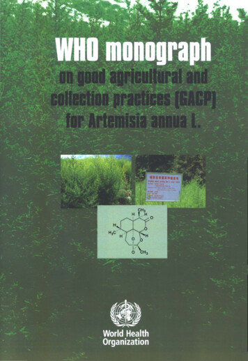
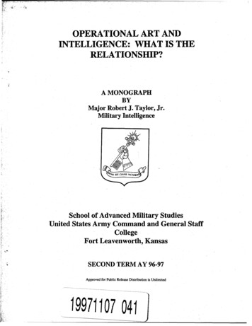
CLINICAL HISTORY OF AMNIOTIC TISSUEAmniotic allograft tissues in the clinical setting have over a century of documented use in many different surgicalapplications. The first reported use of an amniotic allograft tissue as a wound covering was in 1910.1 Amnioticallografts were specifically used as a barrier against adhesion in ophthalmology procedures2 and nerve wraps asearly as the 1940s. Amniotic allograft tissues have been used as a wound covering, including for diabetic woundulcers and burn victims3, spinal and abdominal surgeries, and other applications4-7 where it is desirable to maintaina plane of separation between tissue types in the body during wound repair. Amniotic allografts are used in a widevariety of surgical situations to provide mechanical protection to surgically traumatized tissues.AMNIOTIC ALLOGRAFT TISSUE SOURCESAmniotic allografts are derived from specific amniotic tissues, whichinclude the placental disc, the umbilical cord membrane, and the amnioticsac. The amniotic sac has two main layers, each of which is subdivided intomany layers of different tissue composition. From the perspective of thefetus, the inner layer is called the amnion and the outer layer is chorion.8During consenting healthy full-term births, after the baby has beendelivered, the amniotic tissues are removed from the mother.As shown in Figure 1, the amnion layer is comprised of five sub-layers. Theepithelium, the closest layer to the fetus, is a single layer of epithelial cells,followed by the basement membrane of collagens III, IV, and V coupledwith glycoproteins laminin and fibronectin. The compact layer maintainsstructural integrity of the membrane with collagens I, III, V, and VI. Thefibroblast layer is the thickest part of the amnion section and is a loosecollagen network with abundant proteoglycans and glycoproteins.9-10 Thefinal layer of the amnion portion of the amniotic sac is the intermediatespongy layer. The spongy layer is retained by VIVEX’s Integrity Processing methodology, unlike other cleaning processes where the spongy layeris lost when the amnion layer is separated from the chorion layer. Thispreserved spongy layer contains a meshwork of mostly type III collagen,a protein that has a prominent role in wound repair.11-12The chorion layer, which is 4X thicker than the amnion layer, is comprisedof three sub-layers, of which the reticular layer is the second thickest andcomprised of collagens I and III-VI.13-14 The basement membrane layeranchors the thick trophoblast layer to the reticular layer with collagen IV,fibronectin, and laminin.13-14Amniotic ibroblastLayerIntermediate(Spongy) astsMaternal DeciduaFigure 1: The Layers of the Amniotic SacThe amniotic sac acts as a biologically active and mechanically robust barrier between the mother and the fetusduring pregnancy. The sac contains the developing fetus and amniotic fluid, so this thin membrane must possesssufficient structural integrity to support the growing fetus to term.5 The metabolically active tissue must continuallyremodel to accommodate the increasing volume of fetus and fluid. Tissue remodeling is governed by mechanicalresponse and growth factors, cytokines, and extracellular matrix produced by cells. One of the inherent valuesattributed to amnion and chorion layers comes from the scope of tissue expansion without scar tissue. Thebiologically active nature of this barrier is evidenced in the low levels of antigens.15
PRODUCT DESCRIPTIONVIVEX offers multiple amniotic allograft options to meet the needs of thesurgical community. These products include a single amnion-only layer tissue,which is the thinnest amniotic allograft product. The medium thickness allografttissue is composed of the amnion and chorion membranes, which are neverseparated during processing. Finally, the thickest amniotic allograft tissueoffered by VIVEX is an umbilical cord membrane product.CYGNUS Matrix is an amniotic tissue allograft composed of intact humanamniotic membranes, where the amnion and chorion layers are neverseparated, that preserves and contains multiple extracellular matrix proteins,growth factors, collagen, cytokines, and other specialty proteins that areintrinsic to wound repair.16-17 Extracellular matrix provides three-dimensionalscaffold support for cellular migration and attachment to facilitate proliferationand integration.18-21 Growth factors guide cell signaling processes that affectcellular growth, proliferation, and healing through activities such asangiogenesis and immune modulation.22-23 CYGNUS Solo is an amnion-onlysingle layer membrane ideal for superficial wounds and topical applications.CYGNUS Max is an umbilical cord membrane robust enough to be sutured inplace. CYGNUS Max XL is a fenestrated umbilical cord membrane, increasingthe available allograft size to cover a larger wound while also allowing thewound to drain.CYGNUS amniotic allograft products are intended for homologous use toprovide a mechanical barrier and soft tissue protection for wound repair. VIVEXprocesses this scaffold material with its proprietary Integrity Processing thatprotects the components of the amniotic tissue to leave the matrix intact. Theresult is a durable dehydrated allograft with inherent barrier properties whichmay be stored at ambient conditions for up to 5 years. CYGNUS products haveexcellent handling properties that hydrate in place and sheets are available in awide variety of sizes and thickness to optimize the donor gift. CYGNUSproducts are processed in accordance to guidelines established by the Foodand Drug Administration (FDA) and by the American Association of TissueBanks (AATB).KEY FEATURES AND BENEFITS Effective allograft with excellenthandling characteristics Hydrates quickly in surgical site Ideal for internal or external applications Contains many identified growth factors,cytokines, and extracellular matrix proteins
AMNIOTIC ALLOGRAFT TISSUE, PROCESSING, SAFETY, PACKAGINGVIVEX’s amniotic tissues are supplied through a donation process in multiple states across the US. Industrystandards require that donated tissue only come from healthy and consenting mothers delivering full-term babiesvia live births. Prospective donors learn of the amniotic tissue donation process through their obstetrician.Prior to the birth of their healthy child, mothers are informed of the program and must sign an informed consentform. Each donor must fill out a medical questionnaire and have a blood sample drawn. Serology performed fromthe blood sample include testing for human immunodeficiency virus (HIV), human T-lymphotropic virus (HTLV),syphilis, West Nile Virus, Hepatitis B and Hepatitis C. There are other medical conditions that can disqualify adonor, including active cancer and infectious diseases. All donor interactions and tests are consistent with theguidelines issued by the AATB.After delivery of the baby, VIVEX partners with acquisition specialists who work with the obstetric team to acquirethe donated amniotic tissue. The donor mother has no further requirements, and after the tissue is acquired, it istransported to a VIVEX processing facility. Once reviewed and released by a Medical Director in accordance withboth FDA and AATB regulations and standards, the amniotic allograft is processed using VIVEX’s Integrity Processing .This is an innovative process methodology focused on protecting donated amniotic tissue from harmful cleaningagents to maintain its natural state and characteristics to support the patient's regenerative potential.The goal of the Integrity Processing is to clean the amniotic tissue of any blood components, while retaining theinherent properties of the original tissue. Aseptic processing was developed to minimize risk of contaminationduring preparation and reduce the presence of microorganisms. Integrity Processing is a gentle process thatremoves blood remnants, while preserving the allograft composition without compromising structural integrity.All VIVEX amniotic allograft tissue products are processed within cGTP conditions at VIVEX’s state-of-the-artmanufacturing facility in Miami, Florida. The allografts are sized to be specific for various clinical uses and thenpackaged for storage. Selected dehydrated tissues are terminally sterilized, subjected to a small, validated doseof electron beam irradiation after being packaged.VIVEX’s CYGNUS amniotic allograft tissues are packaged in 5-year, room temperature, shelf stable forms. One ofVIVEX’s amniotic tissues, CYGNUS Cryopreserved Max, is aseptically processed and available in a cryopreservedformat, which should be stored at -65 C or colder and has a shorter shelf life. All tissues undergo multiple,separate quality assurance checks prior to release to ensure utmost quality and safety. Traceability is maintainedfor all tissues from recovery through testing, processing, packaging, and distribution.VIVEX has an impeccable record of quality that exemplifies its robust manufacturing processes with millionsof allografts distributed over its 50 years of service. Measures integrated in processing ensure that regulatorycompliance and the final allograft products maintain the safety record VIVEX has established.
DONOR TESTINGT E STSYMB OLHuman Immunodeficiency Virus (HIV)HIV-1/2 Plus O AntibodiesHIV-1/2 Plus O AbNucleic Acid Test for HIV-1 RNAHIV-1 NATHepatitis B (HBV)HBV Surface AntigenHBsAgHBV Core Antibody (IgG & IgM)HBcAbNucleic Acid Test for HBV DNA (if performed)HBV NATHepatitis C Virus (HCV)HCV AntibodyHCVAbNucleic Acid Test for HCV RNAHCV NATSyphilis*Rapid Plasma Reagin ScreenRPRT. Pallidum IgGT. Pallidum IgGWest Nile Virus (WNV)Nucleic Acid Test for WNV RNAWNV NAT*A donor whose blood specimen is unsuitable for the non-treponemal screening assay, such as the RPR test, or with a reactiveresult from the non-treponemal screening assay, is cleared for transplantation use only when the result from the treponemal-specific(confirmatory) assay is nonreactive.
CYGNUS CONFIGURATIONSVIVEX offers five different configurations of amniotic allograft tissues, all under the CYGNUS product brand. CYGNUSis intended for use as a wound covering or barrier in soft tissue applications. The CYGNUS amniotic allografts canhelp provide mechanical protection to damaged tissues.18-21 VIVEX’s proprietary Integrity Processing retains inherentgrowth factors that are essential for signaling as well as maintaining the intact extracellular matrix of the tissue itself.Multi-layer membrane allograft, maintaining the amnion layer, itsintermediate/spongy layer, and the chorion layer of the amnioticsac, containing inherent growth factors, collagen, cytokines, andextracellular matrix.17,24Multi-layer amniotic membrane allografts, 400µm (0.4mm) thick,up to 4X thicker than the single amnion layer 5-year shelf life at room temperature storage No upfront preparation – hydrates rapidly in surgical site Ideal for both internal and external application Available in a variety of sizes to meet clinical needs and allow usethroughout the course of wound repair Available circular shape saves time by reducing the need totrim and associated potential to waste tissue SIZERECTANGLESSIZEDISKS2 2cm15mm Disk2 3cm25mm Disk3 3cm35mm Disk4 4cm45mm Disk4 6cm55mm Disk7 7cm65mm DiskSingle layer membrane allograft, featuring the amnion layer ofthe amniotic sac, offering inherent growth factors, cytokines,chemokines, and extracellular matrix of the amnion layer.17,24Thin amniotic membrane allograft, 100µm (0.1mm) thick5-year shelf life at room temperature storage No upfront preparation – hydrates rapidly in surgical site Ideal for superficial wounds and topical application Burn treatment, acute wounds, non-healing/chronic wounds,diabetic ulcers SIZESIZE2 2cm4 6cm2 3cm4 8cm3 3cm7 7cm4 4cm
Comprised of the umbilical cord membrane, this tissue is thethickest of the VIVEX dehydrated amniotic allograft products andis robust enough to be sutured in place.Thick umbilical cord membrane, 400µm (0.4mm),up to 4X thicker than the single amnion layer 5-year shelf life at room temperature storage No upfront preparation – hydrates rapidly in surgical site Tissue can be sutured into place Excellent handling properties SIZE2 2cm2 3cm2 4cmComprised of fenestrated umbilical cord membrane, increasingthe available allograft size to cover a larger wound while alsoallowing the wound to drain.Thick umbilical cord membrane, 400µm (0.4mm) thick,up to 4X thicker than amniotic membrane allografts 5-year shelf life at room temperature storage No upfront preparation – hydrates rapidly in surgical site Tissue can be sutured into place Excellent handling properties Fenestrated to allow for wound drainage and increases the sizeof the umbilical cord membrane SIZESIZE2 3cm4x6cm3 3cm4x8cm3 8cm5x7cm4 4cmComprised of the cryopreserved umbilical cord membrane, thistissue is the thickest amniotic allograft of the VIVEX products and isrobust enough to be sutured in place.Thick umbilical cord membrane, 800–2,000μm (0.8-2.0mm) thick,up to 8X thicker than the single amnion layer 9-month shelf life at temperature of -65ºC or colder Thaws quickly in 3-5 minutes Tissue can be sutured into place SIZE2x2cm2x4cm3x4cm
SCIENTIFIC EVIDENCEGROWTH FACTORS FUNCTIONSThe following list of growth factors and cytokines24-40 are present within amniotic allograft tissues. Some of the mostnotable and commonly mentioned are: epidermal growth factor (EGF), transforming growth factors alpha and beta(TGF-α&β), fibroblast growth factor (FGF), platelet derived growth factors (PDGF AA & BB), and vascular endothelialgrowth factors (VEGF).16,25REGULATORS OF WOUND HEALING VIA ANGIOGENESIS, CELLULAR GROWTH,MIGRATION, PROLIFERATION, AND DIFFERENTIATIONABBREVIATIONGROWTH FACTORFUNCTIONAngAngiogeninStimulates migration, proliferation, and vesselformation by endothelial and smooth muscle cellsAng-2Angiopoietin-2Regulates neovascularization in conjunctionwith angiopoeitin-1 and VEGFbFGFBasic Fibroblast Growth FactorRegulates neovascularization in conjunctionwith angiopoeitin-1 and VEGFβFGFBeta Fibroblast Growth FactorHas a role in smooth muscle cell productionand regulationβNGFBeta Nerve Growth FactorImportant for growth and apoptosis cyclemaintenance of neuronsBMP-5Bone Morphogenic Protein 5Has a role in bone and cartilage developmentBDNFBrain Derived Neurotrophic FactorHas a role in bone and cartilage developmentEG-VEGFEndocrine Gland-Derived VascularEndothelial Growth FactorStimulates endothelial cell migration, proliferation,and survival; Potent stimulatorEGFEpidermal Growth FactorStimulates proliferation, differentiation, andsurvival in numerous cell types, includingepithelial cellsFGFFibroblast Growth Factor22 members of FGF family regulate cellproliferation, survival, migration & differentiationin skin, blood vessels, muscle, adipose,tendon/ligament, cartilage, bone, tooth, and nerveGHGrowth HormoneStimulates body growth through IGF-1 production,involved in anabolic activityGRO-αGrowth Related Oncogene - alphaMultiple functions affecting angiogenesisand inflammation systemsHB-EGFHeparin Binding EGF-likeGrowth FactorCauses keratinocytes and fibroblasts tomigrate to the wound and proliferate,promotes angiogenesis
ABBREVIATIONGROWTH FACTORFUNCTIONHGFHepatocyte Growth FactorRegulates cell growth and cell migration ofhematopoietic stem cell and epithelial cells,promotes angiogenesisIGF-1&2Insulin-like Growth FactorPart of a general growth-promoting system,regulates cellular processes and promotesangiogenesisIGFBP (-1 thru 6)Insulin-like Growth FactorBinding ProteinsBinds and stabilizes IGF-1 as a carrier proteinKGFKeratinocyte Growth FactorPromotes proliferation and migration of epithelialcells and keratinocytesPIGFPlacental Growth FactorPromotes angiogenesis through the stimulationof proliferation and migration of endothelial cellsPDGFα, β, AA, BBPlatelet Derived Growth FactorsAttract macrophages, aid in migration,proliferation and differentiation of target cellsto support angiogenesisTGFα, β1, β2Transforming Growth Factorsalpha, betaFamily with multiple roles in stimulatinggrowth, proliferation, migration, and apoptosisof numerous cell types; potent stimulatorof angiogenesisTIMP-1,-2,-3,-4Tissue Inhibitor of MetalloproteinaseBinds to and inactivates metalloproteinasesto halt the degradation of ECM, affects cellgrowth and migrationTNF-αTumor Necrosis Factor alphaProduced by macrophages to regulate cellfunctions including cell proliferation, survival,differentiation, and apoptosisVEGFVascular Endothelial Growth FactorStimulates endothelial cell migration andactivation for the formation of blood vessels
REGULATORS OF INFLAMMATION AND IMMUNE MODULATIONABBREVIATIONGROWTH FACTORFUNCTIONGCSFGranulocyte ColonyStimulating FactorStimulates the proliferation, differentiation,and growth of neutrophilsGM-CSFGranulocyte MacrophageColony-Stimulating FactorStimulates the differentiation and proliferationof monocytes and granulocytesGDF-15Growth Differentiation FactorRegulates the apoptosis and inflammationresponses in injured tissuesIFNyInterferon GammaMacrophage activator, regulates earlyinflammation processIL-1α, 1βInterleukin 1 Alpha & BetaAffects lymphocyte proliferation, differentiation,and apoptosisIL-1raInterleukin 1 receptor antagonistCentral role in regulation of immune andinflammatory responsesIL-4Interleukin 4Promotes proliferation of B & T cellsIL-5Interleukin 5Promotes B cell growth, increasesimmunoglobulin secretionIL- 6Interleukin 6Promotes proliferation and growth ofneutrophils and B cellsIL-7Interleukin 7Promotes proliferation and apoptosis ofB cells, T cells, and natural killer cellsIL-10Interleukin 10Promotes B cell proliferation andantibody productionIL-15Interleukin 15Promotes proliferation of T lymphocytesand natural killer cellsIL-17Interleukin 17Increases and supports proliferationof chemokinesMCP-1Monocyte ChemoattractantProtein-1Regulates migration and of monocytesand macrophagesMCSFMacrophage ColonyStimulating FactorPromotes Monocyte and Macrophageproliferation and differentiationOPGOsteoprotegerinInhibits osteoclast activation, limitsosteo-ECM degradationTGF-β1 & β2Transforming GrowthFactor beta 1&2Growth-enhancing or growth inhibitingimmunomodulator
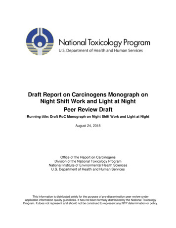
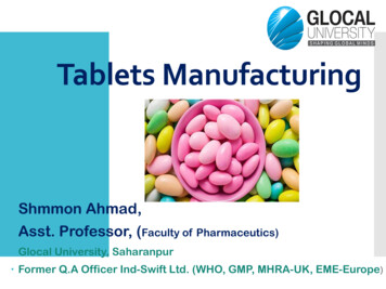
GROUPING SOME WELL-STUDIED GROWTH FACTORS INTO THEIR MULTIPLE FUNCTIONSGROWTH FACTORROLEMCP-139, IL1-RA 22, TGF-β1&2 32, IL6 22Immune Modulation / Anti-InflammatoryTNF-α40, GRO-α38, HGF 28, IGF1&2 29,31, VEGF 33, βFGF 34,PDGFα&β30, Ang 32AngiogenesisEGF 35, FGF 34, TGFβ32, TIMP(1-4) 26, HGF 27Cell GrowthPDGFα&β36, EGF 35, TIMP-2&-326, HGF 27, Ang23, KGF 37Cell MigrationPDGFα&β36, EGF 35, FGF 34, TGF-β1&2 32, IGF1&2 30, Ang23, KGF 37Cell ProliferationPDGFα&β36, EGF1, TIMP-2& -326, TGF-β1&2 32Cell DifferentiationHIGHLIGHTS OF GROWTH FACTORS MEASURED IN CYGNUS ALLOGRAFT TISSUEVIVEX’s proprietary Integrity Processing methodology helps to maintain the inherent levels of key extracellularmatrices, including proteins, carbohydrates, growth factors and cytokines.17,24 Amniotic tissues recovered from livebirths, once reviewed and released by a Medical Director in accordance with FDA and AATB regulations andstandard, were processed via the Integrity Processing gentle cleansing procedure and then dried for long-termroom temperature storage. The propriety Integrity Processing preserves up to 600 signaling proteins inCYGNUS Matrix, Max and Solo.41ELISA assays were used to analyze the content of key growth factors and cytokines released from the amnioticallograft tissues from four donors. Tissue was placed in DPBS media and incubated for 24 hours at 37ºC. Thesupernatant was evaluated to quantify the concentration of growth factors / cytokines. The data was normalized withrespect to the dried weight of the tissue. Testing was performed pre- and post-processing to ensure that selectmolecules were preserved after processing. The figures below and on the next page present the data for the productsin the CYGNUS 2HGFTIMP3IGF1IGF2TGFPD - B1GF-BGRO- 1O-GRTIM3B2P-F-TGM2TIHGTIMFP-0IL6IL1raMCPD P -1GF-BBEGSD FF1TG aFBVE 3GF-ATNFaTGFTG 0020001500332Average pg GF/cm2 (mean s.d.)3,8415000Average pg GF/cm2 (mean s.d.)1,2135500906000CYGNUS SOLO GROWTH FACTORSRELEASED AFTER 24 HOURS AT 37 C1,911CYGNUS MATRIX GROWTH FACTORSRELEASED AFTER 24 HOURS AT 37 C
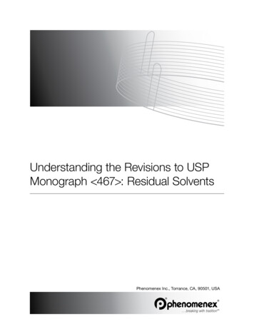
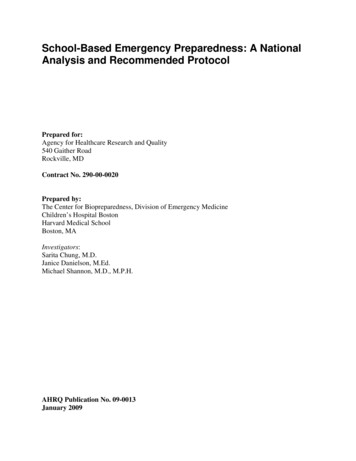
CYGNUS CRYOPRESERVED MAX GROWTH FACTORSRELEASED AFTER 24 HOURS AT 37 CCYGNUS MAX GROWTH FACTORSRELEASED AFTER 24 HOURS AT 37 FVElp-aOF-ha3P-74EXTRACELLULAR MATRIX COMPONENTS & FUNCTIONSAmniotic allografts are natural barriers, and the sheet-like three-dimensional matrix is comprised of many proteinsrecognized as important in the complex wound repair process. The extracellular matrix (ECM) of the amnioticallograft is a biological surface that provides a familiar and natural scaffold for cells to attach, migrate, and proliferateupon during the progression of the wound repair process.22,23 The components of the ECM in the amniotic allograftsare proteins found in a variety of tissue types in the human body.42,43The main protein components in amniotic allografts are collagens (I, II, III, IV, VII), fibronectin, laminin, and hyaluronicacids (HA).42 Additionally, the ECM of amniotic allografts tissues include proteoglycans, cytokines, and amnio acids.Collagen is the main structural component in amniotic allografts and also in the body in general. In fact, 25-35% ofthe whole human body protein content is a type of collagen. In amniotic allografts, type I collagen is 90% of theextracellular matrix. Type III collagen, predominant in the intermediate/spongy layer and expressed in earlygranulation tissue, has been proposed to play a prominent role in cutaneous wound repair.12 Other collagen proteinstructures in amniotic tissues include type IV and type VII collagen. Cells in the human body recognize collagen as aprotein that is advantageous to adhere to, and supportive of cellular activities, such as migration and 3,427F-300003,5403500TGAverage pg GF/cm2 (mean s.d.)31,62835000TIAverage pg GF/cm2 (mean s.d.)40000
ECM COMPONENTDESCRIPTIONCollagen, type IMain structural protein component in the bodyCollagen, type II–VIIStructural protein component of the bodyFibronectinBinding protein agent, supports initial cell attachmentLamininHigh molecular weight protein to which cells easily bind and migrate acrossHyaluronic AcidLubricating hydrophilic protein that coats cells and aids in hydrodynamic movementsProteoglycansConnective proteins that fill the spaces between cells in tissue and affect the stabilityof the proteins and growth factorsCytokinesSmall proteins produced by a broad range of cells and embedded into the ECM of tissuesAmino AcidsSmall proteins produced by a broad range of cells and embedded into the ECM of tissuesFibronectin is another commonly occurring protein in amniotic allografts and plays a role in holding the extracellularmatrix of tissues together by aiding in initial cell attachment. Laminin is a high molecular weight protein of the ECMto which cells easily bind and migrate across. Proteoglycans are connective proteins that fill the spaces betweencells in tissue and affect the stability of the proteins and growth factors in the ECM.43 Cytokines and amnio acids aresmall proteins produced by a broad range of cells and embedded into the ECM of tissues.Hyaluronic acid (HA) is an important component of the ECM in human tissues.44 It is a lubricating smooth proteinthat coats cells, aiding in the cell’s hydrodynamic movements.45 The ability of cells to migrate across an ECM andthus proliferate during the wound healing cascade is aided by this smo
VIVEX operates the oldest civilian tissue bank in the country, which it acquired in 2014. The University of Miami Tissue Bank (UMTB) was founded in 1970 as the US Navy Tissue Bank was curtailing its service to civilian patients. Theodore I. Malinin, MD, who was trained and experienced in tissue banking at the US Navy Tissue Bank