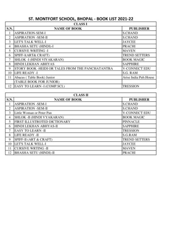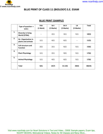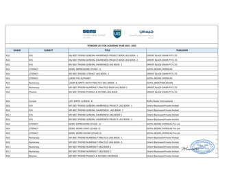
Transcription
Chapter 6TISSUESFrom the last chapter, we recall that all livingorganisms are made of cells. In unicellularorganisms, a single cell performs all basicfunctions. For example, in Amoeba, a singlecell carries out movement, intake of food,gaseous exchange and excretion. But in multicellular organisms there are millions of cells.Most of these cells are specialised to carry outspecific functions. Each specialised functionis taken up by a different group of cells. Sincethese cells carry out only a particular function,they do it very efficiently. In human beings,muscle cells contract and relax to causemovement, nerve cells carry messages, bloodflows to transport oxygen, food, hormones andwaste material and so on. In plants, vasculartissues conduct food and water from one partof the plant to other parts. So, multi-cellularorganisms show division of labour. Cellsspecialising in one function are often groupedtogether in the body. This means that aparticular function is carried out by a clusterof cells at a definite place in the body. Thiscluster of cells, called a tissue, is arranged anddesigned so as to give the highest possibleefficiency of function. Blood, phloem andmuscle are all examples of tissues.A group of cells that are similar in structureand/or work together to achieve a particularfunction forms a tissue.6.1 Are Plants and Animals Madeof Same Types of Tissues?Let us compare their structure and functions.Do plants and animals have the samestructure? Do they both perform similarfunctions?There are noticeable differences betweenthe two. Plants are stationary or fixed – theydon’t move. Since they have to be upright, theyhave a large quantity of supportive tissue. Thesupportive tissue generally has dead cells.Animals on the other hand move aroundin search of food, mates and shelter. Theyconsume more energy as compared to plants.Most of the tissues they contain are living.Another difference between animals andplants is in the pattern of growth. The growthin plants is limited to certain regions, while thisis not so in animals. There are some tissues inplants that divide throughout their life. Thesetissues are localised in certain regions. Basedon the dividing capacity of the tissues, variousplant tissues can be classified as growing ormeristematic tissue and permanent tissue. Cellgrowth in animals is more uniform. So, thereis no such demarcation of dividing and nondividing regions in animals.The structural organisation of organs andorgan systems is far more specialised andlocalised in complex animals than even in verycomplex plants. This fundamental differencereflects the different modes of life pursued bythese two major groups of organisms,particularly in their different feeding methods.Also, they are differently adapted for asedentary existence on one hand (plants) andactive locomotion on the other (animals),contributing to this difference in organ systemdesign.It is with reference to these complex animaland plant bodies that we will now talk aboutthe concept of tissues in some detail.2022-23
Q uestions1. What is a tissue?2. What is the utility of tissues inmulti-cellular organisms?6.2 Plant Tissues6.2.1 MERISTEMATICFrom the above observations, answerthe following questions:1. Which of the two onions has longerroots? Why?2. Do the roots continue growingeven after we have removed theirtips?3. Why would the tips stop growingin jar 2 after we cut them?The growth of plants occurs only in certainspecific regions. This is because the dividingtissue, also known as meristematic tissue, islocated only at these points. Depending onthe region where they are present,meristematic tissues are classified as apical,lateral and intercalary (Fig. 6.2). New cellsproduced by meristem are initially like thoseof meristem itself, but as they grow andmature, their characteristics slowly change andthey become differentiated as components ofother tissues.TISSUEApical meristemJar 1Jar 2Fig. 6.1: Growth of roots in onion bulbsIntercalary meristemActivity 6.1 Take two glass jars and fill them withwater.Now, take two onion bulbs and placeone on each jar, as shown inFig. 6.1.Observe the growth of roots in boththe bulbs for a few days.Measure the length of roots on day 1,2 and 3.On day 4, cut the root tips of the onionbulb in jar 2 by about 1 cm. After this,observe the growth of roots in both thejars and measure their lengths eachday for five more days and record theobservations in tables, like the tablebelow:LengthDay 1Day 2Day 3Lateral meristemFig. 6.2: Location of meristematic tissue in plant bodyDay 4 Day 5Jar 1Jar 2Apical meristem is present at the growingtips of stems and roots and increases thelength of the stem and the root. The girth ofthe stem or root increases due to lateralmeristem (cambium). Intercalary meristemseen in some plants is located near the node.69TISSUES2022-23
Cells of meristematic tissue are very active,they have dense cytoplasm, thin cellulose wallsand prominent nuclei. They lack vacuoles. Canwe think why they would lack vacuoles? (Youmight want to refer to the functions of vacuolesin the chapter on cells.)6.2.2 PERMANENTTISSUEWhat happens to the cells formed bymeristematic tissue? They take up a specificrole and lose the ability to divide. As a result,they form a permanent tissue. This processof taking up a permanent shape, size, and afunction is called differentiation. Differentiationleads to the development of various types ofpermanent tissues. 3. Can we think of reasons why therewould be so many types of cells?We can also try to cut sections of plantroots. We can even try cutting sectionsof root and stem of different plants.6.2.2 (i) SIMPLE PERMANENT TISSUEA few layers of cells beneath the epidermis aregenerally simple permanent tissue.Parenchyma is the most common simplepermanent tissue. It consists of relativelyunspecialised cells with thin cell walls. Theyare living cells. They are usually looselyarranged, thus large spaces between cells(intercellular spaces) are found in this tissue(Fig. 6.4 a). This tissue generally stores lemVascular bundleFig. 6.3: Section of a stemActivity 6.2 Take a plant stem and with the helpof your teacher cut into very thin slicesor sections.Now, stain the slices with safranin.Place one neatly cut section on a slide,and put a drop of glycerine.Cover with a cover-slip and observeunder a microscope. Observe thevarious types of cells and theirarrangement. Compare it with Fig. 6.3.Now, answer the following on thebasis of your observation:1. Are all cells similar in structure?2. How many types of cells canbe seen? In some situations, it contains chlorophyll andperforms photosynthesis, and then it is calledchlorenchyma. In aquatic plants, large aircavities are present in parenchyma to helpthem float. Such a parenchyma type is calledaerenchyma.The flexibility in plants is due to anotherpermanent tissue, collenchyma. It allowsbending of various parts of a plant like tendrilsand stems of climbers without breaking. Italso provides mechanical support. We can findthis tissue in leaf stalks below the epidermis.The cells of this tissue are living, elongatedand irregularly thickened at thecorners. There is very little intercellular space(Fig. 6.4 b).70SCIENCE2022-23
Intercellular spacesWall thickeningsNucleusVacuoleCell wallaThick lignifiedwallsNarrow lumenLignifiedthick wallc (ii)c (i)bFig. 6.4: Various types of simple tissues: (a) Parenchyma (b) Collenchyma (c) Sclerenchyma (i) transverse section,(ii) longitudinal section.Yet another type of permanent tissue issclerenchyma. It is the tissue which makes theplant hard and stiff. We have seen the husk ofa coconut. It is made of sclerenchymatoustissue. The cells of this tissue are dead. Theyare long and narrow as the walls are thickeneddue to lignin. Often these walls are so thickthat there is no internal space inside the cell(Fig. 6.4 c). This tissue is present in stems,around vascular bundles, in the veins of leavesand in the hard covering of seeds and nuts. Itprovides strength to the plant parts.Activity 6.3 GuardcellsStomaEpidermalcell(a)Guardcell(b)Fig. 6.5: Guard cells and epidermal cells: (a) lateralview, (b) surface viewTake a freshly plucked leaf of Rhoeo.Stretch and break it by applyingpressure.While breaking it, keep it stretchedgently so that some peel or skinprojects out from the cut.Remove this peel and put it in a petridish filled with water.Add a few drops of safranin.Wait for a couple of minutes and thentransfer it onto a slide. Gently placea cover slip over it.Observe under microscope.What you observe is the outermost layerof cells, called epidermis. The epidermis isusually made of a single layer of cells. In someplants living in very dry habitats, the epidermismay be thicker since protection against waterloss is critical. The entire surface of a plant hasan outer covering epidermis. It protects all theparts of the plant. Epidermal cells on the aerialparts of the plant often secrete a waxy, waterresistant layer on their outer surface. This aidsin protection against loss of water, mechanicalinjury and invasion by parasitic fungi. Sinceit has a protective role to play, cells ofepidermal tissue form a continuous layerwithout intercellular spaces. Most epidermalcells are relatively flat. Often their outer andside walls are thicker than the inner wall.We can observe small pores here andthere in the epidermis of the leaf. These poresare called stomata (Fig. 6.5). Stomata areenclosed by two kidney-shaped cellscalled guard cells. They are necessary forexchanging gases with the atmosphere.Transpiration (loss of water in the form ofwater vapour) also takes place throughstomata.71TISSUES2022-23
Recall which gas is required forphotosynthesis.Find out the role of transpiration in plants.Epidermal cells of the roots, whose functionis water absorption, commonly bear long hairlike parts that greatly increase the totalabsorptive surface area.In some plants like desert plants,epidermis has a thick waxy coating of cutin(chemical substance with waterproof quality)on its outer surface. Can we think of a reasonfor this?Is the outer layer of a branch of a treedifferent from the outer layer of a young stem?As plants grow older, the outer protectivetissue undergoes certain changes. A strip ofsecondary meristem located in the cortex formslayers of cells which constitute the cork. Cellsof cork are dead and compactly arrangedwithout intercellular spaces (Fig. 6.6). Theyalso have a substance called suberin in theirwalls that makes them impervious to gasesand water.Cork cellsis a distinctive feature of the complex plants,one that has made possible their survival inthe terrestrial environment. In Fig. 6.3 showinga section of stem, can you see different typesof cells in the vascular bundle?Xylem consists of tracheids, vessels, xylemparenchyma (Fig. 6.7 a,b,c) and xylem fibres.Tracheids and vessels have thick walls, andmany are dead cells when mature. Tracheidsand vessels are tubular structures. This allowsthem to transport water and mineralsvertically. The parenchyma stores food. Xylemfibres are mainly supportive in function.Phloem is made up of five types of cells:sieve cells, sieve tubes, companion cells,phloem fibres and the phloem parenchyma[Fig. 6.7 (d)]. Sieve tubes are tubular cells withperforated walls. Phloem transports food fromleaves to other parts of the plant. Exceptphloem fibres, other phloem cells are living cells.NucleusRuptured epidermisPitPitsCytoplasmFig. 6.6: Protective tissue(a) Tracheid6.2.2 (ii) COMPLEX PERMANENT TISSUE(b) Vessel(c) Xylem parenchymaSieve plateSieve tubeThe different types of tissues we have discusseduntil now are all made of one type of cells,which look like each other. Such tissues arecalled simple permanent tissue. Yet anothertype of permanent tissue is complex tissue.Complex tissues are made of more than onetype of cells. All these cells coordinate toperform a common function. Xylem andphloem are examples of such complex tissues.They are both conducting tissues andconstitute a vascular bundle. Vascular tissuePhloemparenchymaCompanion cell(d) Section of phloemFig. 6.7: Types of complex tissue72SCIENCE2022-23
Questions1. Name types of simple tissues.2. Where is apical meristem found?3. Which tissue makes up the huskof coconut?4. What are the constituents ofphloem?6.3 Animal TissuesWhen we breathe we can actually feel themovement of our chest. How do these bodyparts move? For this we have specialised cellscalled muscle cells (Fig. 6.8). The contractionand relaxation of these cells result inmovement.During breathing we inhale oxygen. Wheredoes this oxygen go? It is absorbed in the lungsand then is transported to all the body cellsthrough blood. Why would cells need oxygen?The functions of mitochondria we studiedearlier provide a clue to this question. Bloodflows and carries various substances from onepart of the body to the other. For example, itcarries oxygen and food to all cells. It alsocollects wastes from all parts of the body andcarries them to the liver and kidney fordisposal.Blood and muscles are both examples oftissues found in our body. On the basis of thefunctions they perform we can think of differenttypes of animal tissues, such as epithelialtissue, connective tissue, muscular tissue andnervous tissue. Blood is a type of connectivetissue, and muscle forms muscular tissue.6.3.1 EPITHELIALTISSUEThe covering or protective tissues in the animalbody are epithelial tissues. Epithelium coversmost organs and cavities within the body. Italso forms a barrier to keep different bodysystems separate. The skin, the lining of themouth, the lining of blood vessels, lung alveoliand kidney tubules are all made of epithelialtissue. Epithelial tissue cells are tightly packedand form a continuous sheet. They have onlya small amount of cementing material betweenthem and almost no intercellular spaces.Obviously, anything entering or leaving thebody must cross at least one layer ofepithelium. As a result, the permeability of thecells of various epithelia play an important rolein regulating the exchange of materialsbetween the body and the externalenvironment and also between different partsof the body. Regardless of the type, allepithelium is usually separated from theunderlying tissue by an extracellular fibrousbasement membrane.Different epithelia (Fig. 6.9) show differingstructures that correlate with their uniquefunctions. For example, in cells lining bloodvessels or lung alveoli, where transportationof substances occurs through a selectivelypermeable surface, there is a simple flat kindSmooth muscle fibresNucleusSmooth muscle fibre(Cell)Fig. 6.8: Location of muscle fibres73TISSUES2022-23
of epithelium. This is called the simplesquamous epithelium (squama means scale(a) Squamous(b) Stratified squamousof skin). Simple squamous epithelial cells areextremely thin and flat and form a delicatelining. The oesophagus and the lining of themouth are also covered with squamousepithelium. The skin, which protects the body,is also made of squamous epithelium. Skinepithelial cells are arranged in many layers toprevent wear and tear. Since they are arrangedin a pattern of layers, the epithelium is calledstratified squamous epithelium.Where absorption and secretion occur, asin the inner lining of the intestine, tall epithelialcells are present. This columnar (meaning‘pillar-like’) epithelium facilitates movementacross the epithelial barrier. In the respiratorytract, the columnar epithelial tissue also hascilia, which are hair-like projections on theouter surfaces of epithelial cells. These cilia canmove, and their movement pushes the mucusforward to clear it. This type of epithelium isthus ciliated columnar epithelium.Cuboidal epithelium (with cube-shapedcells) forms the lining of kidney tubules andducts of salivary glands, where it providesmechanical support. Epithelial cells oftenacquire additional specialisation as gland cells,which can secrete substances at the epithelialsurface. Sometimes a portion of the epithelialtissue folds inward, and a multicellular glandis formed. This is glandular epithelium.6.3.2 CONNECTIVE TISSUE(c) CuboidalBlood is a type of connective tissue. Why wouldit be called ‘connective’ tissue? A clue isprovided in the introduction of this chapter!Now, let us look at this type of tissue in somemore detail. The cells of connective tissue areloosely spaced and embedded in anintercellular matrix (Fig. 6.10). The matrix maybe jelly like, fluid, dense or rigid. The natureof matrix differs in concordance with thefunction of the particular connective tissue.Activity 6.4(d) Columnar (Ciliated)Take a drop of blood on a slide andobserve different cells present in itunder a microscope.Fig. 6.9: Different types of epithelial tissues74SCIENCE2022-23
CytoplasmNucleusDifferent whiteblood corpusclesNeutrophil Eosinophil Basophil(polynuclearleucocyte)Lymphocyte MonocytePlatelets(a)Haversian canal(contains blood vesselsand nerve fibres)ChondrocyteHyaline matrix(b)Canaliculus (containsslender process of bonecell or osteocyte)(c)Red bloodcorpuscleReticular fibreFibroblastMacrophageCollagen fibreMast cell(d)Fat dropletPlasma cellNucleusBlood has a fluid (liquid) matrix calledplasma, in which red blood corpuscles (RBCs),white blood corpuscles (WBCs) and plateletsare suspended. The plasma contains proteins,salts and hormones. Blood flows andtransports gases, digested food, hormonesand waste materials to different parts of thebody.Bone is another example of a connectivetissue. It forms the framework that supportsthe body. It also anchors the muscles andsupports the main organs of the body. It is astrong and nonflexible tissue (what would bethe advantage of these properties for bonefunctions?). Bone cells are embedded in ahard matrix that is composed of calcium andphosphorus compounds.Two bones can be connected to each otherby another type of connective tissue called theligament. This tissue is very elastic. It hasconsiderable strength. Ligaments containvery little matrix and connect bones withbones. Tendons connect muscles to bones andare another type of connective tissue. Tendonsare fibrous tissue with great strength butlimited flexibility.Another type of connective tissue,cartilage, has widely spaced cells. The solidmatrix is composed of proteins and sugars.Cartilage smoothens bone surfaces at jointsand is also present in the nose, ear, tracheaand larynx. We can fold the cartilage of the ears,but we cannot bend the bones in our arms.Think of how the two tissues are different!Areolar connective tissue is found betweenthe skin and muscles, around blood vesselsand nerves and in the bone marrow. It fillsthe space inside the organs, supports internalorgans and helps in repair of tissues.Where are fats stored in our body? Fatstoring adipose tissue is found below the skinand between internal organs. The cells of thistissue are filled with fat globules. Storage offats also lets it act as an insulator.Adipocyte6.3.3 MUSCULAR(e)Fig. 6.10: Types of connective tissues: (a) types of bloodcells, (b) compact bone, (c) hyaline cartilage,(d) areolar tissue, (e) adipose tissueTISSUEMuscular tissue consists of elongated cells,also called muscle fibres. This tissue isresponsible for movement in our body.75TISSUES2022-23
Muscles contain special proteins calledcontractile proteins, which contract and relaxto cause movement.NucleiStriations(a)Spindle shapedmuscle cellNucleus(b)Striations[Fig. 6.11(a)]. These muscles are also calledskeletal muscles as they are mostly attachedto bones and help in body movement. Underthe microscope, these muscles show alternatelight and dark bands or striations whenstained appropriately. As a result, they arealso called striated muscles. The cells of thistissue are long, cylindrical, unbranched andmultinucleate (having many nuclei).The movement of food in the alimentarycanal or the contraction and relaxation of bloodvessels are involuntary movements. We cannotreally start them or stop them simply bywanting to do so! Smooth muscles [Fig.6.11(b)] or involuntary muscles control suchmovements. They are also found in the iris ofthe eye, in ureters and in the bronchi of thelungs. The cells are long with pointed ends(spindle-shaped) and uninucleate (having asingle nucleus). They are also called unstriatedmuscles – why would they be called that?The muscles of the heart show rhythmiccontraction and relaxation throughout life.These involuntary muscles are called cardiacmuscles [Fig. 6.11(c)]. Heart muscle cells arecylindrical, branched and uninucleate.Activity 6.5Compare the structures of differenttypes of muscular tissues. Note downtheir shape, number of nuclei andposition of nuclei within the cell inthe Table 6.1.NucleiTable 6.1:FeaturesStriatedSmoothCardiacShapeNumber of nuclei(c)Position of nucleiFig. 6.11: Types of muscles fibres: (a) striated muscle,(b) smooth muscle, (c) cardiac muscleWe can move some muscles by consciouswill. Muscles present in our limbs move whenwe want them to, and stop when we so decide.Such muscles are called voluntary muscles6.3.4 NERVOUSTISSUEAll cells possess the ability to respond tostimuli. However, cells of the nervous tissueare highly specialised for being stimulated and76SCIENCE2022-23
then transmitting the stimulus very rapidlyfrom one place to another within the body. Thebrain, spinal cord and nerves are all composedof the nervous tissue. The cells of this tissueare called nerve cells or neurons. A neuronconsists of a cell body with a nucleus andcytoplasm, from which long thin hair-likeparts arise (Fig. 6.12). Usually each neuronhas a single long part (process), called theaxon, and many short, branched parts(processes) called dendrites. An individualnerve cell may be up to a metre long. Manynerve fibres bound together by connectivetissue make up a nerve.The signal that passes along the nerve fibreis called a nerve impulse. Nerve impulses allowus to move our muscles when we want to. Thefunctional combination of nerve and muscletissue is fundamental to most animals. Thiscombination enables animals to move rapidlyin response to stimuli.NucleusDendriteAxonNerve endingCell bodyFig. 6.12: Neuron-unit of nervous tissueQuestions1. Name the tissue responsible formovement in our body.2. What does a neuron look like?3. Give three features of cardiacmuscles.4. What are the functions ofareolar tissue?Whatyou havelearnt Tissue is a group of cells similar in structure and function. Plant tissues are of two main types – meristematic andpermanent. Meristematic tissue is the dividing tissue present in thegrowing regions of the plant. Permanent tissues are derived from meristematic tissue oncethey lose the ability to divide. They are classified as simpleand complex tissues. Parenchyma, collenchyma and sclerenchyma are three typesof simple tissues. Xylem and phloem are types of complextissues. Animal tissues can be epithelial, connective, muscular andnervous tissue. Depending on shape and function, epithelial tissue isclassified as squamous, cuboidal, columnar, ciliated andglandular.77TISSUES2022-23
The different types of connective tissues in our body includeareolar tissue, adipose tissue, bone, tendon, ligament,cartilage and blood. Striated, unstriated and cardiac are three types of muscletissues. Nervous tissue is made of neurons that receive and conductimpulses.Exercises1. Define the term “tissue”.2. How many types of elements together make up the xylemtissue? Name them.3. How are simple tissues different from complex tissues inplants?4. Differentiate between parenchyma, collenchyma andsclerenchyma on the basis of their cell wall.5. What are the functions of the stomata?6. Diagrammatically show the difference between the threetypes of muscle fibres.7. What is the specific function of the cardiac muscle?8. Differentiate between striated, unstriated and cardiacmuscles on the basis of their structure and site/location inthe body.9. Draw a labelled diagram of a neuron.10. Name the following.(a) Tissue that forms the inner lining of our mouth.(b) Tissue that connects muscle to bone in humans.(c) Tissue that transports food in plants.(d) Tissue that stores fat in our body.(e) Connective tissue with a fluid matrix.(f) Tissue present in the brain.11. Identify the type of tissue in the following: skin, bark oftree, bone, lining of kidney tubule, vascular bundle.78SCIENCE2022-23
12.13.14.15.Name the regions in which parenchyma tissue is present.What is the role of epidermis in plants?How does the cork act as a protective tissue?Complete the following chart:79TISSUES2022-23
It is the tissue which makes the plant har d and stif f. W e have seen the husk of a coconut. It is made of sclerenchymatous tissue. The cells of this tissue are dead. They are long and narrow as the walls are thickened due to lignin. Often these walls are so thick that there is no internal space inside the cell (Fig. 6.4 c). This tissue is .