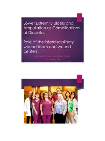
Transcription
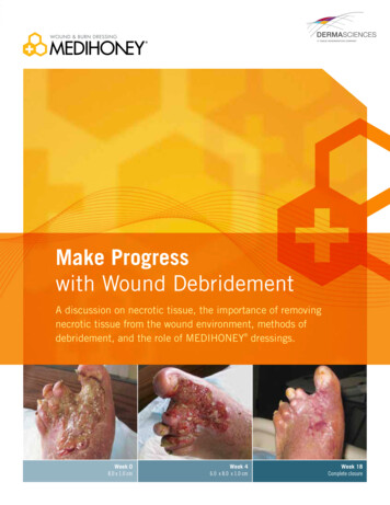
Make Progresswith Wound DebridementA discussion on necrotic tissue, the importance of removingnecrotic tissue from the wound environment, methods ofdebridement, and the role of MEDIHONEY dressings.Week 08.0 x 1.0 cmWeek 46.0 x 8.0 x 1.0 cmWeek 18Complete closure
DebridementOverviewDebridement is the process of removing devitalized or necrotic (dead)tissue from a wound or wound bed to expose healthy tissue, allowing thehealthy tissue to granulate and advance the wound through the healingprocess. Debridement is indicated for any wound, acute or chronic, whennecrotic tissue, foreign bodies or infected tissue is present. Necrotic tissueor foreign bodies within a wound can lead to a negative cascading effectcausing the wound to become chronic. Because this effect can lead tomore serious consequences, including infections or limb amputation, itbecomes imperative that an effective wound management strategy bestructured and implemented.Primary purposes for debridement are to:nC ontrol and remove infectious materials,bioburden and/or biofilm 1, 2, 3nI nterrupt the cycle of the chronic wound sothat protease and cytokine levels more closelyresemble those of the acute wound1, 2, 3n acilitate visualization of the wound edgesFand base for accurate assessment 1, 3Progress
Necrotic Tissue and Necrotic BurdenCauses of Necrotic BurdenNecrotic or avascular tissue is the result of an inadequateblood supply to the tissue in the wound area. It contains deadcells and debris that is a consequence of the dying cells.4S K IN FA ILU R EThere are different types of necrotic tissue including escharand slough. Avascular tissue exposed to the air will form ahard black crust known as eschar. If kept moist, avasculartissue will appear brown, yellow or gray and soft, flimsy orstringy. This tissue is called slough. Slough is fibrinoustissue consisting of fibrin, bacteria, intact leucocytes, celldebris, and serous exudates. After eschar is debrided,slough may be present as the wound is not completelyclean. Thereafter, if a moist wound environment is notmaintained, continued exposure to air may dessicateremaining slough, causing eschar to reform.4, 5Necrotic burden is the combination of necrotic tissue,excess exudate and high levels of bacteria present indead tissue that accumulate in chronic wounds. Necroticburden creates an altered cellular environment (elevatedpH, proteases, biofilm, free radicals) which causes acascading effect that can prolong the inflammatoryphase, obstruct wound contraction and impede thereepithelialization process.2Skin failure happens when skin and underlying tissue diebecause of hypoperfusion, often concurrent with severedysfunction or failure of other organ systems. Determiningskin failure is currently done by gross examination of musclemass, subcutaneous tissue thickness, wound granulation, andtissue necrosis. In addition, stratifying skin failure accordingto the patient’s medical condition can be useful in planninginterventions and setting treatment goals.Skin failure can be typified as acute, chronic and end-stage.Acute skin failure occurs when skin and underlying tissue diebecause of hypoperfusion concurrent with a critical illness.Chronic skin failure is an event in which skin and underlyingtissue die because of hypoperfusion concurrent with a chronicdisease state. It typically happens more steadily over time,and in older individuals. Multiple co-morbidities combinedwith other age-related declines can accelerate degeneration.Deterioration of internal organs can manifest in the externalorgan of skin.End-stage skin failure occurs when skin and underlying tissuedie because of hypoperfusion concurrent with the end of life.Mortality rates range from 33% within 30 days to 73.3% within1 year of onset of skin failure in the intensive care population.6ESCHARSLOUGH
LACK OF BLOOD FLOW OR DECREASED TISSUE PERFUSIONIN FE C T IO N A N D B IO FILMLack of blood flow or decreased tissue perfusion may becaused by occlusion, vasoconstriction, venous hypertension,hypotension, dehydration, medications, radiation, smoking, andinability to transport oxygen. Oxygen fuels the cellular functionsessential to the repair process, making it critical to woundhealing.6 Lack of blood flow causes a decrease in oxygen,slowing or stalling the healing process.An infection is the presence of replicating microorganismsinvading wound tissue and causing damage to the tissue andthe host. Biofilms are created by colonies of bacteria attachedto a substrate.9 These colonies produce an extra-polymericsubstance that holds the bacteria together and protectsthem. Biofilms and their causative organisms are notvisible to the naked eye, and conventional swabbing is ofteninconclusive in identifying them.9 Biofilms may repopulatein toxic wound environments in which cells break downand chronic inflammation occurs. This is exacerbated bythe release of planktonic bacteria from the biofilm, whichstimulates an inflammatory response.10 Persister cells canrepopulate the biofilm despite antibiotic susceptibility andtherapy.E L E VATED PR OTEASE ACTIVITYElevated protease activity may occur in chronic wounds andmay inhibit healing by degrading extra-cellular matrix proteins,growth factors, their receptors and protease inhibitors.2Protease activity can potentially be reduced by loweringthe pH of a wound. This also may result in increased oxygenrelease, enhanced destruction of abnormal wound collagen,and increased macrophage and fibroblast activity.7F R E E RA DI CA L SNon-healing wounds typically display increased reactiveoxygen species (ROS). ROS are deleterious in excess amountsdue to their high reactivity, which causes oxidative stress.This is associated with reperfusion injury, one of the primaryfactors in chronic wound development. In the chronicwound environment, ROS attack DNA, which can lead tocell apoptosis.8 Tight regulation of ROS production anddetoxification is crucial for the repair process in wounds.FIBRINOUS TISSUEFO R E IG N B O D IE SFinally, external factors such as foreign bodies cancomplicate wound management. Debris (projectiles,splinters, glass and shrapnel), and even fragments ofdressing and suture material, must be removed as theycan interfere with healing.
OptimizingImportance of Optimizing and Controlling the Wound Bed EnvironmentA wound management plan should include a thorough wound assessment and selection of appropriateproducts to address the specific needs of the wound. Setting goal- oriented strategies to gain control overthe wound environment will help get the wound back on track towards healing. Appropriate goals such asmaintaining the physiologic wound environment (e.g., debridement, cleansing, prevention/managementof infection)4,11 and providing systemic support (e.g., edema reduction, nutrition, hydration) are thefoundation to the process.When necrotic tissue is present, there are a number of related factors that could be the root cause ofdelayed healing:nNon-resolving inflammationnBacterial infection or 10 5 organisms per gram of wound tissuenElevated levels of proteasesnImpaired perfusion, decreased tissue oxygenation and/or oxidative stressControllingRemoval of necrotic tissue is therefore fundamental to allowing the wound to progress. Proper debridementremoves and reduces the barriers that impede the healing process and provides an environment thatstimulates the growth of healthy tissue needed for wound healing.10Defining Stalled Woundsn If a Pressure Ulcer does not reduce in size at least 39% in two weeks it may not heal in a timely fashion.12n a Venous Leg Ulcer does not show at least 30% reduction in 4 weeks, it is probable that it will not beIfhealed at 6 months.13n 9% of patients with Diabetic Foot Ulcers with 53% closure in 4 weeks, go on to close by Week 12.14GoalPrepare the wound bed and promote moist wound healing.Time Management 11Proper wound bed preparation is essential to promoting the wound healing process. Utilizing the TIMEManagement protocol will help to keep that wound on track.TTissue managementIInfection andinflammation controlMMoisture balanceEEpithelial(edge) advancement3
Types of Debridement2,4,5The standard methods of debridement are autolytic, mechanical, enzymatic, sharp and biologic. The methodof debridement used often depends on the amount of necrotic tissue present in the wound bed, the extent ofthe wound, and the patient’s medical history and overall condition. Clinicians sometimes use more than onedebridement method to achieve the most successful removal of necrotic tissue.Autolytic debridement uses the body’s own enzymesand moisture to re-hydrate, soften and finally liquefy escharand slough. During autolysis, enzymes present in wound fluidhave the effect of liquefying non-viable tissue. Clinicians fosterautolytic debridement by utilizing moist wound dressings. Bymaintaining a moist wound environment, the body is able touse its own processes to eliminate necrotic tissue. Autolyticdebridement can be achieved with the use of occlusive orsemi-occlusive dressings which maintain wound fluid in contactwith the necrotic tissue. It is virtually painless for the patient andsafe, yet is generally slower than other forms of debridement.It can be used on its own, after surgical debridement, or inconjunction with enzymatic or mechanical debridement.Mechanical debridement is a process in which a tool isused on the necrotic tissue to rip, pull, push, cut or abradeaway devitalized tissue from the healthy tissue. Mechanicaldebridement is often non-selective and may remove or causedamage to healthy tissue as well as necrotic tissue. Examplesof mechanical debridement include wet-to-dry dressings, woundirrigation, pulsatile lavage, whirlpool, contact ultrasound andscrubbing the surface with gauze. Wet-to-dry dressings do notprovide a moist wound healing environment and are not optimalfor wound care once the wound is free of necrotic tissue.Sharp debridement, a form of mechanical debridement, isthe removal of devitalized tissue by a skilled clinician, typicallyusing a scalpel, scissors, curette or other sharp instrument.Clinicians use conservative sharp debridement to removeloosely adherent nonviable tissue at the bedside or ina clinical setting. Surgical debridement is done by aphysician usually in the operating room, under anesthesia,with instruments and/or a laser when the tissue removalneeds are extensive, or when the patient has a seriousinfection associated with the wound. Although sharpdebridement is fast, it is non-selective and can be verypainful to the patient.Enzymatic debridement, or chemical debridement,makes use of certain enzymes and other compounds todissolve necrotic tissue. It requires a prolonged periodof enzyme activity, and a moist wound environment withappropriate pH and temperature. Enzymes are inactivatedby metals in some wound care products (e.g. silver, zinc).15The enzyme used in the U.S., collagenase, works bydissolving the collagen “anchors” that secure the avasculartissue to the wound bed. Collagenase has been shown tobe most active within a pH range of 6 to 8. 16Biologic debridement uses maggots grown from thesterilized eggs of Lucilia sericata. The larvae are placedin the wound bed, where it is theorized that they secreteproteolytic enzymes that break down necrotic tissue, whichthey then ingest. This is considered an option when thepatient is not a surgical candidate and has not respondedto other methods of debridement.4Continual vs. intermittent debridementIt has been found that rates of healing increase when wounds are debrided more frequently.17 With the increasedknowledge of biofilms and their ability to repopulate, as well as the damaging effects of elevated proteases (MMP’s)of the chronic wound, more focus has been placed on continual debridement vs. single or intermittent debridement.Continual debridement provides a consistent action of removing necrotic tissue from the wound bed over a period of time,unlike single or intermittent methods. The ability to provide continual and consistent removal of necrotic tissue helps tocreate an optimal environment for healing and allows for less disruption to the wound bed.4
The Role of MEDIHONEY The overall goal for wound bed preparation is to remove factors that may delay healing.1 Thesefactors in a stalled or chronic wound include necrotic tissue and altered levels and composition ofwound exudates.MEDIHONEY dressings, containing Active Leptospermum Honey (ALH), helps to address factorsthat may cause delayed healing in chronic wounds. As demonstrated in multiple RCTs and hundredsof clinical papers, ALH helps to promote autolytic debridement due to its properties which lead tomultiple mechanisms of action. The osmolarity of MEDIHONEY assists with multiple aspects ofdebridement for optimal patient care. The high sugar content of MEDIHONEY aids in the increasedflow of fluid to support the continual cleansing of the wound environment, helping to removedevitalized or necrotic tissue through an osmotic effect.18 Additionally, the low pH of MEDIHONEY helps to lower the pH levels within the wound environment,19 which has been shown to have woundhealing benefits.2012AUTOLYTIC DEBRIDEMENTDuring autolysis, the body breaks down tissue or cells. A moist environment, associated with ALH dressings,aids the body’s own process of moisturizing and re-hydrating, thus loosening and liquefying necrotic tissue.HIGH OSMOTIC POTENTIALALH incites an osmotic effect, which occurs when the high sugar content of honey facilitates movement offluid toward the wound surface. The increased flow of fluid helps the body’s natural processes to cleansethe wound, removing debris and necrotic tissue.REDUCTION IN pH3The failure of a chronic wound to progress has been correlated with alkaline pH levels.7 The surface pH ofchronic wounds has been reported to range from 6.5 to 8.5.21 ALH has a low pH of 3.5 – 4.5, which mayhelp to reduce the pH of the wound environment.19 Lowering the pH of the wound can help aid the body’snatural reparative processes that support the removal of dead tissue and wound healing.20Low pHHigh OsmolarityFigure 15idicAc78Alkalin103pH9e46HealingBreakdownpHpH SPECTRUMHigh Osmotic pull batheswound and helps to liquifynecrotic tissue.The low pH of MEDIHONEY is associated with a lowerpH within the wound environment, which has beenshown to have wound healing benefits.19, 205
Evidence Supporting the Use of MEDIHONEY ebridement of lower extremity wounds withDActive Leptospermum Honey22Becky Strilko RN, BSN, CWOCN, APN; Chris Barkauskas RN, BA, CWOCN, APN;Andrea McIntosh, RN, BSN, CWOCN, APN, Silver Cross Hospital, Joliet, ILC L INI CA L I SS UETopical products containing papain, used for removal ofnecrotic tissue, control of inflammation, reduction of woundodor, and rehydration of the skin, are no longer approvedfor use in the United States.23 With the loss of papain-ureabased debridement agents, there was a need to find a safeand cost-effective alternative for debridement of non-viabletissue and for wound bed preparation.IN C L US I O N CR I TERIAFive patients were asked to participate in an open labelpilot study. Selection criteria was limited to lower extremityulcers with a minimum of 50% devitalized tissue. ActiveLeptospermum Honey (ALH), MEDIHONEY , hasexperienced increased attention in the literature as aneffective agent to promote autolytic debridement.18, 24Many patients in the outpatient setting are not candidatesfor surgical debridement.O UTC OM ETo determine the efficacy of this product for debridementof devitalized tissue as an adjunct to a successful woundtreatment program.T R EATM ENT P ROTOCOLIn each case an ALH calcium alginate dressing was appliedto non-viable tissue and covered with an absorbent coverdressing. Dressings were changed as needed based onwound exudate. Wound photography was performed on aweekly basis to document progression of debridement.R E S U LT SDressings with ALH demonstrated the ability to promotedebridement of necrotic tissue for five patients with lowerextremity non-healing wounds. Several patients who wereunable to tolerate hydrogel therapy reported improveddressing tolerance with the use of ALH. Improvementin all wounds was documented. There was a decreasedpercentage of non-viable tissue, increased percentageof granulation tissue, and increased dressing tolerance.The investigators confirmed that ALH dressings werean effective, first-line choice for promoting autolyticdebridement and wound bed preparation for these cases.C O N C LU S IO NDebridement is a vital part of the successful managementof chronic wounds. ALH’s ability to effectively promoteautolytic debridement was observed in this group of fivepatients with differing wound types. Additional improveddressing tolerance were noted. The dressings were easy touse and well received by caregivers and patients.C A SE 1 - VENOU S U LCERA 63 year-old female with a history of venous ulcer disease and Sjögren’s syndrome presentedwith three full thickness, non-healing ulcers of the right posterior leg. She reported being“terrified” of surgical intervention and steadfastly refused sharp debridement. Persistent,consistent “bad” pain was reported as 4-6 on a 0-10 visual analog scale. Medications includedhydrochlorothiazide and propoxyphene napsylate as needed for pain. Previous treatments withtriple antibiotic ointment and papain-urea-chlorophyllin complex ointment were ineffective.Initially, the wounds measured 1.0 x 1.0 cm, 5.0 x 2.0 x 0.2 cm, and 1.5 x 1.5 cm with scantexudates and 100% eschar. Hydrogel therapy was prescribed to promote autolytic debridement.Increased pain was noted with hydrogel therapy (6-8 on a 0-10 visual analog scale). ALHcalcium alginate was initiated on Day 0, covered with an absorbent cover dressing and changedone to two times per week. Gradual wound improvement was noted; the patient reported, “Thedressings are natural and I did not need surgical debridement.”6Day 0, ALH initiatedDay 116
C A S E 2 - P RES SURE U LCERAn 89 year-old male with a history of coronary artery bypass graft, congestive heartfailure, atrial fibrillation, chronic renal failure, and gout presented to the wound carecenter with an unstageable pressure ulcer on the left heel. The patient was takingthe following medications: amlodipine besylate, levothyroxine sodium, niacininaide,valsartan, finasteride, allopurinol, furosemide, simvastatin, terazosin hydrochloride, andclonidine hydrochloride. Previous treatment with hydrogel therapy to promote debridementwas ineffective. ALH calcium alginate dressings were initiated on Day 0, covered withan absorbent cover dressing and changed one to two times per week. Within one weekprogress toward debridement was noted.Day 0, ALH initiatedDay 8C A S E 3 - TR AUM AT IC W OU N DA 53 year-old female with a history of hypertension, hyperlipidemia and bipolar disordersustained a traumatic wound on the left anterior tibial region in a motor vehicle accidentin May of 2008. Prior topical antimicrobial therapy (silver sulfadiazine) was ineffective forthe promotion of wound management. Her medications included valproic acid, bupropionhydrochloride, a multivitamin, and zinc supplement. She presented to the wound carecenter on Day 0 with a full thickness wound with 90% slough and 10% granular tissuewith a moderate amount of serosanguinous exudates. ALH calcium alginate was initiated,covered with compression bandaging and changed once per week. Within one weekslough was eradicated, exudates decreased, and the patient reported the dressings were“comfortable”. A skin graft was performed for final wound closure.Day 7Day 14C A S E 4 - TR AUM AT IC W OU N DA 60 year-old healthy male taking no medications sustained two full thickness wounds onthe right patella from a motorcycle accident. Initial surgical debridement was performed.He presented to the wound center two weeks later on Day 14 with two full thickness woundswith 100% granulation tissue. Hydrogel therapy was prescribed to promote moist woundenvironment. One week later the base of each wound was covered with 90% slough andmoderate amounts of wound exudates were noted. ALH calcium alginate dressings wereinitiated, covered with an absorbent cover dressing and changed every other day. Thepatient reported wound closure by week 6.Day 36Day 65C A S E 5 - TR AUM AT IC W OU N DA 77 year-old female with hypertension and COPD sustained an injury to her right lateral legon Day 0. The patient was taking the following medications: fluticasonepropionate/salmetrolinhalation powder, multivitamin, tiotropium bromide, albuterol sulfate, furosemide, diltiazemhydrochloride, prednisone, and travoprost ophthalmic solution. She presented to the woundcenter on Day 37 with a 5 week old full-thickness wound surrounded by erythema andedema. The wound measured 6.0 x 2.5 x 0.3 cm with minimal exudates. The base of thewound was completely covered with slough (50%) and eschar (50%). Previous treatmentswere topical antimicrobial therapy, including silver sulfadiazine x 1 week and silver woundgel x 1 week. ALH calcium alginate dressings were initiated on Day 43, covered with anabsorbent conforming gauze and secured with an adherent cohesive bandage once weekly.Within two weeks slough, eschar, and erythema were decreased.Day 37, ALH initiatedDay 577
Evidence Supporting the Use of MEDIHONEY (continued)Use of Active Leptospermum Honey on difficult toheal wounds of various etiologies25Nancy Chaiken, ANP-C, CWOCN, Swedish Covenant Hospital, Chicago, ILP UR PO SE/ RATI O NALER E S U LT SSince ALH has multiple properties that address manycommon, underlying causes of non-healing wounds,it was chosen for use on several wound types toevaluate its therapeutic effects.In each case significant wound improvement was noted asdemonstrated by decreased slough. ALH dressings wereeasy to use, economical, effective, and well tolerated byeach patient, subsequently improving life quality.IN C L US I O N CR I TERIAC O N C LU S IO NThe dressings were used on two patients with chronicnon-healing wounds of varying etiologies, including rheumatoid ulceration and a stage IV sacral pressure ulcer.Patients also had multiple co-morbidities. The presence ofone or more wounds was causing great pain and discomfortfor each patient.Choosing appropriate dressings and treatment modalities forindividuals with chronic, non-healing wounds is challengingdue to many underlying, causative factors. The use ofdressings with ALH simplified the decision process.Patients in this study, with multiple co-morbidities andvarious wound types saw a reduction in slough.T R EATM ENT P ROTOCOLPatients were selected to receive ALH dressings basedon the ability of ALH to aid in promoting autolyticdebridement. ALH dressings were applied, covered withan absorbent cover dressing, and changed based on theamount of exudate.8
CAS E 1 - R HEU MATOID ARTH RITISA 53 year-old male with a history of rheumatoid arthritis, morbid obesity, myocardialinjury, and hepatitis C was admitted to the hospital with a new diagnosis of esophagealcancer. He was referred for an evaluation for a foot wound that he had for 2 ½ years.Prior treatments of silver calcium alginate dressings and compression bandagingwere ineffective. The patient was evaluated by rheumatology, however he refusedsystemic therapy for the rheumatoid ulcer; chemotherapy for esophageal cancer wasin progress. ALH paste was initiated on Day 0, covered with an absorbent calciumalginate dressing, and secured with conforming gauze bandage. Compressionbandaging was refused for edema management. Closure was achieved by Day 120,despite continual chemotherapy for esophageal cancer.Day 0Day 22Day 120CAS E 2 - P RESSU RE U LCERA 56 year-old female with a history of abdominal compartment syndrome, cirrhosisof the liver, acute pancreatitis, congestive heart failure, malnutrition and hepaticencephalopathy, developed a sacral pressure ulcer after an episode of ischemia.Initially the ulcer presented as deep tissue injury which then evolved to a stage IVpressure ulcer. The patient was not a candidate for surgical debridement and progresswith alternative debridement methods was slow. On Day 0 ALH paste was initiated, andcovered with an absorbent calcium alginate dressing daily. Minimal sharp debridementwas performed as needed to remove loosened necrotic slough tissue. Wound closure,with only a small scab visible, was achieved by Day 122.Day 0Day 67Day 1229
MEDIHONEY Dressing Selection GuideFor the Management of Superficial, Partial andFull Thickness WoundsTYPE OF IVEDebrideRemove SloughPromote GranulationMaintain MoistEnvironmentDry to LightEXUDATEDry to LightModerateLight toModerateHeavyLight to ModerateMEDIHONEY DRESSING(Primary sicClassicFoamSiliconeHCSN/AConformingBandage orGauze WrapConformingBandage orGauze WrapConformingBandageConformingBandage orGauze WrapConforming BandageConforming BandageXTRASORB DRESSING(Secondary Dressing)BIOGUARD DRESSING(Tertiary Dressing)GelHCSHydrogelReferences: 1. Shultz, G et al.: Wound bed preparation, a systemic approach to wound bed management, Wound Rep Regen 11(Suppl):1, 2003. 2. Enoch, S, Harding, K, Wound Bed Preparation: The Science Behind the Removal of Barriers toHealing, Wounds, 2003:15(7). 3. Falanga V. Wound Bed preparation and the role of enzymes: a case for multiple actions of theapeutic agens. Wounds 2002; 14 (2) : 47-57. 4. Ramundo J M, Wound Debridement: Acute and Chronic Wounds, R.A. Bryant and D. P. Nix, editors. 2012, Elsevier Mosby, US. p. 279-287. 5. Langemo, Diane, Brown, Gregory, Skin Fails Too: Acute, Chronic, and End stage Skin Failure, Advances in Skin and Wound Care, 19(4). 6. Chambers, A. C., & Leaper, D.J. Role of oxygen in wound healing: a review of evidence. Journal Of Wound Care. April 2011;20(4):160-164. 7. Greener B, Hughes AA, Bannister NP, et al. Proteases and pH in chronic wounds. J Wound Care 2005; 14 (2) 59-61. 8. Telgenhoff,D, Shroot, B, Cellular senescence mechanisms in chronic wound healing, Cell Death and Differentiation, 2005:12, p. 695-698. 9. Wysocki, AB. Evaluating and Managing Open Skin Wound: Colonization Versus Infection. AACN Clin issues2002; 13 (3): 382-397. 10. Cooper, R. Biofilms and wounds: much ado about nothing? WOUNDS UK, 2010:6(4) 84-90. 11. European Wound Management Association (EWMA). Wound Bed preparation in practice. London: MEP Ltd, 2004. 12.van Rijswijk L, Polansky M. Predictors of time to healing deep pressure ulcers. Wounds. 1994;6(5):159–165. 13. Falanga V, Sabolinski ML. Prognostic factors for healing of venous ulcers. Wounds 2000; 12(5 Suppl A): 42A–46A. 14. Sheehanet al. Percent change in wound area of diabetic foot ulcers over a 4-week period is a robust predictor of complete healing in a 12-week prospective trial. Plast Reconstr Surg 2006;117(7 Suppl):239S-244S. 15. Vowden K, Vowden P. Woundbed preparation. d-Preparation.html. 16. Rao DB, Sane PG, Georgiev EL. Collagenase in the treatment of dermal and decubitus ulcers. J Am Geriatr Soc 1975; 23 (1):22–30. 17.Steed DL, Donohoe D, Webster MW et, al. Effect of extensive debridment and treatment on the healing of daibetic foot ulcers. J AM Coll Surg 1996; 183 (1): 61-4. 18. Amaya R. Safety and efficacy of active Leptospermum honey in neonataland paediatric wound debridement. J Wound Care 2015; 24(3):97-103. 19. Milne SD, Connolly P. The influence of different dressings on the pH of the wound environment. J Wound Care 2014;23(2):53-7. 20. Leveen H, Falk G, Borek, et al.Chemical acidification of wounds. An adjuvant to healing and the unfavourable action of alkalinity and ammonia. Ann Surg 1973; 178(6): 745-50. 21. Greener, B et al. Proteases and pH in chronic wounds. J of Wound Care 2005; 14 (2)59-61. 22. Strilko B, Barauskas C, McIntosh A. A safe and effective alternative for debridement of lower extremity wounds: Active Leptospermum honey dressings. Proceedings of Symposium on Advanced Wound Care and Wound HealingSociety Meeting. April 2010, Orlando, FL. 23. US Department of Health and Human Services. Questions and Answers about FDA’s Enforcement Action Regarding UnapprovedTopical Drug Products Containing Papain 2009. http://www.fda.gov/Drugs/ rovedDrugs/ucm119646.htm. 24. Molan P, Rhodes T. Honey: a biologic wound dressing. Wounds 2015; 27 (6): 141-51. 25.Chaiken N. The use of Active Leptopermum Honey on difficult to heal wounds of various etiologies. Proceedings of Symposium on Advanced Wound Care, Orlando, FL 2010.10
A Guideline for CareMEDIHONEY Dressing Application and Removal nWash hands thoroughly.nApply gloves.n Assessthe wound. Look for signs of healing, increased redness, pain, swelling, or heat within or around the wound.*n Cleansethe wound and skin around the wound with sterile saline, sterile water, or other wound cleansers.nDry the skin around the wound by patting gently with gauze.n rotect the skin around the wound to avoid maceration. Apply a skin protectant barrier. (An initial increase in exudates mayPoccur as a result of the highly osmotic effect of MEDIHONEY .)n hoose a MEDIHONEY dressing that is appropriate for the amount of drainage. (MEDIHONEY Paste or MEDIHONEY GelCfor light to moderate exudates, wounds that are hard to dress, or those that require a wound gel or paste; MEDIHONEY Hydrogel and MEDIHONEY HCS for dry to moderate exudates that are superficial to partial thickness wounds;MEDIHONEY Calcium Alginate dressing for moderate to heavy exudates; MEDIHONEY Honeycolloid dressing for light tomoderate exudates.)n pply the appropriate MEDIHONEY dressing to fit the wound. The MEDIHONEY
healthy tissue to granulate and advance the wound through the healing process. Debridement is indicated for any wound, acute or chronic, when necrotic tissue, foreign bodies or infected tissue is present. Necrotic tissue or foreign bodies within a wound can lead to a negative cascading effect causing the wound to become chronic.