Transcription
RESEARCH ARTICLEFigures and figure supplementsArabidopsis heterotrimeric G proteins regulate immunity by directly coupling to theFLS2 receptorXiangxiu Liang et alLiang et al. eLife 2016;5:e13568. DOI: 10.7554/eLife.135681 of 22
Research ArticleCell biology Plant biologyFigure 1. G proteins are required for FLS2-mediated immunity. (A) XLG2/3 and AGB1 play overlapping but notidentical roles in disease resistance to Pst. Plants of indicated genotypes were infiltrated with H2O and flg22 1 daybefore infiltration with P. syringae DC3000, and bacteria number was determined 2 days later (mean SD; n 6;p 0.05, Student’s t-test; different letters indicate significant difference). (B) xlg2/3 and agb1 are similarlycompromised in flg22-induced ROS burst. Leaves of the indicated genotypes were examined for flg22-inducedROS production, and peak RLU values are shown (mean SD; n 6; p 0.05, Student’s t-test; different lettersindicate significant difference). (C) Flg22 treatment disrupts XLG2-AGB1 interaction. Cluc-XLG2 and AGB1-HANluc constructs are transiently expressed in Nb leaves, relative luminescence unit (RLU) was measured 2 days later.Cluc-CPR5 and BAK1-HA-Nluc were used as negative control (mean SD; n 6). (D) Flg22-induced RbohDphosphorylation is impaired in agb1. FLAG-RbohD and/or AGB1-HA constructs were expressed under control ofthe 35S promoter in WT or agb1 protoplasts. The FLAG-RbohD protein was affinity purified and subject to antiFLAG and anti-pSer39 immuoblot analyses. Numbers indicate arbitrary units of RbohD pS39 phosphorylationcalculated from densitometry measurements normalized to total FLAG-RbohD protein. Each experiment wasrepeated three times, and data of one representative experiment are shown.DOI: 10.7554/eLife.13568.003The following source data is available for figure 1:Source data 1. Raw data and exact p value of Figure 1A, B and Figure 1—figure supplement 1.DOI: 10.7554/eLife.13568.004Liang et al. eLife 2016;5:e13568. DOI: 10.7554/eLife.135682 of 22
Research ArticleCell biology Plant biologyFigure 1—figure supplement 1. Flg22-induced ROS burst is compromised in xlg2 plants. Flg22-induced ROSburst is compromised in xlg2 plants. Col-0, xlg2, and xlg2 transgenic lines complemented with the XLG2transgene under control of the XLG2 native promoter were examined for flg22-induced ROS burst. RLU representpeak value of ROS production after flg22 treatment (mean SD; n 6; representative data from threeindependent experiments are shown).DOI: 10.7554/eLife.13568.005Liang et al. eLife 2016;5:e13568. DOI: 10.7554/eLife.135683 of 22
Research ArticleCell biology Plant biologyFigure 1—figure supplement 2. XLG2/3 and AGB1,but not XLG1, are transcriptionally induced by flg22.qRT-PCR analyses of the indicated genes in WT plants0 hr and 3 hr after infiltration with flg22. Representativedata from three independent experiments are shown.DOI: 10.7554/eLife.13568.006Liang et al. eLife 2016;5:e13568. DOI: 10.7554/eLife.135684 of 22
Research ArticleCell biology Plant biologyFigure 1—figure supplement 3. XLG2/3 interact with AGB1 through both N and C termini. (A) Luciferasecomplementation assay for XLG2/3-AGB1 interactions in Nb plants. Nb leaves were infiltrated with Agrobacteriumtumefaciens strains carrying the indicated constructs and luciferase activity was recorded 2 days later (mean SD;n 6; representative data from three independent experiments are shown). (B) Co-IP assay for XLG2/3-AGB1interaction in protoplasts. Three independent experiments were performed with similar results. (C) Both N and Ctermini of XLG2 interact with AGB1. XLG2NT-FLAG contains amino acids 1–458 whereas XLG2CT-FLAG containsamino acids 459–861. The indicated constructs were expressed in WT protoplasts, immunoprecipitated withagarose-conjugated FLAG antibody, and the immune complex was subject to immunoblot with specificantibodies.DOI: 10.7554/eLife.13568.007Liang et al. eLife 2016;5:e13568. DOI: 10.7554/eLife.135685 of 22
Research ArticleCell biology Plant biologyFigure 2. Flg22 regulates interactions between G proteins and the FLS2-BIK1 receptor complex. (A) XLG2 and AGB1 interact with BIK1 and FLS2 in Nbplants. The indicated Nluc and Cluc constructs were transiently expressed in Nb plants for luciferase complementation assay. Relative luminescenceunit (RLU) shows the strength of protein-protein interaction (mean SD; n 6. (B) XLG2 interacts with BIK1 in Arabidopsis protoplasts and theinteraction is dynamically regulated by flg22. (C) XLG2/3 interact with FLS2 in Arabidopsis protoplasts and the interaction is dynamically regulated byflg22. The indicated constructs were co-expressed in WT protoplasts, and Co-IP assays were performed using agarose-conjugated anti-FLAG antibody.BIK1K105E carries a mutation in the ATP-binding site. (D) The C terminus of XLG2 directly interacts with FLS2 kinase domain. XLG2CT-HIS (amino acids459–861) was incubated with GST or GST-FLS2KD (FLS2 kinase domain) for GST pull-down assay and detected by anti-HIS and anti-GST immunoblots.(E) XLG2 primarily interacts with non-phosphorylated BIK1. XLG2CT-HIS was incubated with GST or GST-BIK1 that was untreated or pre-treated with lphosphatase (PPase), and GST pull-down assay was performed. (F) AGB1 interacts with the non-phosphorylated BIK1. Untreated or PPase-treated BIK1HIS was incubated with GST or GST-AGB1, and GST pull-down assay was performed. Each experiment was repeated two (D–F) or three (A–C) times,and data of one representative experiment are shown.DOI: 10.7554/eLife.13568.008Liang et al. eLife 2016;5:e13568. DOI: 10.7554/eLife.135686 of 22
Research ArticleCell biology Plant biologyFigure 2—figure supplement 1. PBL20 interacts with G proteins. (A) Identification of PBL20 as a XLG2-interactingprotein. XLG2-FLAG was expressed in protoplasts, isolated by anti-FLAG immunoprecitation and subject to LCMS/MS. No PBL20 peptides were identified in the control experiment using CPR5-FLAG as bait. (B) PBL20interacts strongly with XLG2 and weakly with AGB1 in Arabidopsis protoplasts. Co-IP assay was performed usingArabidopsis protoplasts transfected with the indicated constructs. Three independent experiments wereperformed with similar results. (C) PBL20 interacts with XLG2 in Nb plants. Nb leaves were infiltrated withAgrobacteria containing the indicated constructs and luciferase activity was recorded 2 days later (mean SD;n 6; representative data from two independent experiments are shown).DOI: 10.7554/eLife.13568.009Liang et al. eLife 2016;5:e13568. DOI: 10.7554/eLife.135687 of 22
Research ArticleCell biology Plant biologyFigure 2—figure supplement 2. XLG3 interacts with BIK1. (A) XLG3 interacts with BIK1 in Nb plants. Agrobacteriacontaining the indicated constructs were infiltrated into Nb leaves, and luciferase activity was recorded 2 days later(mean SD; n 6). (B) XLG3 interacts with BIK1 in protoplasts. Co-IP assay was performed using WT Arabidopsisprotoplasts transfected with the indicated constructs. Representative data from three independent experimentsare shown.DOI: 10.7554/eLife.13568.010Liang et al. eLife 2016;5:e13568. DOI: 10.7554/eLife.135688 of 22
Research ArticleCell biology Plant biologyFigure 2—figure supplement 3. XLG2 interacts with FLS2 and BIK1 primarily through the C terminus. (A) XLG2 Cterminus interacts with FLS2 in Nb plants. Agrobacteria carrying the indicated constructs were infiltrated into Nbleaves, and luciferase activity was recorded 2 days later (mean SD; n 6). (B) XLG2 C terminus interacts with BIK1.(C) XLG2 C terminus interacts with FLS2. WT Arabidopsis protoplasts were transfected with the indicatedconstructs, and total protein was subject to Co-IP assays. Two independent experiments were performed withsimilar results.Figure 2—figure supplement 3 continued on next pageLiang et al. eLife 2016;5:e13568. DOI: 10.7554/eLife.135689 of 22
Research ArticleCell biology Plant biologyFigure 2—figure supplement 3 continuedDOI: 10.7554/eLife.13568.011Liang et al. eLife 2016;5:e13568. DOI: 10.7554/eLife.1356810 of 22
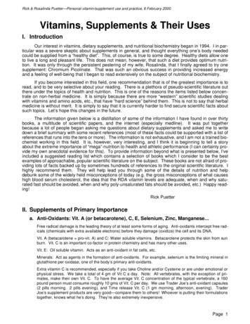
Research ArticleCell biology Plant biologyFigure 3. G proteins positively regulate immunity and BIK1 stability. (A) AGB1 is required for accumulation ofBIK1, but not FLS2 and BAK1. BIK1-HA was introduced into agb1 by crossing, homozygotes of the indicatedgenotypes in F3 generation were used for immunoblot analyses. (B) AGG1/2 are required for BIK1 stability. NP::BIK1-HA was introduced into agg1/2 by crossing, homozygous plants in F3 generation were subject toimmunoblot analyses. (C) XLG2/3 are required for BIK1 accumulation. NP::BIK1-HA, 35S::BIK1-HA and NP::BAK1HA plasmids were transiently expressed in WT and xlg2/3 protoplasts, and accumulation of BIK1 and BAK1 wasdetermined by immunoblot analyses. (D) AGB1 regulates BIK1 accumulation through the proteasome pathway.One-week-old NP::BIK1-HA seedlings of WT (Col-0) or agb1 background were pretreated with DMSO (-) or 100mM proteasome inhibitor PS341( ) for 8 hr before total protein was isolated for immunoblot analysis. (E) The agb1extract shows accelerated degradation of BIK1 in vitro (F) The xlg2 xlg3 extract shows accelerated degradation ofBIK1. Total extracts from WT (Col-0), agb1 and xlg2 xlg3 seedlings were incubated with HIS-BIK1 protein at 22 Cfor the indicated times, and equal amounts of sample were analyzed using anti-HIS immunoblot. Each experimentwas repeated at least three times, and data from one representative experiment are shown.DOI: 10.7554/eLife.13568.012Liang et al. eLife 2016;5:e13568. DOI: 10.7554/eLife.1356811 of 22
Research ArticleCell biology Plant biologyFigure 3—figure supplement 1. G proteins are required for BIK1 stability. (A) agb1 plants are largely normal inBIK1 transcription. Total RNA was isolated from plants of the indicated genotypes, and quantitative real time PCRwas carried out to determine BIK1-HA transcript levels. (B) Accumulation of BIK1 expressed from a transgeneunder control of the constitutive 35S promoter was similarly compromised in the agb1-2 mutant. 35S::BIK1-HAtransgenic lines in WT and agb1-2 background with similar BIK1 transcript levels were identified by Semi-qPCRand used for detection of BIK1 accumulation. (C) Accumulation of BIK1 is not compromised in the gpa1-3 mutant.NP::BIK1-HA was introduced into gpa1-3 by crossing, and BIK1 accumulation was detected by anti-HAimmunoblot. Three independent experiments were performed with similar results.DOI: 10.7554/eLife.13568.013Liang et al. eLife 2016;5:e13568. DOI: 10.7554/eLife.1356812 of 22
Research ArticleCell biology Plant biologyFigure 3—figure supplement 2. AGB1 regulates BIK1 stability through proteasome pathway. (A) Treatment withMG132 allows accumulation of BIK1 in agb1 mutant seedlings. NP::BIK1-HA seedlings of WT (AGB1) and agb1background were treated with DMSO (-) or 100 mM specific proteasome inhibitor MG132 ( ) for 8 hr, BIK1 stabilitywas detected by anti-HA immunoblot. (B) Treatment with PS341 inhibits BIK1 degradation in vitro. Total extractsfrom WT plants pretreated with DMSO or 100 mM PS341 were incubated with the HIS-BIK1 recombinant protein,and equal amounts of sample were withdrawn at the indicated times for anti-HIS immunoblot assays. Threeindependent experiments were performed with similar results.DOI: 10.7554/eLife.13568.014Liang et al. eLife 2016;5:e13568. DOI: 10.7554/eLife.1356813 of 22
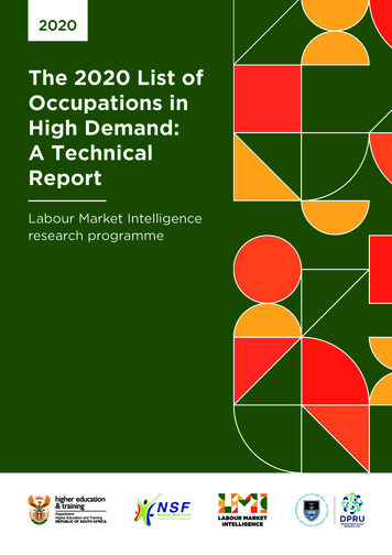
Research ArticleCell biology Plant biologyFigure 3—figure supplement 3. AGB1 and XLG2/3 attenuate PBL20 degradation. (A) Accelerated PBL20degradation in agb1 extracts. (B) Accelerated PBL20 degradation in xlg2 xlg3 extracts. Total extracts from WT,agb1 and xlg2/3 were incubated with the PBL20-HIS recombinant protein, and equal amounts of sample werewithdrawn at the indicated times for anti-HIS immunoblot analyses. Two independent experiments wereperformed with similar results.DOI: 10.7554/eLife.13568.015Liang et al. eLife 2016;5:e13568. DOI: 10.7554/eLife.1356814 of 22
Research ArticleCell biology Plant biologyFigure 4. BIK1 level accounts for G protein-mediated regulation of FLS2 immunity. (A) Transient expression ofBIK1 in agb1 and xlg2/3 mutant protoplasts restores RbohD phosphorylation. FLAG-RbohD and BIK1-HAconstructs are transiently expressed in protoplasts from WT (Col-0), agb1 and xlg2/3. FLAG-RbohD protein wasaffinity purified and detected by anti-FLAG and anti-pSer39 immuoblots. (B) BIK1 transgene restores flg22-inducedROS burst in agb1. (C) BIK1 transgene partially restores flg22-induced resistance to Pst in agb1. NP::BIK1-HA wasintroduced into agb1 by crossing, transgenic lines of agb1 or Col-0 background in the F3 generation were usedfor the assays. (D) BIK1 transgene partially restores flg22-induced ROS burst in xlg2 xlg3 mutant. (E) BIK1transgene partially restores flg22-induced resistance to Pst. The NP::BIK1-HA transgene was introduced into WT(Col-0-L32) and xlg2 xlg3 (xlg2/3-L64 and xlg2/3-L51) plants by Agrobacterium-mediated transformation.Independent T2 transgenic lines were used for the assays. Peak relative luminescence unit (RLU) values wereshown for ROS assays (B and D) and leaf bacterial populations 2 days after bacterial inoculation were shown forflg22-protection assays (C and E). Bars in B-E represent mean SD (n 6; p 0.05, Student’s t-test; different lettersindicate significant difference). Each experiment was repeated two (A) or three (B–E) times, and data of onerepresentative experiment are shown.DOI: 10.7554/eLife.13568.016The following source data is available for figure 4:Source data 1. Raw data and exact p value of Figure 4B—E.DOI: 10.7554/eLife.13568.017Liang et al. eLife 2016;5:e13568. DOI: 10.7554/eLife.1356815 of 22
Research ArticleCell biology Plant biologyFigure 5. Phosphorylation of XLG2 by BIK1 regulates flg22-induced ROS. (A) Flg22-induces phosphorylation ofXLG2 in the N terminus. Protoplasts expressing XLG21-203-FLAG were treated with flg22. The total protein wastreated with ( ) or without (-) l protein phosphatase (PPase) prior to anti-FLAG immunoblot analysis. (B) Flg22induced phosphorylation of XLG2 in protoplasts primarily occurs in Ser141, Ser148, Ser150 and Ser151. Differentmutated form of XLG21-203-FLAG constructs were transiently expressed in WT protoplast, treated with flg22 andthe migration of XLG21-203-FLAG were examined by anti-FLAG immunoblot. (C) BIK1 phosphorylates XLG2 Nterminus in vitro. XLG21-203-HIS was incubated with HIS-BIK1 and HIS-BIK1K105E in the presence of 32P-g-ATP andanalyzed by autoradiography. CBB, coomassie brilliant blue. (D) BIK1 phosphorylates XLG2 at Ser148 and Ser150in vitro. XLG21-203-HIS was incubated with HIS-BIK1 and HIS-BIK1K105E in kinase reaction buffer. Proteinphosphorylation was detected by anti-pSer148 and pSer150 immunoblots. (E) XLG2 phosphorylation is requiredfor flg22-induced ROS. xlg2 mutant plants were transformed with WT (NP::XLG2-L34), non-phosphorylatable (4AL1 and 4A-L7), or phospho-mimicking (4D-L7 and 4D-L9) forms of XLG2 under control of the native XLG2promoter. Independent T2 lines were examined for flg22-induced ROS burst and peak relative luminescence unit(RLU) values are shown. (mean SD; n 6; p 0.05, Student’s t-test; different letters indicate significant difference).(F) XLG2 phosphorylation is required for Pst resistance. xlg2/3 double mutant plants were transformed with WT(XLG2-L3) or non-phosphorylatable (4A-L6 and 4A-L7) form of XLG2 under control of the native XLG2 promoter.Independent T2 lines were inoculated with Pst, and bacterial populations in leaves were measured 3 days postinoculation. (mean SD; n 6; p 0.05, Student’s t-test; different letters indicate significant difference). (G) XLG2interacts with RbohD in Arabidopsis plants. rbohD plants were transformed with the FLAG-RbohD transgeneFigure 5 continued on next pageLiang et al. eLife 2016;5:e13568. DOI: 10.7554/eLife.1356816 of 22
Research ArticleCell biology Plant biologyFigure 5 continuedunder control of the RbohD native promoter. The resulting plants were used for Co-IP assay. Each experiment wasrepeated two (C, G) or three (A, B, D–F) times, and data of one representative experiment are shown.DOI: 10.7554/eLife.13568.018The following source data is available for figure 5:Source data 1. Raw data and exact p value of Figure 5E and F.DOI: 10.7554/eLife.13568.019Liang et al. eLife 2016;5:e13568. DOI: 10.7554/eLife.1356817 of 22
Research ArticleCell biology Plant biologyFigure 5—figure supplement 1. The N terminus ofXLG3, but not XLG1, is phophorylated upon flg22treatment. WT protoplasts were transfected withXLG11-188-FLAG or XLG31-200-FLAG, treated with flg22,and protein was analyzed by anti-FLAG immunoblot.DOI: 10.7554/eLife.13568.020Liang et al. eLife 2016;5:e13568. DOI: 10.7554/eLife.1356818 of 22
Research ArticleCell biology Plant biologyFigure 5—figure supplement 2. Phospho-sites in XLG2 isolated from flg22-treated protoplasts. List of phospho-peptides identified. Protoplastsprepared from WT plants were transfected with XLG2-FLAG and treated with flg22 for 10 min, affinity-purified and subjected to LC-MS/MS forphospho-sites identification.DOI: 10.7554/eLife.13568.021Liang et al. eLife 2016;5:e13568. DOI: 10.7554/eLife.1356819 of 22
Research ArticleCell biology Plant biologyFigure 5—figure supplement 3. Mutations that block or mimic XLG2 phosphorylation do not impact BIK1stability and XLG2-BIK1 interaction. WT or xlg2 xlg3 protoplasts were transfected with BIK1-HA along with WT,non-phosphorylatable (3A, 4A), phospho-mimicking (4D) forms of XLG2-FLAG constructs, treated with ( ) orwithout (-) flg22, and total protein was subject to Co-IP assays and immunoblot analysis. Three independentexperiments were performed with similar results.DOI: 10.7554/eLife.13568.022Liang et al. eLife 2016;5:e13568. DOI: 10.7554/eLife.1356820 of 22
Research ArticleCell biology Plant biologyFigure 5—figure supplement 4. XLG2/3 interact with RbohD in Nb plants. Agrobacteria containing the indicatedconstructs were infiltrated in to Nb leaves, and luciferase activity was recorded 2 days later (mean SD; n 6;representative data from 2 independent experiments are shown).DOI: 10.7554/eLife.13568.023Liang et al. eLife 2016;5:e13568. DOI: 10.7554/eLife.1356821 of 22
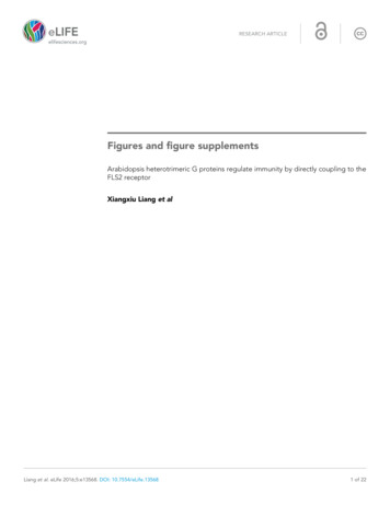
Research ArticleCell biology Plant biologyFigure 6. Model for G protein-coupled FLS2 signaling. In the pre-activation state, the heterotrimeric G proteinscomposed of XLG2/3, AGB1, and AGG1/2 interact with the FLS2-BIK1 complex. Stimulation by flg22 inducesBAK1-FLS2 interaction and activation of the receptor complex. This leads to the activation of the G proteins andphosphorylation of XLG2 in the N terminus. The activated G proteins dissociate from the receptor complex andregulate RbohD and other downstream effectors to positively modulate immune responses.DOI: 10.7554/eLife.13568.024Liang et al. eLife 2016;5:e13568. DOI: 10.7554/eLife.1356822 of 22
Figure 2—figure supplement 2. XLG3 interacts with BIK1. (A) XLG3 interacts with BIK1 in Nb plants. Agrobacteria containing the indicated constructs were infiltrated into Nb leaves, and luciferase activity was recorded 2 days later (mean SD; n 6). (B) XLG3 interacts with BIK1 in protoplasts. Co-IP assay was performed using WT Arabidopsis