Transcription
HindawiBioMed Research InternationalVolume 2020, Article ID 5764017, 19 pageshttps://doi.org/10.1155/2020/5764017Review ArticleSleep Deprivation and Neurological DisordersMuhammed Bishir,1 Abid Bhat ,1 Musthafa Mohamed Essa ,2,3 Okobi Ekpo ,4Amadi O. Ihunwo ,5 Vishnu Priya Veeraraghavan ,6 Surapaneni Krishna Mohan ,7Arehally M. Mahalakshmi ,1 Bipul Ray ,1 Sunanda Tuladhar,1 Sulie Chang ,8Saravana Babu Chidambaram ,1 Meena Kishore Sakharkar ,9 Gilles J. Guillemin ,10M. Walid Qoronfleh ,11 and David M. Ojcius 121Department of Pharmacology, JSS College of Pharmacy, JSS Academy of Higher Education & Research, Mysuru, IndiaDepartment of Food Science and Nutrition, CAMS, Sultan Qaboos University, Muscat, Oman3Ageing and Dementia Research Group, Sultan Qaboos University, Muscat, Oman4Department of Medical Bioscience, Faculty of Natural Science, University of the Western Cape, Robert Sobukwe Road,Bellville 7535, South Africa5School of Anatomical Sciences, Faculty of Health Sciences, University of the Witwatersrand, Johannesburg, York Road, Parktown,2193 Johannesburg, South Africa6Department of Biochemistry, Saveetha Dental College, Saveetha Institute of Medical and Technical Sciences, Saveetha University,600 077, Chennai, India7Department of Biochemistry, Panimalar Medical College Hospital & Research Institute, Varadharajapuram, Poonamallee, 600123,Chennai, India8Department of Biological Sciences and Institute of Neuroimmune Pharmacology, Seton Hall University, 400 South Orange AveSouth Orange, NJ 07079, USA9College of Pharmacy and Nutrition, University of Saskatchewan, 107, Wiggins Road, Saskatoon, SK, Canada S7N 5C910Neuroinflammation Group, Faculty of Medicine and Health Sciences, Macquarie University, NSW 2109, Australia11Research & Policy Department, World Innovation Summit for Health (WISH), Qatar Foundation, P.O. Box 5825, Doha, Qatar12Department of Biomedical Sciences, University of the Pacific, Arthur Dugoni School of Dentistry, San Francisco, CA, USA2Correspondence should be addressed to Saravana Babu Chidambaram; csaravanababu@gmail.comReceived 19 July 2020; Accepted 10 November 2020; Published 23 November 2020Academic Editor: Alessandro MartoranaCopyright 2020 Muhammed Bishir et al. This is an open access article distributed under the Creative Commons AttributionLicense, which permits unrestricted use, distribution, and reproduction in any medium, provided the original work isproperly cited.Sleep plays an important role in maintaining neuronal circuitry, signalling and helps maintain overall health and wellbeing.Sleep deprivation (SD) disturbs the circadian physiology and exerts a negative impact on brain and behavioural functions.SD impairs the cellular clearance of misfolded neurotoxin proteins like α-synuclein, amyloid-β, and tau which are involvedin major neurodegenerative diseases like Alzheimer’s disease and Parkinson’s disease. In addition, SD is also shown toaffect the glymphatic system, a glial-dependent metabolic waste clearance pathway, causing accumulation of misfoldedfaulty proteins in synaptic compartments resulting in cognitive decline. Also, SD affects the immunological and redoxsystem resulting in neuroinflammation and oxidative stress. Hence, it is important to understand the molecular andbiochemical alterations that are the causative factors leading to these pathophysiological effects on the neuronal system.This review is an attempt in this direction. It provides up-to-date information on the alterations in the key processes,pathways, and proteins that are negatively affected by SD and become reasons for neurological disorders over a prolongedperiod of time, if left unattended.
21. IntroductionSleep is a ubiquitous phenomenon occurring in life formsof the animal kingdom and has been shown to be presentfrom Drosophila melanogaster (fruit fly) to human beings.Sleep is a vital component for healthy brain function, andsleep deprivation (SD) is the reduction in sleep time belowan individual’s baseline requirement while sleep restriction(SR) refers to partial loss of sleep. SD and SR have beenreported to affect overall wellness and health, including,but not limited to lowering in the immune system,decrease in cognitive function and memory, learning, anddisruption in emotional wellbeing [1]. National SleepFoundation, USA, suggests that 7–8 h of sleep is essentialfor maintenance and restoration of metabolic homeostasis[2]. There are two stages of sleep: (i) nonrapid eye movement(NREM) and (ii) rapid eye movement (REM). NREM is subdivided into four different stages based on the depth andwave patterns, movement of the eye, and muscle strengthduring sleep. REM sleep is characterized by uneven brainwave activity, muscle atonia, and increased eyeball movements [3]. Sleep is regulated by two processes that work independently and influences sleep and sleep-related variables inconjunction “Rheostat” [4]: (1) Circadian rhythm—a processmaintained by the biological clock in the suprachiasmaticnucleus (SCN) in the hypothalamus, which regulates sleep–wake cycles in response to the input from retina [5]; (2)homeostatic process—loss of sleep is compensated byextending subsequent sleep which is a function of wakingduration and intermittent naps during the wake period. During wakeful hours, the tendency to sleep gradually increaseswith time and attains a critical threshold; this urge to sleepis referred to as homeostatic.Sleep helps to maintain metabolic homeostasis throughneural, hormonal, and immune supports [6]. NREM sleepis characterized by a low metabolic rate and an increase inbrain temperature which helps to overcome the damages thatare introduced during the wake cycle [7]. A study conductedon shift workers revealed SD alters glucose and lipid metabolism [8], which suggests the role of sleep in metabolicdysfunction. Krause et al. reported that SD affects attentionand working memory, positive and negative emotions, andhippocampal learning [9].The increasing number of people across the globe arebeing affected with the epidemic of SD [10]. SD impairssympathetic functions which in turn causes metabolic dysregulation [11, 12]. Furthermore, SD modulates immunefunctions and increases the release of proinflammatorycytokines such as Interleukin 6 (IL-6), Tumour necrosisfactor (TNF-α), and C-reactive protein (CRP) [13]. Sleeprestriction increases prolactin and oxytocin and decreasesbody weight in pregnant rats. Offsprings from these ratsshow decreased hippocampal brain-derived neurotrophicfactor (BDNF) expression, suggesting the detrimental roleof SR on neuronal growth factors during the developmentalstage [14]. Sleep loss during developmental stages reducesbrain size and alters behaviour and neural homeostasis[15]. Electroencephalogram studies have revealed that SDpromotes interictal epileptiform discharges and neuronalBioMed Research Internationalexcitability causing activation of seizure episodes [16]. SDhas also been associated with the impairment of cognitivefunction in humans. A recent study showed that onenight SD increased amyloid-β burden in the hippocampalregion [17]. Thus, mounting evidences reveal that SD islinked with many neurodegenerative diseases and neurological disorders. In this review, we attempt to provide information on the links between sleep deprivation/restrictionon the changes in gene expression and pathophysiologicalmechanism associated with neurological disorders.2. Sleep and Brain Anatomical StructuresSleep and its mechanism are controlled by defined regions inthe brain. Microanatomically, cell bodies of neurons thatproduce neurotransmitters playing a role in sleep mechanisms are usually located in one region while the terminalends of the neuronal axons project elsewhere [18]. In themammalian brain, the cell bodies of the neurons involvedin sleep are located in the brainstem while the axons end incentres located in the cerebral hemisphere. Sleep entails apatterned interaction between the cerebral cortex, thalamus,and subcortical areas like the brainstem. According to [19],“The ebb and flow of neurotransmitters switches our brainsbetween sleep and wakefulness in carefully regulated cyclesin several brain regions.”The hypothalamus is located deep in the brain, proximalto the pituitary gland. It contains thousands of nerve cellbodies called the suprachiasmatic nuclei (SCN), whichreceive information about light exposure to control the sleepand arousal cycle [20]. The pineal gland lies in the depressionbetween the superior colliculi. Through its numerous connections, the production of the sleep-promoting neurohormone melatonin is regulated; hence, it plays a key functionin regulating the circadian rhythm. The amygdala, a structureknown in the processing of emotions, has been suggested tobe very active during rapid eye movement (REM) sleep,which explains the co-occurrence of mood disorders withsleep abnormalities [21].Neurons of the reticular activating system are central tothe regulation of wakefulness. The brain stem components(midbrain, pons, and medulla oblongata) have connectionswith the hypothalamus to control the wake and sleep cycle.The midbrain is associated with vision, hearing, motorcontrol, sleep and wake cycles, alertness, and temperatureregulation. The pons and medulla oblongata in particularhave connections with descending neural pathways thatmaintain muscle activity and body posture and limb movements at the relaxed state. Brainstem nuclei that are involvedin sleep processing include the cholinergic nuclei at thepons–midbrain junction, the raphe nuclei, tuberomammillary nuclei, and locus coeruleus [18]. The thalamus acts as arelay centre for information from the main sense organs tothe cortex. It is very active during REM sleep as the ascendingbrainstem reticular activating system (ARAS) relays at multiple intermediary sites (including the thalamus), to activatethe forebrain during waking and REM sleep [22]. Damageto the thalamus may affect proper brain function during bothwakefulness and sleep in humans [23]. Cholinergic neurons
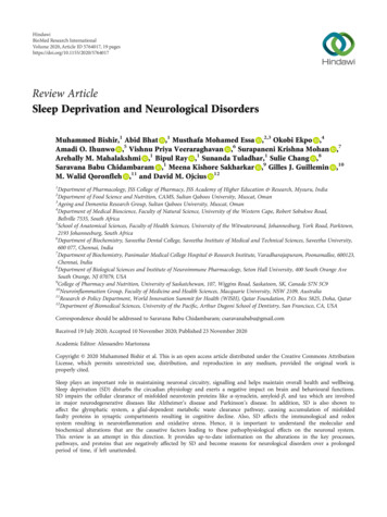
BioMed Research Internationalin the basal forebrain region have been reported to promotesleep via the release of the cellular energy by-product, adenosine. Caffeine and some medications are known to counteract sleepiness by blocking the actions of adenosine [24].3. Sleep and Neural CircuitsThe involvement of neural circuits in sleep-wake cycles wasfirst studied by Magoun and Moruzzi in the year 1949 [25].They showed that stimulation of cholinergic neurons nearpons and midbrain causes wakefulness and arousal. In addition, stimulation of the thalamic region with a low-frequencypulse produces a slow-wave sleep. This study provided aninsight on the interaction between the thalamus and cortexduring sleep (Figure 1).3.1. Neural Circuits Involved in NREM Sleep3.1.1. Preoptic Area (POA). POA is the rostral part of thehypothalamus and is mainly involved in the thermoregulation of the body. In 1968, McGinty and Sterman’s researchon cats and rats revealed that POA contains neurons thatpromote sleep [26]. They also showed that certain neuronsin the POA and Basal Forebrain (BF) are active duringREM and NREM sleep [27]. The analysis of the Fos expression revealed that the ventrolateral preoptic area (VLPO)and median preoptic nucleus (MnPO) consist of neuronsessential for NREM sleep [28]. Lesions in the VLPO andMnPO produced a long-lasting decrease in sleep [29]. Furthermore, these neurons are GABAergic. Interestingly,VLPO neurons release a neuropeptide called “galanin,”which inhibits cholinergic neurons of the locus coeruleus,Basal Forebrain, TMN, orexin neurons, and causes sleeparousal [30].3.1.2. Basal Forebrain (BF). BF is innervated by cholinergicneurons which govern cortical activation during wakefulness.The inhibition of cortical activation is governed by theGABAergic neurons and promotes slow-wave sleep.GABAergic neurons initiate firing at NREM onset and havetheir maximal output throughout the NREM cycle [31]. BFneurons produce somatostatin and are active during NREMsleep, and optogenetic stimulation of these neurons is shownto increase NREM sleep [32]. Somatostatin promotes NREMsleep by inhibiting wake active neurons in mice [32, 33]. Onthe other hand, prolonged wakefulness increases GABAAreceptors in BF cholinergic neurons, and the GABAmediated inhibition of cholinergic neurons decreases thecortical activity [34].3.1.3. Parafacial Zone (PZ). Caudal brainstem neuronspromote NREM sleep [35]. Most of the neurons in theparafacial zone are GABAergic and glycinergic. Theyexpress Fos during sleep, and lesions of these neuronsare shown to increase wakefulness. PZ neurons releaseGABA onto parabrachial neurons which in turn releaseglutamate onto cortically projecting neurons of the BFthereby potentiating slow-wave sleep and regulate corticalEEG [36]. Chemogenetic inhibition of PZ neurons3markedly declines NREM sleep even after SD, indicatingthat PZ neurons play key roles in NREM sleep [37].3.1.4. Cortical Neuronal Nitric Oxide Synthase (nNOS). Nitricoxide synthase neurons regulate sleep homeostasis and cortical rhythm through the release of GABA and nitric oxide(NO). Interestingly, the nNOS knockout mice showed adecrease in NREM sleep [38], which indicates the major roleof nNOS in NREM. Majority of the neurons present in thecortical regions are wake-promoting and are GABAergicneurons, wherein the slow-wave activity during the NREMsleep directly correlates with the expression of nNOS [39].3.2. Neural Circuits Involved in REM Sleep. Evidence fromrecent studies suggests that GABAergic/glycinergic neuronsin the medullary reticular formation release melatonin, andGABAergic neurons present in the hypothalamus regulateREM sleep. Pedunculopontine and Laterodorsal TegmentalNuclei—cholinergic LDT/PPT neurons and glutaminergicneurons of the pons play a crucial role in regulating REMsleep. These reports suggest the existence of a strong neuralcircuit in the regulation of REM sleep3.2.1. Pedunculopontine and Laterodorsal Tegmental Nuclei(PPT/LDT). The administration of carbachol, a cholinergicagonist, by intracerebroventricular injection into the laterodorsal region of the pons produces a high degree of REMsleep in cats and rodents, indicating that acetylcholine levelsare high during sleep [40]. These neurons initiate firingduring the beginning of REM sleep, suggesting that they helpin switching between REM and NREM sleep [41].3.2.2. GABAergic Neurons in the Hypothalamus. Anincreased number of c-Fos immunopositive cells wererecorded in various regions of the hypothalamus includingzona increta, perifornical area, and lateral hypothalamic areafollowing REM sleep [42]. GABAergic neurons present inthis region are immunoreactive to melanin-concentratinghormone (MCH) and nesfatin both of which regulate REMsleep [42, 43]. The optogenetic activation of MCH neuronsfacilitates the switching between NREM and REM sleep andalso increases the duration of REM sleep [43, 44]. The inhibition of lateral hypothalamic (LH) neurons with muscimol, aGABAA receptor agonist, is shown to completely inhibitREM sleep and increase the NREM sleep episodes. The bilateral administration of muscimol increases the expression ofcFOS /GABA and GABA in vlPAG and dorsal deep mesencephalic reticular nucleus (dDpMe) regions [45]. These resultsindicate that MCH /GABA and MCH-/GABA neurons inthe LH promote REM sleep by the inactivation ofvlPAG/dDpMe neurons.3.2.3. Sublaterodorsal Nucleus (SLD) Region. Chemicallesions and optogenetic stimulation studies have shown thatSLD contains neurons responsible for the initiation andmaintenance of REM sleep [46]. In addition to cFO expression following REM sleep, the neurons in SLD also showthe existence of glutamatergic markers, which reveal thatREM sleep-promoting neurons in SLD are glutaminergic innature [47]. SLD sends direct efferent projections to
4BioMed Research InternationalBrain structures involved in REM sleepLTD & PPTLTD & talgray areaLocus coeruleusHypothalamusGABA, MCH,nesfatinSublaterodorsaltegmental nucleus(SLD) glutaminergicActivationInhibition(a)Brain structures involved in NREM sleepVentrolateralpreoptic areaReleaseGABA, galaninReleaseMedian preoptic areaCortical nitric oxidesynthase neuronsParafacialzoneReleaseGABAActivatesBasal forebrainGABA, somatostatinSWSLocus coeruleusWake activatingneuronsGABA, galanin inhibits cholinergicneurons in the locus coeruleus. Basalforebrain. TMN, orexin neuron(b)Figure 1: (a) Brain structures involved in REM sleep. PPT/LDT initiates firing during REM sleep and helps in switching between NREM andREM sleep. GABAergic neurons in the hypothalamus promote REM sleep by inactivating vlPAG/dDpMe REM-off GABAergic neurons. SLDconsists of projections to glycinergic neuron Raphe magnus, ventral and alpha gigantocellular nuclei, lateral paragigantocellular nucleus, andalso spinal, facial, trigeminal neurons and thereby produce muscle atonia during REM sleep. (b) Brain structures involved in NREM sleep.Ventrolateral preoptic area and median preoptic nucleus contain GABAergic neurons that release galanin which inhibit cholinergicneurons in regions like locus coeruleus, Basal Forebrain, TMN, and orexin neurons, thereby inhibiting arousal. The basal forebrainconsists of GABAergic neurons that inhibit cortical activation and somatostatin inhibits wake active neurons in the basal forebrain.Parafacial zone releases GABA onto parabrachial neurons which in turn release glutamate onto cortically projecting neurons of the BF,hence promoting SWS. Cortical nitric oxide synthase neurons release GABA and promote SWS.
BioMed Research Internationalglycinergic neurons in the Raphe magnus, ventral and alphagigantocellular nuclei, lateral paragigantocellular nucleus,and also spinal, facial, trigeminal neurons [48, 49]. On theother hand, the ventromedial medulla-glycinergic/GABAergic is populated near rostral to inferior olive in the ventralgigantocellular reticular (GiV) and the alpha gigantocellularreticular (GiA) nuclei [45]. These neurons fire very fast inREM, slower in NREM, and moderately in the wake cycle,indicating their role in muscle atonia [50], and lesions tothese neurons are shown to disrupt atonia of REM sleep[51]. Another study proposes that neurons in SLD directlytrigger spinal interneurons through glycinergic/GABAergiccomponents, which further supports their role in muscleatonia [52].3.2.4. Medullary Reticular Formation. GABAergic and glycinergic neurons regulate REM sleep. They receive inputsignals from SLD and innervate the motor neurons ofthe brainstem and spinal motor neurons. Upon stimulation, they produce glycinergic Inhibitory PostsynapticPotential (IPSP) in motor neurons [49]. Neurons of thedorsal paragigantocellular reticular (DPGi) and lateralparagigantocellular (LPGi) promote REM sleep by inhibiting REM sleep suppressing neurons of the pons like LC,DRN, and vlPAG/LPT [53]. Photostimulation of neuronsin LPGi and vlPAG promotes REM sleep, and inhibitionof these neurons produces opposite effects [54]. Thesefindings suggest that medullary neurons not only promoteatonia but are also involved in REM sleep regulation.Further studies are required to understand whether REMsleep is driven by the medulla or pons or both the regionsof the brain.4. Genomics and Sleep DeprivationHippocampus is the key brain structure involved in spatial,contextual, and declarative memory. The formation of memory depends upon the expression of various genes/proteins[55, 56]. Hippocampal dependent memory consolidation iscritically affected in SD [57, 58]. SD is shown to affect thesignalling mechanisms that regulate transcription and translation processes involved in memory [59]. A genome-widemicroarray analysis by Vecsey et al. [60] showed the groupof genes that are differentially regulated in SD animals incomparison to animals that have normal sleep patterns.Tsc22d3, Prkab2, Adamts2, Htr1a, Kcnv1, and Sirt7 are thegenes that show upregulation. Tsc22d3 negatively regulatesmemory consolidation and neural plasticity, and its expression is reported to be upregulated after SD. Prkab2, a subunitof AMPK, undergoes hyperphosphorylation following SDand alters homeostatic response. In addition, the expressionof certain genes in the cortex such as Arc/Arg3.1, Fos,Hnrpdl, Rbm3, and the chaperones Hspa5/Bip and Hspa8 isalso altered by SD [60].SD impairs protein synthesis; it downregulates the geneslike Eif4e2 and Eif5, which are involved in the initiation oftranscription and those genes like Rprd2, Rbm3, Hnrpdl,Cirbp, RbmX, and Denr involved in the process of translation[60]. Please refer to the supplementary materials (available5here) for the table and graphical presentation of genes upand downregulated following SD.5. Sleep and Neurological Disorders5.1. Alzheimer’s Disease. Alzheimer’s disease (AD) is a progressive neurodegenerative disease pathologically characterized by the deposition of extracellular amyloid β- (Aβ-)plaques, intracellular tangles, and neuronal loss [61]. Studieshave shown a direct correlation between SD and neuropathological events associated with AD [62]. Sleep plays a crucialrole in clearing the toxic metabolites produced in the brain.Two investigations using photon microscopy have shownthat amyloid β in the brain is cleared by astroglial-mediatedinterstitial fluid (ISF) bulk flow called “Glymphatic Pathway”and also by γ-oscillations during REM sleep [63]. Aβ clearance is increased by 25-30% during sleep in comparison towake state [64]. Sleep enhances interstitial fluid (ISF) to cerebrospinal fluid (CSF) bulk flow, thereby increasing the Aβclearance [65]. On the other hand, SD increases the Aβ burden and its clearance leads to AD [65, 66]. Clinical reportsindicate that the levels of Aβ in CSF are high before sleepand low after sleep [67]. Positron emission tomography(PET) analyses show that SD increases Aβ in the hippocampus, precuneus, thalamus, and cortex of human subjects [17].This deposition of Aβ brings structural and functionalchanges in AD brains. The aggregation of Aβ in the hippocampal region leads to the formation of amyloid plaques,which inhibit neurogenesis and lead to cognitive dysfunction[17]. SD upregulates the expression of BACE1 and APP processing, which also play critical roles in AD [68]. SD increasescortisol level, a stress maker which was shown to impaircognition in AD patients [69]. Stress directly affects the brainin 3 different ways: (a) it enhances tau aggregation throughhyperphosphorylation and causes genetic alterations in theDNA [71]; (b) it declines synaptic density and number ofneurons and increase the deposition of Aβ; and (c) it alsoimpairs cardiovascular, metabolic, and GI functions andweakens the immune system [70]. Chronic stress causeshypothalamic–pituitary–adrenal (HPA) axis imbalance,which leads to the accumulation of Aβ and tau proteins, cognitive impairment, and neuronal death, resulting in AD [71].Aquaporin 4 (AQP4) is the most abundant water channelin the brain which regulates water homeostasis. AQP4 ishighly expressed in astrocytes, lining of the ventricles, andin perivascular astrocytes end feet enclosed by blood vesselsfrom the CNS [73, 74]. It also plays a crucial role in removingtoxic metabolites including Aβ and tau and prevents theiraggregation. SD impairs the glymphatic system which resultsin the aggregation of Aβ [17, 75]. A paravascular pathwayfacilitates CSF flow through the brain parenchyma and theclearance of interstitial solutes, including amyloid β. Studiesreport that glymphatic function declines over mid to terminal stages of AD due to impaired polarity of AQP4 at theastrocyte end feet [75, 76]. A recent report indicates a geneticmutation in AQP4 in patients with less sleep or SD [77].Phosphorylated Tau proteins bind to the microtubuleand provide structural stability to the neurons. Hyperphosphorylation of Tau is a hallmark of AD, which causes
6aggregation and formation of filamentous structures calledNeurofibrillary tangles (NFT). Preclinical evidences revealthat an impaired sleep-wake cycle can increase the hyperphosphorylation of Tau proteins [78]. Apolipoprotein E (APOE)is one of the major constituent of chylomicrons andintermediate-density lipoproteins (IDLs) and is involved inregulating catabolism of lipoproteins [79]. APOE genes consist of three variants of genes -ε2, ε3, and ε4 [80]. APOE/ε4variant gene is a risk factor of AD; ε4 allele also increasesthe deposition of Aβ and cognitive impairment [81]. Leoniet al. have shown that ApoE/ε4 is associated with sleep disorders and increases the levels of LDL and triglycerides. Brainremoves excess cholesterol by converting it into polar 24hydroxycholesterol (24-OHC) that is cleared through APOE.CSF samples from AD patients have shown an increase in theconcentration of ApoE, tau, NFTs, and 24-OHC. Increasedlevels of these components lead to increased NFT loadingcells [82]. It is suggested that ApoE/ε4 is less efficient in clearing cholesterol from the neurons and promotes NFT load inneurons. Lim et al. have also shown that adequate sleepinhibits the effects of APOE in the formation of NFT anddelays the progression of AD [81].Cholinergic neurons are essential in regulating sleepwake states, memory, and learning [83]. Cholinergic neuronsare active during REM sleep and wakefulness, and they areless active during NREM sleep [84]. SD increases adenosinelevels in the basal forebrain, thereby inhibits the cholinergicsystem and disrupts the switch between sleep and wakefulness [85]. The stimulation of the muscarinic receptorsenhances long-term potentiation and synaptic plasticity inthe CA1 region of the hippocampus [86]. Experiments onanimal models have also shown cognitive impairment following treatment with muscarinic antagonist [87]. BDNFexpression is also regulated by the muscarinic receptors.These results suggest that equilibrium in cholinergic activityis essential for memory encoding and recall. Cognitivedysfunction, inability to learn new things, and difficulty inperforming the known tasks all are the primary symptomsof AD. According to WHO, 30 million people across theglobe are suffering from Alzheimer’s dementia [88]. It isknown that sleep is essential for cognition, and SD leads tomemory deficits, impaired attention decision making, andretrieval [89]. REM sleep is essential for the LTP, whereasNREM sleep deprivation does not affect LTP [90]. SDincreases the adenosine and cholinergic functions in variousregions of the brain including the hippocampus and basalforebrain [91]. These results suggest the negative impact ofSD on cholinergic neurons and cognitive functions.Excitatory neurotransmission in the mammalian CNS isprimarily regulated by the ionotropic glutamate receptors(iGLURs). These receptors play a crucial role in LTP, learning, and neuronal survival [92]. Abnormal signalling ofthese receptors is involved in various neurological disorderslike Alzheimer’s disease, Parkinson’s disease, Huntingtondisease, and multiple sclerosis [93]. NMDA receptors, adivision of iGLURs, due to its unique properties likevoltage-dependent activation, enhanced Ca2 influx andslow ligand-gated kinetics make them the key players inLTP [94, 95]. NMDA receptors are also crucial for neuronalBioMed Research Internationalsurvival. These receptors activate survival pathways likeERK, PI3K/AKT, and CaMK and also inhibit proapoptotickinases like GSK3β [96]. Many studies suggest that overexcitation of glutaminergic neurons or excitotoxicity leads toneurodegeneration. This is due to an increased influx ofCa2 through the NMDA receptors [97–99]. Increased levelsof Ca2 influx lead to impaired synaptic function andeventually to neuronal cell death. This is closely associatedwith cognitive decline and pathological neural anatomysimilar to AD patients.SD has a negative impact on LTP and synaptic plasticityand hippocampal glutaminergic NMDA receptors. SDreduces the expression of the GluN1 subunit of the NMDAreceptors, thereby lowering the excitatory postsynaptic current in the CA1 region [100]. Studies also report that 12 hof SD leads to a drastic decrease in the phosphorylation ofhippocampal AMPA receptors at the GluR1-S845 site anddecreases the levels of AKAP150 scaffolding proteins [101].All these data suggest that SD leads to impaired spatial andworking memory by decreasing AMPA receptor phosphorylation through the decreasing levels of AKAP150 scaffoldingproteins. Thus, SD is closely connected to the pathologicalmarkers and that it plays a crucial role in the pathogenesisof AD (Figure 2).5.2. Parkinson’s Disease. Parkinson’s disease (PD) is a distinctive motor disease that involves neuromuscular rigidity,bradykinesia, and tremor. The accumulation of α-synuclein,Aβ, TDP-43, and tau is the major hallmarks of PD [102–105]. Sleep improved the cognitive and mental ability in PDpatients [106]. Conversely, the destruction of nigrostriataldopaminergic neurons or dorsal striatum has been reportedto disrupt the sleep–wake cycle [107].Paravascular clearance pathway-“Glymphatic system”is responsible for the clearance of the toxic metabolitesfrom the brain [75, 104]. As noted in AD, the clearanceof Aβ proteins through the glymphatic pathway and theclearance of α-synuclein are not reported as of today andneed to be investigated. Hence, we propose that interventions promoting glymphatic clearance might be beneficialin PD. This could be a potential target at least in the earlystages of the disease as it can prevent the accumulation ofα-synuclein beyond mesencephalic and limbic regions.Postmortem analysis of PD brains has shown to containhigher levels of oxidized proteins and lipids. SirT3 is aNAD-dependent deacetylase sirtuin-3 protein present inthe inner mitochondrial membrane and is involved in ATPproduction and redox processes [108]. Metabolic homeostasis requires SirT3. Chronic SD impairs SirT3 activity thateventually leads to locus coeruleus neurons (LCns) superoxide
Review Article Sleep Deprivation and Neurological Disorders Muhammed Bishir,1 Abid Bhat ,1 Musthafa Mohamed Essa ,2,3 Okobi Ekpo ,4 Amadi O. Ihunwo ,5 Vishnu Priya Veeraraghavan ,6 Surapaneni Krishna Mohan ,7 Arehally M. Mahalakshmi ,1 Bipul Ray ,1 Sunanda Tuladhar,1 Sulie Chang ,8 Saravana Babu Chidambaram ,1 Meena Kishore Sakharkar ,9 Gilles J. Guillemin ,10 M. Walid Qoronfleh ,11 and David .