Transcription
Journal of Translational MedicineBioMed CentralOpen AccessResearchWhole tumor antigen vaccination using dendritic cells: Comparisonof RNA electroporation and pulsing with UV-irradiated tumor cellsFabian Benencia1,2,3, Maria C Courrèges1 and George Coukos*1,2Address: 1Center for Research on Early Detection and Cure of Ovarian Cancer, University of Pennsylvania, Philadelphia, PA, USA, 2AbramsonFamily Cancer Research Institute, University of Pennsylvania, Philadelphia, PA, USA and 3Department of Biomedical Sciences, College ofOsteopathic Medicine and Biomedical Engineering Program, Russ College of Engineering and Technology, University of Ohio, Athens, OH, USAEmail: Fabian Benencia - benencia@oucom.ohiou.edu; Maria C Courrèges - Courrege@mail.med.upenn.edu;George Coukos* - gcks@mail.med.upenn.edu* Corresponding authorPublished: 29 April 2008Journal of Translational Medicine 2008, 6:21doi:10.1186/1479-5876-6-21Received: 15 January 2008Accepted: 29 April 2008This article is available from: 21 2008 Benencia et al; licensee BioMed Central Ltd.This is an Open Access article distributed under the terms of the Creative Commons Attribution License (http://creativecommons.org/licenses/by/2.0),which permits unrestricted use, distribution, and reproduction in any medium, provided the original work is properly cited.AbstractBecause of the lack of full characterization of tumor associated antigens for solid tumors, wholeantigen use is a convenient approach to tumor vaccination. Tumor RNA and apoptotic tumor cellshave been used as a source of whole tumor antigen to prepare dendritic cell (DC) based tumorvaccines, but their efficacy has not been directly compared. Here we compare directly RNAelectroporation and pulsing of DCs with whole tumor cells killed by ultraviolet (UV) B radiationusing a convenient tumor model expressing human papilloma virus (HPV) E6 and E7 oncogenes.Although both approaches led to DCs presenting tumor antigen, electroporation with tumor celltotal RNA induced a significantly higher frequency of tumor-reactive IFN-gamma secreting T cells,and E7-specific CD8 lymphocytes compared to pulsing with UV-irradiated tumor cells. DCselectroporated with tumor cell RNA induced a larger tumor infiltration by T cells and produced asignificantly stronger delay in tumor growth compared to DCs pulsed with UV-irradiated tumorcells. We conclude that electroporation with whole tumor cell RNA and pulsing with UV-irradiatedtumor cells are both effective in eliciting antitumor immune response, but RNA electroporationresults in more potent tumor vaccination under the examined experimental conditions.IntroductionBecause tumor-associated antigens are not well characterized for the majority of human tumors, polyvalent vaccines prepared with whole tumor antigen are an attractiveapproach to induce tumor vaccination [1,2]. Recentadvances in generation and manipulation of DCs provideopportunities to design powerful tumor vaccines. DCs areideal vehicles for polyvalent tumor vaccination, as theyreadily process and present tumor antigen taken up fromdying tumor cells.DCs pulsed with apoptotic tumor cells have been usedsuccessfully to induce tumor vaccination [3-6,6-12].Although controversy surrounds the ability of necroticversus apoptotic tumor cells to serve as a source of multivalent antigen to pulse DCs [10,13-15], UVB irradiationhas been shown to result in a mixed population of apoptotic and necrotic tumor cells [16]. Tumor cells exposed tolethal ultraviolet-B (UVB) radiation have been shown toprovide a suitable source of tumor antigen for DCs[16,17]. For example, UV-irradiated primary tumor cellsprovide sufficient tumor antigen to elicit expansion oftumor-reactive autologous T cells ex vivo in patients withPage 1 of 14(page number not for citation purposes)
Journal of Translational Medicine 2008, 6:21advanced ovarian cancer [17], suggesting that thisapproach can be used clinically to induce therapeutic vaccination.Several reports have described the use of tumor-extractedRNA as source of tumor antigen for the preparation ofDCs and have indicated its potential use for antigen-specific or polyvalent tumor vaccination in the absence ofidentified tumor antigens [18-23]. Such approach mayaddress important limitations in the procurement oftumor antigen, as primary tumor cell cultures are not feasible for a large number of patients. Although the feasibility and efficacy of electroporation of DCs with RNA for thepreparation of polyvalent tumor vaccines has been convincingly demonstrated, a direct comparison of DC vaccines prepared with tumor RNA versus dying whole tumorcells has not been performed.In this study, we compared tumor RNA to apoptotictumor cells as a source of tumor antigen to generate a DCbased vaccine against tumors expressing the early geneproducts E6 and E7 of the human papilloma virus (HPV).We report that the use of tumor RNA as a source of tumorantigen is valuable alternative and superior to UV-irradiated tumor cells.Materials and methodsCell linesTC-1, cell line was a generous gift from Dr. Yvonne Paterson, University of Pennsylvania. TC-1 cells were maintained in RPMI (Invitrogen, Carlsbad, CA) supplementedwith L-glutamine (2 mM); penicillin (100 U/ml); streptomycin (100 µg/ml); 10% fetal bovine serum (FBS), andgeneticin (1 mg/ml).ID8, a cell line derived from spontaneous in vitro malignant transformation of C57BL/6 mouse ovarian surfaceepithelial cells, was a generous gift from Dr. Paul F. Terranova, University of Kansas [24]. ID8 cells were maintained in DMEM medium (Invitrogen) supplementedwith 4% FBS, penicillin, streptomycin, insulin (5 µg/ml),transferrin (5 µg/ml), and sodium selenite (5 ng/ml, allRoche, Indianapolis, IN) in a 5% CO2 atmosphere at37 C. An ID8 cell line expressing the HPV16 E6 and E7antigens was generated by transducing ID8 cells with theretroviral vector LXSN16E6E7 (American Type CultureCollection, Rockville, MD, donated by Dr. D. Galloway),which encodes the HPV16 E6 and E7 genes, as well as theneomycin phosphotransferase gene. The PA317 cell linewas used to generate the retroviral vectors as previouslydescribed [25]. Selection of ID8 cells transduced with E6and E7 (ID8-E6/7) or ID8 cells transduced with a controlretroviral vector (LXSN) was achieved under neomycinpressure (1 mg/ml) [25]. The murine L929 (ATCC)immortalized cell line was grown in RPMI 1640 with /1/21FBS and penicillin/streptomycin. All lines tested negativefor Mycoplasma by PCR.Animals and tumorsSix to eight week old female C57BL/6 (H-2Kb) and BALB/c (H-2Kd) mice (Charles River Laboratories, Wilmington,MA) were used in protocols approved by the InstitutionalAnimal Care and Use Committee and the University ofPennsylvania. TC-1 tumors were generated in C57BL/6mice by s.c. inoculation of 2 104 TC-1 cells in 0.2 ml ofPBS. Tumors were detectable ten days later and weremeasured weekly using a Vernier caliper. Tumor volumeswere calculated by the formula V 1/2 L W2, where L islength (longest dimension) and W is width (shortestdimension). For some in vivo studies, CD8 cells weredepleted with rat anti-mouse CD8 (MCA1768XZ) andCD4 cells with rat anti-mouse CD4 (MCA1767XZ) (bothSerotec, Raleigh, NC). The antibodies were administeredintravenously (100 µg/animal) on the day of tumor injection and a second dose one week later.Generation of bone marrow-derived DCsMurine dendritic cells were generated from bone marrowprecursor cells with recombinant murine granulocytemacrophage colony-stimulating factor (GM-CSF; Peprotech, Rocky Hill, NJ; 20 ng/ml) as described previously[26]. Cells were counted using Trypan blue. Differentiation into immature DCs was documented through flowcytometry detection of CD80, CD86 and major histocompatibility complex class II (MHC-II). DC maturation wasinduced by culturing cells in RPMI media under standardconditions in the presence of 10 ng/ml murine GM-CSFsupplemented with 0.1 µg/ml lipopolysaccharide (LPS,Sigma Chemical Co, Saint Louis, MO) and 20 ng/mltumor necrosis factor-alpha (TNF-α, Peprotech).DC electroporation with tumor RNATotal cellular RNA was extracted from TC-1 cells usingTRIzol Reagent (Invitrogen). Cells grown in 75 cm2 flaskswere resuspended and lysed using TRIzol reagent. Chloroform (0.2 ml per ml of TRIzol reagent) was added andincubated at room temperature for 2 min. The sampleswere centrifuged at 12,000 g for 15 min at 4 C, and theaqueous phase was transferred to a new tube. Cold isopropanol was added at 0.5 ml per ml TRIzol reagent to precipitate RNA. Following 10 min incubation at roomtemperature the samples were centrifuged at 12,000 gfor 10 min at 4 C. The RNA pellet was washed once with70% DEPC-ethanol and centrifuged at 7500 g for 5 min.The pellet was briefly dried and dissolved in DEPC water.The quality and quantity of the total RNA was checkedusing RNA Nano LabChip (Agilent Technologies, PaloAlto, CA) according to the protocol provided. Two millionDCs were resuspended with the appropriate amount oftotal TC-1 RNA in a 0.2-cm cuvette and electroporatedPage 2 of 14(page number not for citation purposes)
Journal of Translational Medicine 2008, 6:21using Gene Pulser II (BIO-RAD Laboratories, Hercules,CA) under different voltage and capacitance settings. DCselectroporated in the absence of TC-1 RNA (mock) wereused as controls for some experiments.DC pulsing with apoptotic tumor cellsSubconfluent cultures of TC-1 cells were rinsed twice inphosphate buffer saline (PBS) and exposed to ultravioletB (UVB) radiation at various doses up to 1500 µW/cm2 forvarious times. Apoptosis at 24 hours was quantified byflow cytometry detection of annexin-V staining using theTACS Annexin-Biotin Apoptosis detection kit (R&D Systems, Minneapolis, MN) and confirmed with the ApopTagperoxidase in situ detection kit (Intergen, Purchase, NY)and the Apoptotic DNA-Ladder Kit (Roche), according tothe manufacturers' instructions. Twenty-four hours afterUVB radiation tumor cells were incubated with immatureDCs at a 2:1 ratio (tumor cells, DCs). Twenty-four hourslater, TNF-α (Peprotech; 20 ng/ml) and LPS (0.1 µg/ml)were added for additional 48 hours. DCs were harvested,rinsed and counted by trypan blue exclusion. In someexperiments, radiated tumor cells were labeled withPKH26 fluorescent dye (Sigma; 5 µM) for 5 min at roomtemperature. RPMI supplemented with 10% FBS wasadded to stop the reaction and cells were rinsed threetimes prior to using them for pulsing DCs.Animal vaccinationAnimals received one intraperitoneally (i.p.) and thentwice subcutaneously (s.c.) seven days apart, DCs (5 105per dose) electroporated with TC-1 RNA or loaded withTC-1 cells killed by UVB and incubated with TNF-α (20ng/ml) and LPS (0.1 µg/ml) were injected once. DCs.Control animals were injected with DCs mock electroporated and matured with TNF-α and LPS. Animals werechallenged with tumor cells seven days after the last DCvaccination.Flow cytometryCells were subjected to four-color flow cytometry on aFACSCalibur flow cytometer using CellQuest 3.2.1f1 software (Becton Dickinson, San Jose, CA). Non-specificstaining was blocked with anti-CD16/CD32 antibody (Fcblock, 2.4G2; BD Pharmingen; San Diego, CA). Fluorochrome-conjugated monoclonal antibodies against CD3(17A2), CD8 [53–67], CD11c (HL3), CD80 (16-10A1),CD86 (GL1), MHC-I (H-2 kb/H-2Db), MHC-II (KH74; allBD Pharmingen, San Diego, CA) were used at 1:100 dilution. PE-conjugated H2-Db RAHYNIVTF tetramer recognizing a dominant MHC-I epitope of E7 antigen [27] wasa kind gift of Dr. Yvonne Paterson. Rabbit anti-HPV-16 E6(N-17) and E7 (ED-17) antibodies (Santa Cruz Biotechnology, Santa Cruz, CA) were used at 1:100 tent/6/1/21Proliferation assaysFor mixed leukocyte reactions C57BL/6 DCs were electroporated with TC-1 RNA (50 µg/106 DCs) or loaded withTC-1 cells killed with UVB; incubated with TNF-α and LPSfor 48 h; washed twice with PBS; and subjected to gammairradiation (20 Gy). DCs were seeded in 96-well roundbottom plates at various dilutions in RPMI containing10% FBS. BALB/c spleen lymphocytes were procured asdescribed [28]. Constant number of BALB/c lymphocytes(1 105 cells/well) were incubated with irradiated C57BL/6 DCs at increasing ratios (DC: lymphocytes) for 5 days at37 C. [3H]thymidine (NEN Life Science, Boston, MA, 1µCi/well) was added for 18 hours at 37 C. Samples wererecovered on glass fiber filters and analyzed with aMicrobeta Trilux (Perkin Elmer Wallac, Inc., Gaithersburg,MD). Each experimental value was determined threetimes. For some experiments, splenocytes from vaccinatedanimals were cultured with gamma-irradiated (20 Gy)tumor cells in the presence of 20 U/ml of recombinantmurine IL-2 (Peprotech). After 5 days 1 µCi/well of[3H]thymidine was added for 18 hours at 37 C.Chemotaxis assayMigration of DCs towards murine macrophage inflammatory protein (MIP)-3α or MIP-3β (R&D Systems, Minneapolis, MN) was assessed in 96-well chemotaxis chambersusing an 8 µm-pore nitrocellulose membrane (Neuroprobe, Gathersburg, MD) [29]. Pyrogen-free RPMI 1640containing 1% BSA was used as chemotactic media.Results are presented as chemotactic index (CI), definedas fold increase in cell migration in the presence of chemotactic factors compared to chemotactic media alone.Each experiment was performed in triplicate.IFN-γ ELISPOTELISPOT was performed as previously described [28]. Weused purified anti-mouse IFN-γ (R4-6A2) for capture andbiotin anti-mouse IFN-γ (XMG1.2) (both BD Pharmingen) for detection.ImmunostainingSolid tumor samples (n 6 for each experimental group)were snap-frozen in ornithine carbamyltransferase (OCT,Tissue Tek, Sakura, Torrance, CA). For immunofluorescense analysis, sections were fixed in cold acetone for 10minutes and sequentially incubated with anti-mouseCD16/32 antibody (1:100 dilution), FITC-anti-mouseCD3 (17A2) and biotin-anti-mouse CD8 (53–67, 1:100dilution, both BD Pharmingen). After incubation withstreptavidin tetramethylrhodamine conjugate (MolecularProbes, Eugene, OR) (1:200 dilution), sections werecounterstained with 4',6'-diamidino-2-phenylindolehydrochloride (DAPI) and inspected under the fluorescent microscope.Page 3 of 14(page number not for citation purposes)
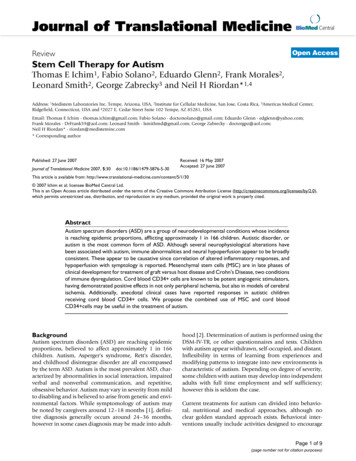
Journal of Translational Medicine 2008, 6:21For immunofluorescense of DCs following electroporation with TC-1 RNA, electroporated DCs were seeded onglass coverslips and cultured in RPMI in the presence of10% FBS and 10 ng/ml GM-CSF for two days. Cells werefixed with acetone and consecutively stained with antiHPV-16 E7 antibody (ED-17, Santa Cruz Biotechnologies) and anti-rabbit FITC (BD Pharmingen). Slides werecounterstained with DAPI. Images were acquired throughCool SNAP Pro color digital camera (Media Cybernetics,Carlsbad, CA). Ten different fields for each sample at 400 magnification were evaluated for cell counting.Immunoprecipitation and Western blotFor E7 immunoprecipitation and Western blot analysis,ID8-E6/7 and ID8 control cells were lysed in M-Per mammalian protein extraction kit (Pierce, Rockford, IL) plusprotease inhibitor cocktail (HALT Protease inhibitor kit,Pierce). Lysate concentration was determined by the Bradford assay (Biorad; Hercules, CA). Two hundred micrograms of extract was subjected to immunoprecipitationwith rabbit anti-HPV16 E7 (ED-17, Santa Cruz Biotechnologies). Immunoconjugates were collected on proteinA-agarose (Invitrogen), washed with lysis buffer, andresolved on 15% acrylamide gels. Proteins were transferred to polyvinylidene difluoride membranes (Immobilon-P, Millipore) and probed with mouse anti-E7antibody (clone 8C9, Zymed Labs Inc., San Fransisco,CA). Sheep anti-mouse IgG-HRP (NA93IV, AmershamBiosciences, Buckinhamshire, UK) was used as the secondary reagent. Proteins were visualized by enhancedchemiluminescence (Lumigen PS-3, Amersham) andexposure to Kodak X-Omat Blue film.RT-PCR and Real-Time Quantitative ReverseTranscription-PCRThe expression of E6 and E7 was demonstrated using RTPCR with the following primers: E6 forward primer (F),5'-AAA GCA GAC ATT TTA TGC ACC A-3'; E6 reverseprimer (R), 5'-TCA TGC AAT GTA GGT GTA TCT CC -3';E7 F 5'-CAC GTA GAG AAA CCC AGC TGT A -3'; E7 R 5'GTA CCC TCT TCC CCA TTG TT-3'. The RT portion of theRT-PCR was conducted using SuperScript reverse transcriptase (Invitrogen) at 50 C (10 min). The PCR cyclingwas conducted with Taq polymerase for both sequences at94 C (3 min), 50 C (1 min), and 72 C (1 min) for 40cycles.The AbiPrism 7700 Sequence Detection System and SYBRgreen I PCR kits (both Applied Biosystems, Foster City,CA) were used for Real-Time PCR as described previously[30]. The following primers were used: E6: F 5'-GAC TTTGCT TTT CGG GAT TTA TGC -3', R 5'-TCA CAC AAC GGTTTG TTG TAT TGC-3'; E7: F 5'-CTG GAC AAG CAG AACCGG ACA-3', R 5'-TGC TTT GTA CGC ACA CCG AA-3'.We normalized the cDNA load to mouse om/content/6/1/213-phosphate dehydrogenase (GAPDH) with primersGAPDH F 5'-CCT GCA CCA CCA ACT GCT TA-3' andGAPDH R 5'-CAT GAG TCC TTC CAC GAT ACC A-3'. Datawere expressed as relative units to GAPDH mRNA molecules. Molecules were considered to be present if morethan five copies of mRNA were detected for every 106 copies of GAPDH mRNA.Statistical analysisA two-tailed Student's t-test was applied to determine differences between two groups. For multiple comparisonswe performed ANOVA with post-analysis comparisons bythe Tukey-Kramer multiple comparisons test. Non-parametric studies were performed by using the Mann-Whitney U test. A value of p 0.05 was considered significant.Data are expressed as mean SD. Data was analyzed usingGraph Pad Instat software (GraphPad Software, Inc., SanDiego, CA).ResultsPreparation of DCs electroporated with TC-1 cell RNA(RNA-DCs)To test the efficacy of loading DCs with tumor antigen byRNA electroporation we used TC-1 cells, a mouse adenocarcinoma cell line generated by cotransfection of lungepithelial cells with HPV-16 E6 and E7 genes and H-Ras[31], which has been used to test E6 and E7-targetedtumor immunotherapy [32-34]. Reproducible amounts oftotal RNA ranging between 20–35 µg RNA/106 cells wereobtained from cultured TC-1 cells (Mean 27.05, SD 9.183, N 10 independent samples). High quality RNAwas procured (Figure 1A), with rRNA ratio (28S/18S)reproducibly greater than 2.00 (Figure 1B).To determine the best conditions for RNA electroporationin our system, bone marrow-derived DCs were electroporated with tumor cell RNA using different capacitance andvoltage settings and 25 µg RNA per 106 DCs, which represents RNA from approximately one tumor cell per DC.The reaction was performed in a total volume of 200 µL.As shown in Figure 1C, the highest expression of E7 antigen in live electroporated DCs, was obtained at 300 V and150 µF. These settings yielded 50% viability in electroporated DCs, as determined by flow cytometry PI exclusionanalysis (Figure 1D). Similar viability levels were obtainedin DCs electroporated in the absence of RNA (mock, notshown).To optimize DC electroporation, we electroporated DCswith different amounts of TC-1 RNA (5–50 µg/106 DCs)using the previously determined settings (300 V, 150 µF).E6-transcripts are longer than E7. As shown in Figure 1E,high E6 expression was observed only after electroporating 50 µg TC-1 RNA/106 DCs. Higher RNA amounts didnot result in increased E6 expression (not shown). Elec-Page 4 of 14(page number not for citation purposes)
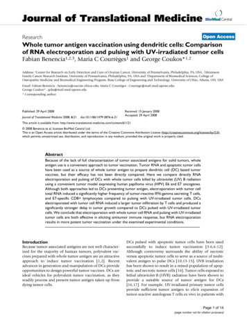
Journal of Translational Medicine 2008, 6/1/21BA28S18S28S18SL 1 2 3 45 6 7 8 9 10TC-1 RNADnon-electroporated300 V, 150 µFC34.60%26.05%300 V, 150 µF300 V, 300 µF81%450 V, 150 µF73%76%450 V, 150 µF300 V, 300 µF50%57.09%E7-FITCPIE300 V, 150 µF (5-50 µg TC-1RNA/10E6 DCs)24 hE6-FITCF0 µg5 µg10 µg25 µg50 µgE6-FITCE7-FITCGRNA300 V, 150 µF (50 µg TC-1 RNA/10E6 DCs)4 daysRNAE7-FITCRNAmockE7-FITCExpressionFigure 1 of tumor-associated HPV E6 and E7 antigens by bone marrow-derived DCs after RNA electroporationExpression of tumor-associated HPV E6 and E7 antigens by bone marrow-derived DCs after RNA electroporation. (A) Gellike image obtained from 10 independent samples of TC-1 RNA analyzed by the Agilent bioanalyzer. (B) Electropherogram ofsample 1 from A showing ribosomal RNA peaks. (C) Flow cytometry analysis of intracellular E7 expression in CD11c cells 24hours after electroporation with TC-1 RNA at different voltage and capacitance conditions. Electroporation was performedwith 25 µg TC-1 RNA/106 DCs (grey): mock electroporated (white). (D) Flow cytometry analysis of DC viability 24 hours afterelectroporation at different conditions of voltage and capacitance. Electroporation was performed with 25 µg TC-1 RNA/106DCs. X-axis reflects propidium iodide (PI) staining. (E) Flow cytometry analysis of intracellular E6 and E7 expression in CD11c cells 24 hours after electroporation with different amounts of TC-1 RNA at 300V and 150 µF. The experiment was repeatedtwo times with similar results. (F) Flow cytometry analysis of intracellular E6 and E7 expression in CD11c cells 4 days afterelectroporation with 50 µg of TC-1 RNA/106 DCs at 300V and 150 µF. RNA electroporated (white); mock electroporated(grey). (G) Immunofluorescence of DCs stained with anti-HPV E7 antibody 24 hours after electroporation with TC-1 cell RNAor mock electroporated. Cells were counterstained with DAPI. 200X magnification. All experiments were repeated twice withsimilar results.Page 5 of 14(page number not for citation purposes)
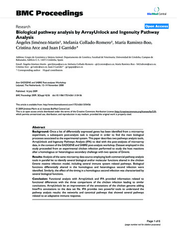
Journal of Translational Medicine 2008, 6/1/21troporation with 50 µg RNA/106 DCs also ensured highexpression of E7-antigen in DCs (Figure 1E). As shown inFigure 1F, high expression of E6 and E7 protein wasdetectable by flow cytometry for at least 4 days after RNAelectroporation with 50 µg of TC-1 RNA/106 DCs.Immunofluorescence staining confirmed the presence ofE7 protein in the cytoplasm of DCs 24 hours after electroporation with 50 µg TC-1 RNA/106 DCs (Figure 1G).cells were cocultured for 18 hours to allow the uptake oftumor cell material by DCs. Cells were then stained withantibody against CD11c, and CD11c DCs were analyzedfor PKH26 expression with flow cytometry. More than60% of DCs had taken up fluorescent tumor cells (Figure2D), compared with a background level of 1% in the control sample containing DCs and UV-irradiated cellsadmixed just before analysis (not shown).Preparation of DCs pulsed with UV irradiated TC-1 cells(UV-DCs)We tested various doses of ultraviolet-B (UVB) light andexposure times to identify UVB conditions that kill greaterthan 95% of TC-1 cells (not shown). Irradiation with1500 µW/cm2 UVB for 10 min induced apoptosis in TC-1cells as assessed by DNA fragmentation detectable byTUNEL assay (Figure 2A) and DNA laddering (Figure 2B),while by flow cytometry the majority of cells wereannexin-V positive or propidium iodide and annexin-Vdouble positive within 24 hours (Figure 2C).Maturation of RNA-DCs and UV-DCsWe assessed whether DCs prepared by RNA electroporation or pulsing with UV-irradiated tumor cells responddifferently to maturation stimuli such as TNF-α and LPS.Significant surface expression of CD86 and CD80 andMHC-II molecules was noted in DCs 48 hours post-electroporation with tumor cell RNA, as described above, inthe presence of TNF-α and LPS (Figure 3A). Similarly,immature DCs incubated with TC-1 cells exposed 24 hourearlier to lethal dose of UVB radiation upregulated CD80,CD86 and MHC-II 48 hours post-phagocytosis, in thepresence of TNF-α and LPS (Figure 3B).To verify the uptake of UV-irradiated cells by DCs, UVirradiated TC-1 cells were labeled with PKH26 fluorescentdye prior to pulsing of DCs. DCs and UV-irradiated tumorABTC-1 DNACUVB irradiated TC-1 cellsANNEXIN VcontrolDCD11cTC-1 cellsPIUVB irradiatedNext, we assessed whether DCs electroporated with tumorcell RNA (RNA-DCs) or pulsed with UV-irradiated tumor35%19% UVBirradiated40%PKH26Figure 2UVB-inducedapoptosis of TC-1 cells and maturation of TC-1 antigen-loaded DCsUVB-induced apoptosis of TC-1 cells and maturation of TC-1 antigen-loaded DCs. (A) TUNEL assay of UVB-treated and control TC-1 cells 24 hours post-irradiation. (B) DNA ladder assay of TC-1 cells 24 hours after irradiation with UVB light. (C)Flow cytometry analysis of annexin V in UVB-treated cells 24 hours post-irradiation. (D) Percentage of DCs that have engulfedtumor cells, as determined by flow cytometry. DCs were pulsed with cells killed by UVB. Apoptotic tumor cells were labeledwith PKH26 prior to pulsing. DCs were stained with antibody against CD11c.Page 6 of 14(page number not for citation purposes)
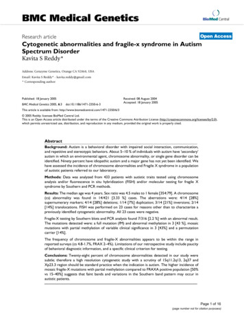
Journal of Translational Medicine 2008, m/content/6/1/21CD80UVBCD86MHC-IIallogeneic proliferationCD80MHC-IID20MIP 3 alphaMIP 3 betaChemotactic index181614121086420immature DCUV-ApoptoticElectroporatedcells)Figure3(A) Expressionof maturation markers in E7 CD11c gated cells 72 hours after electroporation with TC-1 RNA (50 µg/106(A) Expression of maturation markers in E7 CD11c gated cells 72 hours after electroporation with TC-1 RNA (50 µg/106cells). LPS and TNF-α were added to the culture medium 24 hours after electroporation. (white): specific antibody; (grey): isotype control. The experiment was repeated two times with similar results. (B) Expression of markers in CD11c gated cells 72hours after phagocytosis of UVB-irradiated TC-1 cells. LPS and TNF-α were added to the culture medium after 24 hours ofcoincubation. (white): specific antibody; (grey): isotype control. (C) Proliferation of allogeneic BALB/c splenocytes induced byC57BL/6 DCs electroporated with TC-1 cell RNA (RNA) or pulsed with UV-irradiated TC-1 cells (UVB). DCs were treatedwith TNF-α and LPS. Data were obtained 48 hours after stimulation of splenocytes and are expressed as the mean SEM oftwo experiments with triplicate observations per experiment. (D) Migration of DCs towards MIP-3α or MIP-3β 72 hours afterelectroporation with TC-1 RNA (RNA) or pulsing with UV-irradiated TC-1 cells (UVB). LPS and TNF-α were added to theculture medium after 24 hours of coincubation. Chemotactic index is defined as the fold increase in cell migration caused bythe chemotactic factors. Data are expressed as the mean SD of two experiments with quadruplicate observations per experiment.cells (UV-DCs) and matured with TNF-α and LPS differ invitro in their ability to induce proliferation of allogeneiclymphocytes or to migrate towards lymphoid organchemokines. Immature DCs were incubated for 48 withTC-1 cells exposed 24 hours earlier to lethal UVB radiation or were electroporated with tumor cell RNA, asdescribed above. To minimize differences in the amountof original tumor antigen used with both loading proce-dures, DCs were incubated with UVB-irradiated cells at1:2 ratio (DC, tumor cells) or electroporated with anequivalent amount of total RNA (50 µg RNA corresponding to 2 106 tumor cells per 106 DCs). RNA-DCs stimulated proliferation of allogeneic spleen lymphocytes in asimilar manner to UV-DCs. As expected, unpulsed immature DCs did not stimulate allogeneic reaction (Figure3C).Page 7 of 14(page number not for citation purposes)
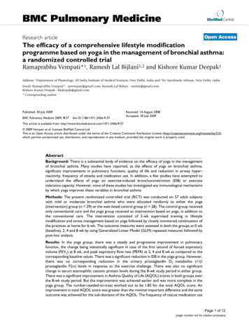
Journal of Translational Medicine 2008, 6/1/21A switch in chemokine receptors is a hallmark of DC maturation. Among others, this entails upregulation of CCR7and downregulation of CCR6 [35]. RNA-DCs and UVDCs matured with TNF-α and LPS exhibited similarmigratory properties. As shown in Figure 3D, both DCslost the ability to migrate towards macrophage inflammatory protein 3-alpha (MIP3-α), the ligand for CCR6, andacquired the ability to migrate towards MIP-3β, the ligandfor CCR7.Development of additional tumor targets expressing HPV16 E6 and E7To further compare tumor vaccination with RNA-DCs andUV-DCs, we engineered an additional tumor cell lineexpressing HPV E6 and E7. We used the syngeneic C57BL/6 murine ovarian cancer cell line ID8. ID8 cells transduced with the retroviral vector LXSN16E6E7, whichencodes the HPV16 E6 and E7 genes, as well as the neomycin phosphotransferase gene (ID8-E6/7) stablyexpressed E6 and E7 mRNAs under genetecin pressure,BTC-1100000Relative expressionID8ID E6/8 7ID8EID 6/78AID8-E6/7ID8100001000100101E6E7HPV antigensME6 E6 E7 E7primersM E6 E7primersDCIntracellular stainingEE6MID8 ID8E6/7E6-FITCE7-FITCMHC-IFigure 4 of HPV-16 E6 and E7 antigens in retrovirus-transduced ID8 cellsExpressionExpression of HPV-16 E6 and E7 antigens in retrovirus-transduced ID8 cells. (A) RT-PCR analysis for HPV-16 E6 and E7 transcripts in ID8 cells transduced with HPV-16 E6 and E7 genes; ID8 cells transduced with empty vector (negative control); andTC-1 cells (positive control). M: molecular markers. (B) Real-time quantitative PCR analysis of E6 and E7 transcripts in ID8cells transduced with HPV-16 E6 and E7 genes and ID8 cells carrying empty vector. (C) Western blot analysis of cell lysateshowing presence of E6 protein in samples obtained from ID8 E6/7 cells but not in ID8 cells carrying empty vector. M: molecular markers. (D) Flow cytometry analysis of intracellular E6 and E7 proteins in ID8 cells transduced with HPV-16 E6 and E7genes. (grey): ID8 E6/7 cells; (white): ID8 cells transduced with empty vector, (dotted line): isotype control. (E) Expression ofMHC-I by ID8-E6/7 cells. (grey): ID8 E6/7 cells; (white): isotype control.Page 8 of 14(page number not for citation purposes)
Journal of Translational Medicine 2008, 6:21compared to ID8 cells transduced with the empty vector(ID8) (Figure 4A and 4B). Immunoprecipitation followedby Western blot analysis revealed the presence of E6 protein in ID8-E6/7 cells but not in ID8 cells transduced withempty vector (Figure 4C). Moreover, intracellular stainingof ID
Roche, Indianapolis, IN) in a 5% CO 2 atmosphere at 37 C. An ID8 cell line expressing the HPV16 E6 and E7 antigens was generated by transducing ID8 cells with the retroviral vector LXSN16E6E7 (American Type Culture Collection, Rockville, MD, donated by Dr. D. Galloway), which encodes the HPV16 E6 and E7 genes, as well as the