Transcription
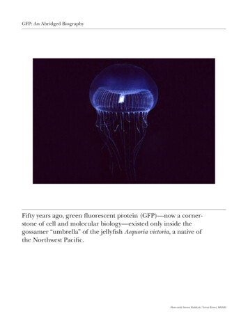
GFP: LIGHTING UP LIFENobel Lecture, December 8, 2008byMartin ChalfieDepartment of Biological Sciences, 1012 Fairchild, Columbia University, NewYork, NY 10027, USA.“You can observe a lot by watching.”Yogi Berra“My companions and I then witnessed a curious spectacle The Nautilusfloated in the midst of truly living light[,] an infinite agglomeration ofcolored globules of diaphanous jelly ”Twenty Thousand Leagues Under the Sea – Jules Verne“Now it is such a bizarrely improbable coincidence that anything so mind-bogglinglyuseful could have evolved purely by chance that some thinkers have chosen to see it as afinal and clinching proof of the nonexistence of God.”The Hitchhiker’s Guide to the Galaxy – Douglas AdamsI want to thank the Royal Swedish Academy of Sciences and the NobelFoundation for this amazing and surprising honor. At first I wondered why I,a biologist and a person with less than enviable college grades in Chemistry,had been selected. Then I realized that this prize had actually been given tothe GFP molecule, and I am one of its assistants. Thank you for letting me bepart of the celebration of a wonderful tool for visualizing life.Scientific inquiry starts with observation. The more one can see, themore one can investigate. Indeed, we often mark our progress in science byimprovements in imaging. The first Nobel Prize, the Physics prize of 1901,was an imaging prize, given to Wilhelm Röntgen for his discovery of X-raysand their astonishing ability to allow the noninvasive viewing of the humanskeleton. A few years later the Nobel Prize in Physiology or Medicine wasawarded for the development of silver nitrate staining to visualize nerve cellsby Camillo Golgi and its improvement and use by Santiago Ramón y Cajal todemonstrate the cellular nature of the nervous system. This research laid thegroundwork of modern neurobiology.Over the years several other imaging techniques and their developershave been honored with the Nobel Prizes including x-ray crystallography(William and Lawrence Bragg, Physics, 1915), the ultramicroscope (RichardZsigmondy, Chemistry, 1925), nuclear magnetic resonance (Felix Bloch andE. M. Purcell, Physics, 1952), the phase contrast microscope (Frits Zernike,150
Physics, 1953), large-array radio telescopes (Martin Ryle, Physics, 1974), theelectron microscope (Ernst Ruska, Physics, 1986), the scanning tunnelingmicroscope (Gerd Binnig and Heinrich Rohrer, Physics, 1986), computer assisted tomography (Allan M. Cormack and Godfrey N. Hounsfield, Physiologyor Medicine, 1979), and, most recently, magnetic resonance imaging (PaulC. Lauterbur and Sir Peter Mansfield, Physiology or Medicine, 2003).My road to imaging was not direct. I had been interested in science fromwhen I was very young, but after a disastrous summer lab experience in whichevery experiment I tried failed, I decided on graduating from college that Iwas not cut out to be a scientist. Instead I did a series of somewhat randomjobs including teaching high school chemistry. During the summer breakfrom teaching, I tried laboratory research one more time, working with JoséZadunaisky at Yale Medical School (Figure 1). The successful experimentsof that summer and his support gave me confidence to apply to graduateschool, and I entered the Physiology Department at Harvard in 1972 whereI did my thesis with Bob Perlman. Bob and I had a wonderful relationship,which continues to this day. He is one of the warmest, kindest, and smartestpeople I know, and a great person to talk over ideas with.Figure 1. Influences on my career. The success I had working for José Zadunaisky convincedme that maybe I could be a scientist. Bob Perlman was an outstanding Ph.D. advisor whoalways had time to listen to my (often crazy) ideas. Working with Sydney Brenner, JohnSulston, and Bob Horvitz during my postdoctoral years started me on my continuedresearch with C. elegans. I am convinced that working with this transparent animal was amajor reason why I was excited about the possibilities of GFP as a biological marker. Photoof José Zadunaisky from Bronner, F. (2006) Obituary: José A. Zadunaisky. J. Exp. Zool. 305A:103; reprinted with permission of John Wiley & Sons, Inc. Photo credits for the remainingphotos are M. Chalfie for Bob Perlman, the Nobel Foundation for Sydney Brenner, JohnSulston, and Bob Horvitz, and Adam Antebi for C. elegans.151
My current studies, however, started when I was accepted as a postdoctoralfellow by Sydney Brenner at the MRC Laboratory of Molecular Biology andbegan working on the nematode Caenorhabditis elegans. In 2002 Sydney, BobHorvitz, and John Sulston won the Nobel Prize in Physiology or Medicinefor their work on C. elegans. All three shaped the direction of my research.Sydney gave me the opportunity to work with him and an amazingly giftedgroup of scientists, Bob, a friend since high school, gave me several crucialpieces of advice, collaborated on several projects, and served as an exampleof what one can achieve in science (I am still following in his footsteps), andJohn, with whom I collaborated the most and who taught me most about howto act honorably as a scientist, started me on the project that still occupiesmost of my time: the study of mechanosensation.My colleagues and I often call their Nobel Prize the first worm prize. Thesecond went in 2006 to Andy Fire and Craig Mello for their discovery ofRNA interference. I consider this year’s Prize to be the third worm prize,because if I had not worked on C. elegans and constantly told people that oneof its advantages was that it was transparent, I am convinced I would haveignored GFP when I first heard of it. These three prizes speak to the geniusof Sydney Brenner in choosing and developing a new organism for biologicalresearch.The year before I learned about GFP, my lab had begun looking at geneexpression in the C. elegans nervous system. We were studying the differentiation and function of nerve cells needed for mechanosensation.Mechanosensors respond to physical perturbation; they underlie many ofour senses, including touch, hearing, and balance. These senses are poorlyunderstood; in particular the transduction molecules, the molecules thatdetect the mechanical signal, are virtually unknown. The genetic studiesthat I had done with John Sulston were directed, in part, at discovering suchtransduction molecules. We thought that by obtaining mutants that were defective in touch, which was sensed by six cells in the animal, we could identifygenes that were needed both for the production and differentiation of theseparticular cells and for transduction. In the late 1980’s my lab began cloningseveral touch sensitivity genes and testing whether they were expressed in theanimal’s touch receptor neurons. At this time three general methods wereused to look at gene and protein expression. The first was the use of labeledantibodies, whose specificity created outstanding protein-specific markers.The second was the use of B-galactosidase from the Escherichia coli lacZ gene,which could be expressed as transcriptional and translation fusions and visualized by the cleavage and subsequent oxidation of X-gal to an insolubleblue product. The third was in situ hybridization to mRNA. We used all threemethods to monitor gene expression (Figure 2).152
ACDFigure 2. Gene expression methods used before (and after) GFP. A. Positions of the sixtouch receptor neurons in C. elegans. B. Antibody staining to the MEC-7 B-tubulin (takenfrom Savage et al., 1994). C. B-galactosidase expression of a transcriptional fusion for mec-9(taken from Du et al., 1996). D. In situ hybridization to mec-7 mRNA (Shohei Mitani).All three methods had considerable limitations; they required extensive andtime-consuming tissue preparation. The animals had to be fixed and thenpermeabilized so either the antibody, the X-gal substrate, or the DNA probecould enter the tissue. This preparation, which needed to be done with eachbatch of animals, meant that we could only look at dead tissues, giving us astatic picture of expression. If we wanted to understand changes during development, we had to compare images from many different individuals.I first had the idea to put GFP into worms a little after noon on Tuesday,April 25, 1989. My department has a lunchtime seminar series on Tuesdaysfor those of us interested in neurobiology, and the speaker that day was PaulBrehm, who at the time was at Tufts University. He began his talk with adescription of light production by jellyfish and similar animals, work that Isubsequently learned had been begun by my co-Laureate Osamu Shimomura(Shimomura et al., 1962; Johnston et al., 1962) and then by Jim Morin andWoody Hastings (Morin and Hastings, 1971a, b). He first spoke about aequorin, which I had heard of as a calcium indicator. But then he talkedabout a protein new to me that fluoresced and allowed the jellyfish to produce green light instead of blue. Being primed from years of talking aboutthe transparency of C. elegans and having just seen the work involved usingantibodies and lacZ fusions, I immediately started to fantasize about how153
this Green Fluorescent Protein, GFP, could be used as a biological marker.I must admit that I didn’t pay any attention to the rest of the seminar; I wastoo excited.Figure 3. Notes I took as I was tracking down the people who were working on GFP.I recently found my notes from that time and they have allowed me to reconstruct what happened next (Figure 3). I spent the next day on the phone,learning about GFP and eventually talking with Douglas Prasher (Figure 4),who was at the Woods Hole Oceanographic Institute and who was cloningthe cDNA for gfp. We had a wonderful conversation, found we had similarideas about what to do with GFP, and decided to collaborate – as soon asDouglas had finished cloning the gene.154
Figure 4. The GFP team. Douglas Prasher cloned the gfp cDNA, Ghia Euskirchen expressedthe cDNA in E. coli, Yuan Tu expressed the cDNA in C. elegans, and Bill Ward comparedthe excitation and emission spectra of the native and recombinant GFP. (Photo credits:Douglas Prasher and Bill Ward for their own photos, M. Chalfie for the photos of Ghiaand Yuan.)During that day I learned that GFP had several features that made it a very attractive candidate for a biological marker. 1) It was a relatively small proteinof only 238 amino acids. 2) It was active as a monomer. 3) It could be excitedby ultraviolet or blue light. 4) It was a stable protein that had high quantumefficiency and did not photobleach easily. And 5) the active protein did notneed a cofactor or other small molecule to fluoresce.GFP had one feature, however, that might make it unsuitable for expression in organisms other than the jellyfish: the chromophone was formed bythe cyclization of the peptide backbone between Ser65 and Tyr66 (Figure 5).No one knew how this cyclization occurred, but the prevailing hypothesiswas that one or more converting enzymes were needed to change what wasreferred to as apoGFP to the fluorescent product (Cody et al., 1993). If otherproteins were needed, GFP would not be a very good marker.155
Figure 5. Formation of the GFP fluorophore. A. The primary amino acid sequence of GFP.B. Sequence after cyclization. Data from Cody et al. (1993).The next important event in this story was my marriage in late 1989 to TulleHazelrigg, a scientist who lived and worked 2000 miles away from New YorkCity at the University of Utah. Given the distance, I felt fortunate to be eligiblefor a sabbatical leave, which I took to work in her lab. Unfortunately, whileI was away, Douglas finished cloning the gfp cDNA, tried to contact me, butfailed to do so. He concluded that I had dropped out of science. For my part,having not heard from Douglas, I imagined that he had not found the cDNA.We remained in mutual ignorance until September, 1992. At that time oneof the new graduate students, Ghia Euskirchen (Figure 4), decided to do arotation project with me. I was particularly happy about her joining the lab,because she had just finished a Masters degree in our Engineering Schoolworking on fluorescence. I told her about my idea of using a fluorescentprotein to mark cells, and then bemoaned the fact that I had not heard fromDouglas. But when we searched for “fluorescent protein” in the Medline database that the University had just installed on our computers, the first paperwe saw was Douglas’ February, 1992 paper describing the isolation of the gfpcDNA (Prasher et al., 1992). We ran down to the library, found the journalwith the paper, discovered that the article included his phone number, and156
rushed back to my office to call him. After we cleared up our misconceptionsof each other’s careers, Douglas and I renewed our collaboration.Six days later Douglas sent the DNA to us. At this point, we had two choicesas to how to do the experiment. Douglas had cloned the cDNA as an EcoRIfragment into a lambda vector. We could either obtain the fragment by cutting it out of the vector with the same restriction enzyme but this would giveus additional non-coding jellyfish DNA, or we amplify only the coding sequence using the polymerase chain reaction (PCR), which was risky becauseit tended, at that time, to introduce base changes. I decided we should usethe latter strategy, a decision that turned out to be fortunate, since we latterlearned that other labs using the restriction enzyme strategy failed to getfluorescence. Presumably, the extraneous jellyfish DNA interfered with theexpression. Given the prevailing assumption that GFP needed one or moreconverting enzymes to fluoresce, the failure of bacteria to fluoresce could beinterpreted as a need for other jellyfish components.Figure 6. The first expression of GFP in heterologous organisms. A. The page in GhiaEuskirchen’s laboratory notebook where she noted that E. coli expressing GFP fluoresced.The microscope she used was not in our laboratory. B. A picture of those first fluorescingbacteria taken by Ghia. C. GFP expressed in the C. elegans touch receptor neurons fromChalfie et al. (1994). Reprinted with permission from AAAS.In any event, one month after receiving the DNA from Douglas, Ghia had E.coli that fluoresced green (Figure 6), although we had to use the microscopein her previous lab to see them. We were ecstatic. No other protein from thejellyfish was needed to convert the protein to the fluorescent form. Ghia tookseveral pictures of the bacteria and I quietly started to show them to people.I couldn’t contain my excitement. Soon afterward Ghia left my lab to doanother lab rotation, and I asked Yuan Tu (Figure 4), my technician at the157
time, to put GFP into C. elegans. Again the experiment succeeded, and for thefirst time we had GFP expressed in the touch receptor neurons of C. elegans(Figure 6). Bill Ward (Figure 4), a biochemist who had studied GFP for several years, then joined the project and showed that the protein produced inE. coli had the same optical properties as the native protein (Figure 7).Figure 7. Excitation (left) and emission (right) spectra for native (dotted line) and recombinant (solid line) GFP (from Chalfie et al., 1994). Reprinted with permission from AAAS.We had one problem doing these experiments, as indicated by that note inGhia’s lab notebook that the bacteria were viewed in her previous laboratory:we did not have a working fluorescence microscope. This problem continuedto plague us. I solved this problem by using the departmental confocal microscope and when I could not use that, by asking microscope sales representatives to bring demonstration microscopes to our lab, so that I could test thembefore purchasing them. In reality we were using the microscopes to do theexperiments.From the late 1970s through the 1990s C. elegans researchers had a tradition of notifying each other of research progress before publication, so thefirst written description of our work with GFP was in the October, 1993 issueof the C. elegans newsletter, The Worm Breeders Gazette (Chalfie et al., 1993). Thisarticle started the flood of requests for the GFP vectors. Approximately 50people asked for them before our official publication in Science in February,1994 (Chalfie et al., 1994).Publication, however, was not without its difficulties. The first problem wasthe title. When we submitted the paper for the editors’ consideration, thetitle proclaimed that we had found “A New Marker for Gene Expression.”The editors told us that we had to change the title by removing that word“new” because every paper published in Science reported novel results. Wewere also asked to make the title more descriptive of the results. Partly in an158
noyance, I changed the title to a much longer one for the manuscript sentto the reviewers (“The Aequorea victoria Green Fluorescent Protein NeedsNo Exogenously-Added Component to Produce a Fluorescent Product inProkaryotic and Eukaryotic Cells”). After the paper was accepted, however,the copy editor asked us to shorten the title. We submitted a new title thatwas almost identical to the original (“Green Fluorescent Protein as a Markerfor Gene Expression”), and it was accepted.We had a second problem, this time with the cover art. I had sent in apicture that I was quite proud of, since it showed a neuronal growth cone ina living animal. The art director, however, informed me that green was thecolor most difficult to reproduce on the cover, and asked if the color couldbe changed. Luckily, I convinced her that a color change in this instancewould not be appropriate (Figure 8).Figure 8. The cover of Science for February 11, 1994 showing GFP in C. elegans neurons.From Science volume 263, number 5148. Reprinted with permission from AAAS.The third problem was that I had difficulty with one of the people who hadalready used GFP and whose unpublished data I had wanted to cite. Mostpeople were happy to have their results discussed, but this person put specialconditions on our use of her data (Figure 9). The fact that she was my wifemay have had something to do with the added requirements. She is shownwith our daughter Sarah, who is prize enough for anyone. Both have mademe a better person and a very happy one.159
Figure 9. Conditions set by my wife, Tulle Hazelrigg, if I wanted to cite her unpublishedwork on GFP in my paper. We still debate whether these conditions were actually met. Sheis shown with our daughter Sarah (Photo credit: Roger Tsien).The work that Tulle finally allowed me to cite was actually the next reallyimportant advance in the use of GFP because she and her graduate studentShengxian Wang made the first protein fusion with GFP, showed that it couldfunctionally replace the original protein, and demonstrated that it couldbe used to show where in the cell the normal protein resided (Wang andHazelrigg, 1994). Typical of her modesty, Tulle didn’t use the words “greenfluorescent protein” or “GFP” in her title.These papers demonstrated the usefulness of GFP as a biological markerfor both gene expression and protein localization. And GFP had severaldistinct advantages over past markers. First, GFP, like β-galactosidase, washeritable. Because organisms could be transformed with DNA encodingGFP, strains could be established that could be studied at latter times. Thisproperty not only allowed repeated observations without extensive tissuepreparation, but also the strains, with particular cells or proteins labeledcould be used for a variety of studies. Second, visualizing GFP was essentiallynon-invasive; the protein could be detected by simply shining blue light onto the specimen. Third, GFP was a relative small and inert molecule that didnot seem to interfere with the biological processes that were being studied.Moreover the active protein was a monomer, which allowed it to diffuse read160
ily throughout cells, particularly nerve cells, and outline their entire shape.In contrast, β-galactosidase monomers are four times larger than GFP monomers and enzymatic activity requires the formation of a tetramer, makingdiffusion difficult. Fourth, the fluorescence from GFP could be observed inliving organisms, allowing a dynamic view of biological events. In addition,biological activities could be monitored for proteins outside as well as insidethe cell (a property not shared by β-galactosidase).Although native GFP was very useful for a variety of experiments, for itto be really established as a useful tool for biological studies, its propertiesneeded to be improved. The person who started improving GFP and whocontinued to lead its development was my co-Laureate Roger Tsien. The firstthings he and his laboratory did were to devise ways to change the emissioncolor and to greatly enhance the fluorescence output of the protein whenirradiated by blue light. But these and the other inventive adaptations thathis laboratory produced are the subjects of his talk, and I want to turn to howresearchers reacted to our paper.After the publication of our paper, people requested and we sent out approximately 1500 sets of the GFP vectors we had produced before turningover the distribution to others. Two aspects of the early requests for GFPstruck me as interesting. First, more often than not an investigator asking forthe GFP vectors would say that he or she had heard about GFP from one oftheir graduate students or postdocs, an indication that these people were thereal drivers of innovation in the laboratories. Second, several people wouldstart their requests by asking if I knew if GFP had been used in their favoriteorganism. I expected that they asked because they wanted to be first to usethis method, so I was surprised that when I said “no,” that some would saythey would wait until someone else had worked out the method. I am stillsomewhat dismayed by this reaction, although I suspect it does mean that wehave fewer real competitors than I would have assumed.In any event GFP was soon put into a staggering array of organisms from allthree domains of life: archaea, bacteria, and eukarya (Figure 10). Moreover,a search of PubMed using the terms GFP or green fluorescent protein netsover 30,000 publications, a number that has been increasing exponentiallysince 1994 and is undoubtedly a lower bound. GFP has also entered the artworld with Eduardo Kac’s GFP bunny Alba and even the movies as evidentby the opening credits of the film Hulk by the director Ang Lee that wasreleased in 2003. The implication is that the Hulk is the first human GFPtransgenic.161
Figure 10. A gallery of GFP images. Photo credits by columns left to right: C. elegans(John Kratz), Drosophila (Ansgar Klebes, Freie Universitaet, Berlin), Alba the GFP bunny(Eduardo Kac), canola [M.D. Halfhill (St. Ambrose University) and H.A. Richards,R.J. Millwood, and C.N. Stewart, Jr. (University of Tennessee)], mice (Ralph Brinster,University of Pennsylvania), zebrafish (Brant Weinstein, NIH), cultured HeLa cells (JerryKaplan and Michael Vaughn, University of Utah), Drosophila embryonic cells (JenniferLippincott-Schwartz, NIH), Arabidopsis thaliana hypocotyl cells (David Ehrhardt, CarnegieInstitution of Washington), mouse Purkinje cell (National Center for Microscopy andImaging Research, University of California, San Diego).After publishing our GFP paper, I went back to the work on mechanosensation, thus turning from a developer to a user of GFP. For the next fewparagraphs I want to describe how we use GFP in our research to provideexamples of the many ways this protein can aid scientific discovery.First, of course, we use GFP in transcriptional fusions to characterize theexpression pattern of genes (Figure 6). Second, we used GFP translationalfusions to examine both gene expression and protein localization (Figure11). With the advent of different colored fluorescent proteins, we could alsotest for the co-expression of different genes. By studying mutants that aredefective in touch, we have found that touch sensitivity in C. elegans requiresa channel complex of at least four proteins and specialized extracellular matrix and microtubules. Using a MEC-4::GFP translational fusion, we showedthat the channel complex is localized to discrete spots along the length ofthe neuronal process (Figure 11D).162
Figure 11. Uses of fluorescent protein translational fusions. A. Diagram of the six touchreceptor neurons (green). B. The six touch receptor neurons are completely filled witha MEC-17::GFP fusion (taken from Zhang et al., 2002). C. GFP protein fusions for thetranscription factors EGL-44 and EGL-46 both localize to the nucleus of an FLP neuron (JiWu cited in Wu et al., 2001). D. Localization of MEC-4::YFP to puncta along the length of atouch receptor neuron process (taken from Chelur et al., 2002).Once cells have been labeled with GFP they can be put to a wide variety ofuses. Because of the strong genetics available in C. elegans, we have used suchanimals as the basis of screens for mutants defective in several aspects ofcellular development. For example, mutating animals in which expressionof GFP has labeled the entirety of the touch receptor neurons, we foundmutants with more or fewer fluorescing cells (allowing us to study genesneeded to control cell fate and number), mutants with cells in abnormallocations (allowing us to study genes that influence the positioning of cells),and mutants with cells (Figure 12A) with additional processes or abnormalbranches (allowing us to study genes involved in neuronal outgrowth andmorphology).163
Figure 12. Using GFP for gene discovery and characterization. A. Mutations in the genesunc-51 and mec-7 affect process outgrowth of touch receptor neurons labeled with MEC2::GFP (from Du and Chalfie, 2001). B. Mutations in mec-15 reduce synapses, visualizedby GFP::RAB-3, in touch receptor neurons (A. Bounoutas, M. Nonet, and M. Chalfie,unpublished data). C. Touch receptors neurons can be cultured from embryos, isolated by cell sorting, and used to identify cell-specific genes (e.g., Zhang et al., 2002). D.Electrophysiology of touch neurons from wild type and mutants is possible because thecells can be identified by their fluorescence (taken, in part, from O’Hagan et al., 2005).By using animals making GFP fused to proteins that localize to particular regions of the cells, one can obtain mutants with even more specific defects. Forexample, Michael Nonet at Washington University in St. Louis is interested ingenes needed for the formation of the chemical synapses that connect nervecells. He has created fusions of GFP with proteins found at synapses that labelthese connections (Figure 12B). These fusion proteins label the two regionsin touch receptor neurons that contain chemical synapses. We are collaborating with him to study a gene needed for touch neuron synapses.Mutageneses are not the only uses for animals with labeled cells. The animals can also be used to isolate and study the labeled cells. For example, wehave disrupted embryos and grown the embryonic cells in culture. The touchreceptor neurons can be detected among all the other cells because of theirfluorescence (Figure 12C). Remarkably, these cells have the same morphology they do within the animal; other labeled nerve cells look entirely different. The cells can be isolated using a fluorescence-activated cell sorter andused to identify messenger RNAs that are specific to the cells. Using thesemethods, we have identified 200 additional genes that are overexpressed inthese cells.Labeling the cells has also allowed us to study the electric properties of thetouch receptor neurons, and to my particular joy identify the first transduction molecules for a eukaryotic mechanical sense. C. elegans is a wonderful164
organism for a wide range of studies, but one of its disadvantages is thatits nerve cells are tiny. Fortunately, Miriam Goodman working with ShawnLockery at the University of Oregon developed a GFP-based method to makeelectrophysiological recording from C. elegans (Goodman et al., 1998), whichshe then adapted to the touch receptor neurons as a postdoctoral fellow inmy lab (Figure 12D). Using this method Bob O’Hagan, a graduate student inmy lab, Miriam, now at Stanford, and I found that specific mutations affecting the channel subunits in the touch neurons change the normal inwardcurrent elicited by touch to an outward current, a demonstration that thesechannel proteins are directly required for transduction.None of the results that I have mentioned would have been possible without the use of GFP in living animals. Despite my enthusiasm, however, GFPdoes have some limitations that have led us to devise modified proteins. Oneproblem is that GFP expression is dependent on gene regulatory elements.We have been fortunate in studying the touch neurons because we know ofgenes that are expressed only in these cells and we can use their regulatorysequences. But such cell-specific expression is not common in C. elegans orother organisms, so specific labeling with GFP can be difficult. We havetaken advantage, however, of a remarkable finding by Lynne Regan of YaleUniversity to overcome this difficulty.Lynne’s lab found that GFP split into two pieces could be reconstituted ifthe two parts were linked by interacting peptides (Ghosh et al., 2000; Figure12A). Her lab and others have used this property to study protein interactions. Two members of my lab (Shifang Zhang and Chuck Ma) realized thatthis two component GFP, which we named recGFP, could help solve thespecificity problem, if the two parts were expressed from different promoters. This two-component system should enable the labeling of 80% ratherthan 20% of the existing neuronal cell types in C. elegans (Figure 13).165
Figure 13. Use of recGFP to label the C. elegans FLP neurons (modified from Zhang et al.,2004).Another feature of GFP that can be both an advantage and a disadvantage isthat the protein is very stable. This feature
The GFP team. Douglas Prasher cloned the gfp cDNA, Ghia Euskirchen expressed the cDNA in E. coli, Yuan Tu expressed the cDNA in C. elegans, and Bill Ward compared the excitation and emission spectra of the native and recombinant GFP. (Photo credits: Douglas Prasher and Bill Ward for their own photos, M. Chalfie for the photos of Ghia and Yuan.)