Transcription
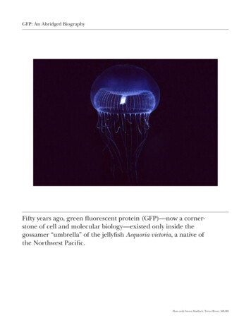
GFP: An Abridged BiographyFifty years ago, green fluorescent protein (GFP)—now a cornerstone of cell and molecular biology—existed only inside thegossamer “umbrella” of the jellyfish Aequoria victoria, a native ofthe Northwest Pacific.Photo credit: Steven Haddock/Trevor Rivers, MBARI
GFP: An Abridged BiographyMartin Chalfie (left), Osamu Shimomura (center), andRoger Y. Tsien (right) appear after delivering their NobelLectures at the Aula Magna at Stockholm University onDecember 8, 2008. The three were awarded the Nobel Prizein Chemistry for the discovery and development of GFP.Photo credit: Copyright Nobel Web AB 2008 Photo: Annalisa Andersson
GFP: An Abridged Biography“If there’s anyone who’s underappreciated, it’s Douglas Prasher,”Tsien has said. Prasher (center) was first to clone GFP andmade the DNA available for further study by Tsien and Chalfie(left and right, respectively). Though Prasher did not sharein receiving the Nobel, Tsien and Chalfie flew him and his wifeVirginia Eckenrode to Sweden for the Nobel ceremony.Photo credit: The Nobel Foundation 2008 Photographer: Mia Åkermark
GFP: An Abridged BiographyThis image shows the barrel-shaped structure of GFP.The chromophore, which is responsible for the protein’sfluorescent properties, is located in the middle of thebarrel, and is sometimes referred to as the “light in the can.”Photo credit: Courtesy of Tsien Lab
GFP: An Abridged BiographyThe use of fluorescent proteins has transformed biologicalresearch by allowing scientists to see when individual proteinsare made, and to watch where they go. In a mutant C. elegansworm (left), a GFP-labeled histone H2B protein fluorescesbright green under ultraviolet light.Photo credit: Jessica Vasale/Mello Lab
GFP: An Abridged BiographyUsing GFP, HHMI investigator Erol Fikrig and colleaguesat Yale University generated this laser scanning confocalfluorescent microscopy image of an Ixodes scapularis tick.Photo credit: Utpal Pal, Ruth R. Montgomery, Erol Fikrig
GFP: An Abridged BiographyA variety of new and old methods are used to see the componentsof this cultured human adenocarcinoma (HeLa) cell. Thenucleus is labeled with a small-molecule dye (blue), the Golgiapparatus is immunolabeled with quantum dots (yellow),microtubules are tagged with GFP (green), and the actincytoskeleton is labeled with a tetracysteine/biarsenical dye (red).Photo credit: From Science Vol 312, Issue 5771, 14 April 2006. Reprinted with permission from AAAS.
GFP: An Abridged BiographyRoger Tsien’s laboratory has created a vibrant palette offluorescent proteins with names such as plum, tangerine,cherry, and tomato.Photo credit: Courtesy of Roger Tsien Lab
GFP: An Abridged BiographyScientists at Harvard University have activated multiplefluorescent proteins in neurons, rendering the cells in adazzling array of colors.Photo credit: Livet, Sanes, Lichtman Center for Brain Science, Harvard University, Cambridge MA
GFP: An Abridged BiographyA petri dish marked with green fluorescent protein.Photo credit: Courtesy of Roger Tsien Lab
" If there's anyone who's underappreciated, it's Douglas Prasher," Tsien has said. Prasher (center) was first to clone GFP and made the DNA available for further study by Tsien and Chalfie (left and right, respectively). Though Prasher did not share in receiving the Nobel, Tsien and Chalfie flew him and his wife