Transcription
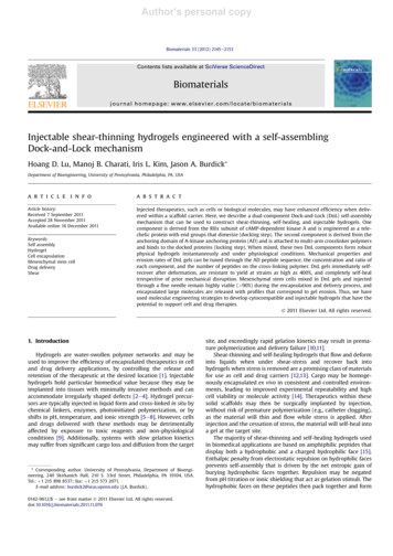
Author's personal copyBiomaterials 33 (2012) 2145e2153Contents lists available at SciVerse ScienceDirectBiomaterialsjournal homepage: www.elsevier.com/locate/biomaterialsInjectable shear-thinning hydrogels engineered with a self-assemblingDock-and-Lock mechanismHoang D. Lu, Manoj B. Charati, Iris L. Kim, Jason A. Burdick*Department of Bioengineering, University of Pennsylvania, Philadelphia, PA, USAa r t i c l e i n f oa b s t r a c tArticle history:Received 7 September 2011Accepted 28 November 2011Available online 16 December 2011Injected therapeutics, such as cells or biological molecules, may have enhanced efficiency when delivered within a scaffold carrier. Here, we describe a dual-component Dock-and-Lock (DnL) self-assemblymechanism that can be used to construct shear-thinning, self-healing, and injectable hydrogels. Onecomponent is derived from the RIIa subunit of cAMP-dependent kinase A and is engineered as a telechelic protein with end groups that dimerize (docking step). The second component is derived from theanchoring domain of A-kinase anchoring protein (AD) and is attached to multi-arm crosslinker polymersand binds to the docked proteins (locking step). When mixed, these two DnL components form robustphysical hydrogels instantaneously and under physiological conditions. Mechanical properties anderosion rates of DnL gels can be tuned through the AD peptide sequence, the concentration and ratio ofeach component, and the number of peptides on the cross-linking polymer. DnL gels immediately selfrecover after deformation, are resistant to yield at strains as high as 400%, and completely self-healirrespective of prior mechanical disruption. Mesenchymal stem cells mixed in DnL gels and injectedthrough a fine needle remain highly viable ( 90%) during the encapsulation and delivery process, andencapsulated large molecules are released with profiles that correspond to gel erosion. Thus, we haveused molecular engineering strategies to develop cytocompatible and injectable hydrogels that have thepotential to support cell and drug therapies.Ó 2011 Elsevier Ltd. All rights reserved.Keywords:Self assemblyHydrogelCell encapsulationMesenchymal stem cellDrug deliveryShear1. IntroductionHydrogels are water-swollen polymer networks and may beused to improve the efficiency of encapsulated therapeutics in celland drug delivery applications, by controlling the release andretention of the therapeutic at the desired location [1]. Injectablehydrogels hold particular biomedical value because they may beimplanted into tissues with minimally invasive methods and canaccommodate irregularly shaped defects [2e4]. Hydrogel precursors are typically injected in liquid form and cross-linked in situ bychemical linkers, enzymes, photoinitiated polymerization, or byshifts in pH, temperature, and ionic strength [5e8]. However, cellsand drugs delivered with these methods may be detrimentallyaffected by exposure to toxic reagents and non-physiologicalconditions [9]. Additionally, systems with slow gelation kineticsmay suffer from significant cargo loss and diffusion from the target* Corresponding author. University of Pennsylvania, Department of Bioengineering, 240 Skirkanich Hall, 210 S. 33rd Street, Philadelphia, PA 19104, USA.Tel.: þ1 215 898 8537; fax: þ1 215 573 2071.E-mail address: burdick2@seas.upenn.edu (J.A. Burdick).0142-9612/ e see front matter Ó 2011 Elsevier Ltd. All rights e, and exceedingly rapid gelation kinetics may result in premature polymerization and delivery failure [10,11].Shear-thinning and self-healing hydrogels that flow and deforminto liquids when under shear-stress and recover back intohydrogels when stress is removed are a promising class of materialsfor use as cell and drug carriers [12,13]. Cargo may be homogenously encapsulated ex vivo in consistent and controlled environments, leading to improved experimental repeatability and highcell viability or molecule activity [14]. Therapeutics within thesesolid scaffolds may then be surgically implanted by injection,without risk of premature polymerization (e.g., catheter clogging),as the material will thin and flow while stress is applied. Afterinjection and the cessation of stress, the material will self-heal intoa gel at the target site.The majority of shear-thinning and self-healing hydrogels usedin biomedical applications are based on amphiphilic peptides thatdisplay both a hydrophobic and a charged hydrophilic face [15].Enthalpic penalty from electrostatic repulsion on hydrophilic facesprevents self-assembly that is driven by the net entropic gain ofburying hydrophobic faces together. Repulsion may be negatedfrom pH titration or ionic shielding that act as gelation stimuli. Thehydrophobic faces on these peptides then pack together and form
Author's personal copy2146H.D. Lu et al. / Biomaterials 33 (2012) 2145e2153b-sheet sandwiches that grow into entangled fibrils, resulting ina solegel transition. Strain fractures the hydrogel into smallernetworks, resulting in shear-thinning behaviour, and fracturednetworks re-intertwine over time, resulting in self-healing behaviour [14]. Other well designed shear-thinning and self-healingsystems based on self-repulsive but mutually attractive peptides,multi-block amphiphilic peptides, oppositely charged particles, andprotein-ligand associations have been engineered; however, noindividual system is without some limitation (e.g., poor gelationkinetics, slow recovery) [10,16e24].To this end, we strived to develop a hydrogel system that (i) gelsquickly under physiological conditions, (ii) is shear-thinning, (iii)recovers rapidly after thinning, (iv) is based on strong and highlyspecific cross-linking interactions, and (v) has tunable mechanicaland erosion properties. The inherently specific interactionsbetween the docking and dimerization domain (DDD) of cAMPdependent protein kinase A (PKA) with the anchoring domain(AD) of A-kinase anchoring proteins (AKAP) were exploited todevelop such hydrogel materials. DDD-AKAP structural biology hasbeen well studied, which provides us with a strong molecularunderstanding of DDD-AD interactions and a library of associationdomains to engineer; however, these precise interactions have notbeen previously used to engineer hydrogels and materials [25e33].DDD forms a type-X four-helix bundle that binds to the a-helicaland amphipathic AD with strong 1e100 nM affinity [25,26]. Thenon-covalent associations between AD and DDD may be disruptedwith physical force, making hydrogels cross-linked by these interactions speculated to exhibit shear-thinning characteristics. Owingto the rapid nature of AD and DDD binding kinetics, such gels werealso expected to self-heal quickly after being thinned. Also, thebinding of AD to DDD may be initiated by simply mixing togetherthe two components, allowing gelation to proceed under constantphysiological conditions.Specifically, we designed a recombinant DDD (rDDD) polypeptide consisting of end-blocks derived from RIIa [2e45] PKAlinked together with a random-coil spacer mid-block containingRGD integrin binding sites [27,28]. The second component wasderived from the anchoring domain of A-kinase anchor protein 13[1247-1264] (AKAP-LBC) conjugated to the ends of a four or eightarm PEG (4aPEG-AD, 8aPEG-AD) [29]. When mixed together, crosslinking was achieved when multivalent AD domains “lock” ontorecombinant dimerization and “docking” domains, in this “Dockand-Lock” (DnL) self-assembling gelation mechanism. Here, wereport on the design of this DnL gel system, as well as characterization of the material tunability, shear-thinning and recoverybehavior, and cytocompatability.2. Material and methods2.1. PEG-AD synthesisAD peptide with the sequence H2N-(ESESLIEEAASRIVDAVIEQVKSESECGGG)-COOHwas synthesized on a Gly-OH functionalized 2-chlorotrityl chloride resin (Novabiochem San Diego, CA) using an automated solid phase peptide synthesizer (PS3,Protein Technologies, Inc, Tucson, AZ). The amino acid residues were activated forcoupling with HBTU in the presence of 0.4 M methyl morpholine in DMF anddeprotection was carried out in 20% piperidine in DMF for approximately 30 min.Standard cycles of 60 min were used for coupling. The peptide was cleaved from theresin in 95:2.5:2.5 Trifluoroacetic acid (TFA): Triisopropylsilane (TIPS): water for3e4 h, precipitated with the addition of excess ether, and pelleted by centrifugationat 4,000 g for 10 min. Ether was decanted and removed by vacuum, and the peptidewas used directly or frozen and lyophilized for storage. The identity of the peptidewas confirmed with MALDI-TOF mass spectroscopy (ABI PerSeptive Voyager, Framingham, MA; Philadelphia Wistar Proteomics Facility). The AD peptide wasconjugated to 10 kDa four arm PEG (4aPEG)-maleimide or 20 kDa eight arm PEG(8aPEG)-maleimide (Jenkem Technology USA) via Michael-type addition reactionbetween peptide cysteine and PEG maleimide in degassed buffer at pH 7.0 overnight,using a 3-fold molar excess of peptide to maleimide groups. The product was isolated by dialysis (6 kDa MWCO dialysis tubing) and lyophilization, and the extent ofthe reaction between PEG-maleimide and AD peptide was determined by 1H NMR(Bruker), through comparison and calibration of hydrogen shifts on L, V, and Iat w1 ppm with the hydrogen shifts on PEG at w3.75 ppm. A similar approach wasused to synthesize an alternate peptide, termed AD-0, with a sequence ofESESQIEYLAKQIVDNAIQQASESECGG.2.2. Cloning and expression of rDDDThe DNA sequence for rDDD, optimized for expression in E. Coli, was synthesizedand cloned in pJExpress411 expression plasmid by DNA2.0 Inc. (CA, USA). rDDD wasexpressed in lactose induced cultures with the Studier autoinduction method(Fig. S1) [34]. For expression of the polypeptide, the recombinant plasmid, rDDDpJExpress411, was transformed into rne131 E. coli expression strainBL21StarÔ(DE3) (Invitrogen). A single colony BL21StarÔ(DE3) containing rDDDpJExpress411 was inoculated in 5 ml sterile LB media containing 30 mg/ml kanamycin and grown overnight. 1 ml of the overnight culture was subsequently used toinoculate 500 ml of ZYP-5052 media for autoinduction of protein expression. The500 ml cultures were grown in a shaker at 37 C for 4e5 h until the OD600 reached0.8 and then grown at 25 C with constant shaking for an additional 24 h. Proteinexpression was monitored by sodium dodecyl sulfate polyacrylamide gel electrophoresis (SDS-PAGE) of samples with normalized OD600 and visualized via Coomassie blue staining. Cells were harvested by centrifugation (10,000 g for 15 min at4 C), stored at 80 C until use, and then thawed and lysed by resuspension in100 mM NaH2PO4, 10 mM Tris·Cl, 8 M urea, 1% triton X-100, pH 8.0 (buffer A) anddisruption on ice using an Ultra Sonic Dismembrator (10 mm tapered horn) for15 min with cycles of 30 s bursts and 30 s cooling time. The cell lysate was centrifuged at 16,000 g for 60 min and the supernatant was carefully decanted. Clarifiedlysate was passed through a Ni-Sepharose HisTrap FF column (GE Healthcare)equilibrated with buffer A. Unbound protein was washed with three columnvolumes of buffer A. Denaturant was removed by three column volume washes of50 mM sodium phosphate, 300 mM NaCl, 20 mM imidazole, pH 8.0 native buffer.Protein was eluted by five column washes of native buffer containing 500 mMimidazole. Purified protein was concentrated and buffer exchanged to deionizedwater via ultrafiltration across a 10 kDa NMWL regenerated cellulose membrane(Amicon Ultracel, Millipore). Protein was frozen and lyophilized to yield a whitefluffy solid. The molecular weight of the purified protein was confirmed via SDSPAGE and MALDI-TOF mass spectrometry.2.3. Gel formationStock PEG-AD and rDDD were separately dissolved in PBS or hMSC growthmedium (aMEM supplemented with 16.67% FBS, 1% L-glutamine, and 1% Penicillin/Streptomycin). To initiate gelation with the DnL self-assembly mechanism, the twocomponents were simply mixed together (Fig. 1). To provide insight into the DnL gelmolecular architecture, gels were flash frozen in liquid nitrogen and imaged withscanning electron microscopy (FEI Quanta 600 Environmental FE-SEM, Oregon).2.4. RheologyDynamic oscillatory time, frequency and strain sweeps were performed using anAR2000 stress-controlled rheometer (TA Instruments, New Castle, DE) with 20 mmdiameter cone and plate geometry, 59 min 42 s cone angle, and at a 27 mm gapdistance. To form the gels, stock suspensions of PEG-AD and rDDD were made in PBSat the desired pH. These solutions were then mixed together to form gels withspecified compositions and the gel was applied between the 2 plates of therheometer. The top plate was lowered to a 27 mm gap distance and excess gel wasscraped off. Care was taken to achieve a homogeneous distribution of gel within thetop and bottom plates of the rheometer. Dynamic oscillatory time sweeps werecollected at angular frequencies of 6.3 rad s 1 and 0.5% strain. An initial strainamplitude sweep was performed at 25 C at different frequencies to determine thelinear viscoelastic range for the gel. Rheological properties were examined byfrequency sweep experiments (u ¼ 0.1e100 rad s 1) at a fixed strain amplitude of0.5%. Experiments were repeated on 3 to 4 samples and representative data ispresented. For shear recovery experiments at 6.3 rad s 1, shear thinning wasinduced via application of 500% strain for 2 min. The strain was released to 0.5% for3 min to allow the gel to recover.2.5. Circular dichroism spectroscopyCircular dichroism (CD) spectra were obtained with a Jasco (Easton, MD) J-810Spectropolarimeter connected with a Peltier temperature controller. Samples wereanalyzed in a 1 mm quartz cuvette in pH 7.4 or 4.2 PBS at 25 C and 50 nm min 1,with 1 nm pitch, 1 nm bandwidth, and 4 s response time. AD and rDDD weremeasured at 0.5 mg mL 1, and mixed samples contained 0.125 mg mL 1 AD with0.5 mL 1 rDDD (yielding approximately 3 AD domains per 2 rDDD domains). A meanresidue weight of 106.86 was used to convert measurements into mean residueelipticities for mixed AD þ rDDD samples. Reported spectra are the average of threescans.
Author's personal copyH.D. Lu et al. / Biomaterials 33 (2012) 2145e21532147Fig. 1. Dock-and-Lock self-assembly mechanism. Docking domains dimerize and lock with the anchoring domain (left). Engineered anchoring domains and docking domains selfassemble to form dynamic, shear-thinning, self-healing hydrogels when mixed (right).2.6. Hydrogel erosionAcrylamide molds were fabricated with a 12 mm high and 15 mm diameter widecavity and containing a 6 mm high and 5 mm wide depression in the center. Moldswere washed with PBS and dried, and stock solutions of the crosslinker and rDDDwere mixed into the depression to form 40 mL gels of the desired composition.Solutions of 8aPEG-AD containing 0.1 wt% fluorescein isothiocyanate conjugateddextran (FITC-dex, MW 500,000; Sigma) were used for mock drug encapsulationand release studies. Gels were gently centrifuged at 500 g for 1 min to level gelsurfaces. Gels were covered in 1 mL PBS and allowed to erode for specified timeperiods, whereupon solutions above the intact gels were gently aspirated and storedat 20 C. Solutions were then replaced with 1 mL fresh PBS, and this process wasrepeated until gels were completely eroded. Care was taken to ensure sterilitythroughout. Protein concentrations in collected solutions were determined byabsorbance at 280 nm, and the FITC-dex concentration was determined by emissionfluorescence at 520 nm after excitation at 490 nm. FITC absorbance at 495 nm wasFITCtaken and used to correct protein absorbance (AFITC280 ¼ A495 /3) for samples containingboth dextran and eroded gel. Reported profiles are based on erosion averages ofthree gels for each specified composition.NMR (Fig. S3). rDDD was successfully expressed with a T7 promotersystem and was isolated to over 95% purity with yields as high as75 mg L 1 culture (representative MALDI-TOF spectra and SDSPAGE shown in Fig. S4).3.2. DnL hydrogel gelationNeither 4aPEG-AD nor 8aPEG-AD formed gels alone in pH 7.4PBS at 6 wt% (Fig. 2). However, these components formed hydrogelswith neutralization of the AD peptide surface charge, indicated bythe increase of material storage modulus (G0 ) over material lossmodulus (G00 ) at decreased pH (Fig. S5eS7). rDDD did not form gelsalone at 6 wt% or in the presence of 4 wt% unmodified 4aPEG (Fig. 2,S8). However, upon mixing rDDD with PEG-AD to a final 3 wt% 4/8aPEG-AD and 4.5 wt% rDDD composition (resulting in approximately 2 AD domains per one rDDD domain) gelation was observed2.7. Hydrogel cytocompatibilityhMSCs (Lonza) were expanded to passage 4 in standard hMSC growth medium.hMSCs were resuspended in hMSC growth media and mixed with PEG-AD dissolvedin the same media. The hMSC/PEG-AD solution was pipetted into a barrel ofa syringe, and rDDD in hMSC growth media was then pipetted into the same barreland gently mixed to induce gel formation. All encapsulation studies were performedwith 5 106 cells mL 1 in 3 wt% 8aPEG-AD - 4.5 wt% rDDD gels. Gels were extrudedthrough a 21-gauge needle directly into a 96-well plate or into a pre-formedcollagen gel (PurCol; Advanced Biomatrix), covered with excess hMSC media, andimaged either 24 h or 3 days later. To assess viability, constructs were rinsed withPBS, stained with a live/dead kit consisting of calcein AM and ethidium homodimer(Invitrogen), and visualized using confocal microscopy. Constructs were excited at488 nm and 543 nm wavelengths to visualize live and dead cells, respectively, and zstacks were taken through the depth of the gels to validate an even distribution ofcells throughout. Experiments were performed in triplicate and viability is reportedas the percentage of calcein stained cells compared to total cells.3. Results3.1. DnL component productionAD peptide was successfully synthesized (representativeMALDI-TOF spectra shown in Fig. S2) and conjugated to multi-armPEG. Molar conjugation efficiencies were similar for both four arm(4aPEG-AD, 0.76 peptides per 1 PEG arm) and eight arm PEG(8aPEG-AD, 0.81 peptides per 1 PEG arm), as determined by 1HFig. 2. Oscillatory time sweeps of DnL materials. Storage modulus (filled symbols) andloss modulus (empty symbols) of PEG-AD components alone, rDDD with unmodified4aPEG, and rDDD with PEG-AD (4aPEG-AD and 8aPEG-AD). Sweeps were performed at0.5% strain and 6.3 rad s 1.
Author's personal copy2148H.D. Lu et al. / Biomaterials 33 (2012) 2145e2153immediately ( 1 s). The pH of the DnL components and assembledgel was ensured to be at 7.4 from pH paper tests, to make suregelation was not an artifact of an unintended pH shift. rDDD alsoformed hydrogels when mixed with multi-arm PEG conjugated toother anchoring domains (AD-0) (Fig. S8), but studies with AD-0 arenot emphasized here because AD-0 exhibited limited solubility andtended to form weak gels by itself at high concentrations. Flashfrozen and lyophilized DnL gels exhibited a networked and porousstructure (Fig. S9).AD at pH 7.4 exhibited a combination of random-coil and ahelical secondary structures, shown by the double minima near208 nm and 222 nm (indicative of a-helical conformation) and netnegative elipticity below 200 nm (indicative of random-coilconformation) in the CD spectra (Fig. S10). At pH 4.2, AD wasmore helical, shown by the decreased negative elipticity between200 nm and 260 nm. rDDD also showed both random-coil and ahelical conformations. When mixed with AD (to approximately 1.4AD per 1 rDDD), the resultant CD spectra was not the simpleaddition of AD and rDDD spectra (especially evident at wavelengthsbelow 200 nm) and indicated an overall increase in helical content.3.3. DnL gel mechanics and erosionMixing PEG-AD and rDDD together resulted in the immediateformation of soft and elastic hydrogels, for both 4aPEG-AD and8aPEG-AD (Fig. 2). Gel moduli were highly dependent on oscillatorydeformation frequency, and were more elastic at high shear rates,but were more viscous at low shear rates (Fig. 3). DnL gels withvariable rheological properties could be constructed by mixing DnLcomponents to different final compositions. Increasing the concentration of DnL components, while keeping the ratio of DnL to PEG-AD(for both 4aPEG-AD and 8aPEG-AD) constant, led to an increase in G0and G00 with minimal effect on gel relaxation time and modulusfrequency dependence (Fig. 3A and B). Gels with plateau G0 as low asw10 Pa (5 wt%) and as high as w1000 Pa (10 wt%) were formed. Inreference to regimes where the AD to rDDD molar ratio is not close toone (2 wt% PEG-AD and 3 wt% rDDD; w2 AD per rDDD), increasingthe molar ratio of rDDD to PEG-AD while keeping PEG-AD wt%constant (2 wt% PEG-AD and 4.5 wt% rDDD; w1.33 AD per rDDD),increases plateau G0 and G00 , lowers the relaxation time of hydrogels,and rendered them vulnerable to deformation at lower shear rates(Fig. 3C and D). In reference to regimes where the AD to rDDD molarratio is near one (2 wt% PEG-AD and 4.5 wt% rDDD), further varyingthe gel composition to have one to one AD to rDDD ratio (2 wt% PEGAD and 6 wt% rDDD) had relatively little effect on moduli, relaxationtimes, and shear-thinning properties. Relaxation times of DnL gelswere highly variable, and ranged from around 0.1e1 s 1.Increasing the PEG-AD arm valency from four to eight led to anincrease in both hydrogel moduli and relaxation times (Fig. 3A andB). However, DnL gels made with 8aPEG-AD had yield strains(w100%) that were approximately half that of gels made with4aPEG-AD (w200%) (2 wt% PEG-AD and 3 wt% rDDD gels) (Fig. 4AFig. 3. Oscillatory frequency sweeps of DnL hydrogels. (A, C) storage modulus (filled symbols) and loss modulus (empty symbols) of DnL gels at various 4a-PEG-AD:rDDD. (B, D)modulus of analogous of DnL gels constructed with 8a-PEG-AD. Sweeps were performed at 0.5% strain.
Author's personal copyH.D. Lu et al. / Biomaterials 33 (2012) 2145e2153and B). Increasing the PEG-AD to rDDD molar ratio resulted ina proportionally increased yield strain for both 8aPEG-AD (w200%)and 4aPEG-AD (w400%) (2 wt% PEG-AD and 6 wt% rDDD gels)(Fig. 4). Regardless, all DnL gels still displayed extraordinary resistance to strain-induced deformation. When subjected to 500% strainover time, all DnL gels underwent gelesol transition and behaved asliquids, as expected from recorded strain sweep curves. However,upon lowering oscillatory strain from 500% to 0.5%, thinned DnL gelsrapidly underwent solegel transition and recovered back to theirinitial moduli immediately ( 6 s, the minimum time between eachdata point on the rheometer) (Fig. 5, S11). All DnL gels were capableof self-healing to their original state without showing any signs thatmechanical fidelity was compromised, irrespective of the number oftimes they were previously shear-thinned. Hydrogels formed withAD-0 were similarly shear-thinning and self-healing, but exhibitedhigher moduli and pseudoplasticity (Fig. S12-S14). A summary of gelrigidity for all formulations investigated is provided in supplementary information (Table S1).DnL gels eroded initially at relatively high rates, followed byslower and nearly linear rates (Fig. 6). Erosion rates of DnL gels werealso tunable. Increasing the locking domain molecular valencydecreased erosion rates; 3 wt% 4aPEG-AD e 4.5 wt% rDDD gelsrequired w5 days to erode 90%, while 3 wt% 8aPEG-AD e 4.5 wt%rDDD gels required w15 days to erode 90% (Fig. 6A). By adjusting2149the molar ratio of AD to rDDD domains closer to one, erosion ratesalso decreased (e.g., 2 wt% 8aPEG-AD e 4.5 wt% rDDD gelsrequired w 35 days to completely erode). However, the erosionrates of DnL gels were minimally affected by changing the total gelweight if AD to rDDD molar ratios remained constant (5 wt% totalcompared with 7.5 wt% total). The erosion rates were also onlyminimally affected when gels were supplemented with dextranand the release of encapsulated dextran correlated with the erosionrates of DnL gels (Fig. 6B).3.4. Cell encapsulation and delivery with DnL gelsPEG-AD and rDDD dissolved in hMSC growth medium(containing serum) behaved the same as when dissolved in PBS,and still formed hydrogels when mixed together. This was takenadvantage of to rapidly encapsulate cells under constant physiological conditions, by dissolving DnL components in hMSC containing media and mixing them together. Cells encapsulated in thismanner were then delivered by thinning the gel through a 21gauge needle from injection. Importantly, cells were highly viable(93.0 3.1%), and remained homogenously encapsulated afterinjection and gel-recovery (Fig. 7A). When delivered into a collagengel (acting as a mock collagenous tissue), hMSCs remained similarlyhighly viable (91 4.8%) and homogenously encapsulated evenFig. 4. Oscillatory strain sweeps of DnL hydrogels. (A, C) storage modulus (filled symbols) and loss modulus (empty symbols) of DnL gels at various 4a-PEG-AD:rDDD wt%, (B, D) andthat of gels at various 8a-PEG-AD:rDDD wt%. Sweeps were performed at 6.3 rad s 1.
Author's personal copy2150H.D. Lu et al. / Biomaterials 33 (2012) 2145e2153Fig. 5. DnL hydrogel deformation and recovery. Storage modulus (filled symbols) andloss modulus (empty symbols) of 3 wt% 8aPEG-AD e 4.5 wt% rDDD gels evolving overtime from repeated cycles of 3 min low 0.5% strain and 2 min high 500% strainoscillations at 6.3 rad s 1.modifier because it is also negatively charged, displays steric flexibility, and could be titrated to remove electrostatic repulsion ifdesired, resulting in the design of AD. This AD construct did notform gels in PBS at physiological pH (Fig. 2). If the full solubility ofPEG-AD was endowed due to our design of utilizing surface chargeto limit undesired peptide interactions, then these peptide interactions could be reinstated when surface charge was removed. Thisallows PEG-AD to be cross-linked by triggering ADeAD associationsby charge removal from pH titration. Indeed, we observed gelationof PEG-AD with decreasing pH, and sol to gel and gel to sol transitions were repeatedly cycled by cycling between acidic andneutral pH (Fig. S5-S6). CD spectra indicated that AD has a mixtureof a-helical and random-coil structure, as is expected from thecombination of the native helical structure of AD and its unstructured flanking modifications (Fig. S10). When titrated to lower pH,the peptide shows increased helicity, consistent with the postulation that helical bundles formed at lower pH. b-sheet forming selfassembling peptides whose gelation can be similarly triggered bycharge removal via pH titration have already been engineered;however, we have developed a pH sensitive gelation mechanismbased on modifying a naturally found a-helical motif, and thismodified peptide has a dual functionality of both triggerable self-after culture for three days (Fig. 7B). Note that detector gain andamplifier offset needed to be increased when visualizing cells incollagen gels to accommodate for the presence of collagen, whichled to the visual artifact of vertically stretched cells in constructedZ-stacks.4. DiscussionWe have constructed a DnL hydrogel system using molecularengineering to meet strict criteria for a self-assembling and shearthinning material that has potential utility for applications in drugand cell delivery. This two-component system was designedthrough the interactions of the RIIa subunit of cAMP-dependentkinase A and the anchoring domain of A-kinase anchoring protein(AKAP). Our original design consisted of AKAP-IS, an anchoringdomain engineered with bioinformatics methods to bind rDDDwith an exceptional 0.45 nM affinity [26]; however, AKAP-ISexhibited poor solubility in solution. The peptide was then modified with flanking ESES amino acids to improve solubility (termedAD-0); however, when conjugated to multi-arm PEG, this peptidetended to form weak gels at high concentrations at pH 7.4. Furtherstudies of gels formed with AD-0 were performed, (Fig. S8,S12eS14) but we sought to engineer DnL components that didnot gel in isolation at physiological conditions, but rather onlywhen the two components were mixed.Analysis of the AKAP-IS crystal structure revealed that AKAP-ISexhibits an a-helical structure with a neutral hydrophilic face anda hydrophobic face (Fig. S15) [25]. This amino acid arrangementmay lead to the stacking of hydrophobicehydrophobic andhydrophilicehydrophilic faces, which likely resulted in weakpeptide association, cross-linking, and weak gelation of PEG-AD-0.Substitution of hydrophobic amino acids into hydrophilic ones toimprove solubility was not possible, as such changes eliminatedbinding affinity to DDD [25]. Structural analysis of other anchoringdomains led us to AKAP-LBC, which has an a-helical structure witha negatively charged hydrophilic face that still bound to DDD with2.2 nM affinity (Fig. S15) [26]. We postulated that the net charge onthe peptide’s face would limit peptide stacking, and the addition ofthe same charged hydrophilic flanking amino acids would furtherassist in preventing peptide aggregation and undesired gelationafter conjugation to multi-arm PEG. ESES was chosen as a flankingFig. 6. DnL hydrogel erosion. (A) fraction of gels eroded over time, with DnL gelsconstructed with various PEG-AD:rDDD wt%, using both 4aPEG-AD (blue circles) and8aPEG-AD (all other groups). (B) fraction of 3 wt% 8aPEG-AD e 6.0 wt% rDDD hydrogelcontaining dextran eroded (blue circles) and fraction of encapsulated dextran released(red diamonds) over time. (For interpretation of the references to colour in this figurelegend, the reader is referred to the web version of this article.)
Author's personal copyH.D. Lu et al. / Biomaterials 33 (2012) 2145e21532151Fig. 7. Cell delivery in DnL hydrogels. (A) top-down and side views of confocal microscopy images, showing viability (viable cells: green, dead cells: red) and spatial distribution ofhMSCs encapsulated in DnL gels and injected into a 96 we
AR2000 stress-controlled rheometer (TA Instruments, New Castle, DE) with 20 mm diameter cone and plate geometry, 59 min 42 s cone angle, and at a 27mm gap, Author's personal copy. Author's personal copy a-a-. of b a.-b a .