Transcription
OSCE-Aid Presents:Common Cases:Orthopaedics
Orthopaedic Stations1)2)3)4)Shoulder pathologyHand pathologyKnee pathologyHip pathology
Key Shoulder PathologiesChronic stable conditions Osteoarthritis Rotator Cuff Injury/Impingement Frozen ShoulderAcute problems Dislocation Fracture
Rotator Cuff InjuryPatient Demographics:– Young, athletic man– Older person with a recent history of minor traumaSIGNS– Pain on palpation of the joint, non specific location– Pain on active movement of the joint, further passivemovement– Unable to move joint all together due to pain
Impingement “Painful Arc Syndrome” Decreased active range of movement Further passive movement is painful Low/mid/high arc impingement can indicate pathology
Subacromial impingementThe most commonly injured/impinged structure isthe supraspinatus under the acromion
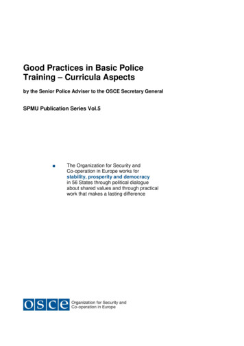
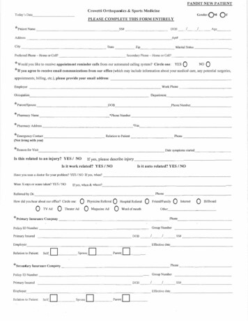
Special testsSupraspinatus Pain is exacerbated with theempty can test: Arms abducted to 90 andmoved 20 towards midline Arms internally rotated sothe thumbs are pointingdownward Push “up” against theexaminers hands which areplaced on top of the wrist
Special tests Subscapularis– Gerber’s lift off/bellypress test Infraspinatus & Teresminor– External rotation againstresistance
Present your findings:“This 20 year old cricket player has pain on activemovement of the shoulder joint, and is unable toinitiate abduction unaided by other muscles. Thispain is exacerbated by the empty can test. My topdifferential is a supraspinatus injury.”
Top questions: (1)What are the four muscles ofthe rotator cuff and what aretheir actions?Supraspinatus - AbductionSubscapularis - InternalrotationInfraspinatus and TeresMinor - External rotation
Top questions: (2)How do you investigate and manage this type ofinjury?Plain XRs to rule out fractureMRI or MR arthrogramConservative, Medical and Surgical– Analgesia– Physio– Cease high intensity exercise– Arthroscopic or open repair
Orthopaedic Stations1)2)3)4)Shoulder pathologyHand pathologyKnee pathologyHip pathology
Rheumatoid ArthritisPatient demographics: Middle aged to elderly woman Smoker Signs of steroid treatment
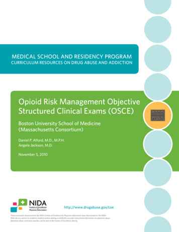
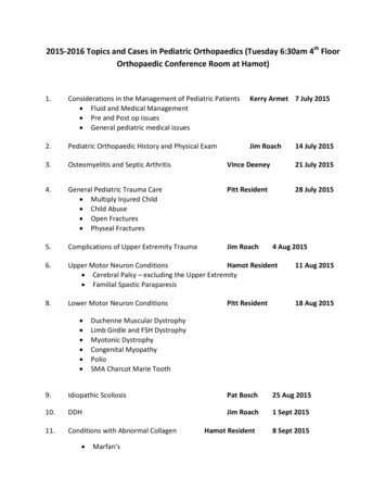
Rheumatoid Arthritis - signs:1. Boutonniere deformity –flexed PIP with hyperextendedDIP2. Swan neck deformity –hyperextended PIP with flexedDIP3. Z–thumb deformity – fixedflexion and subluxation of theMCP thumb joint, andhyperextension of the thumbIP joint
Rheumatoid Arthritis - signs:4. Ulnar deviation and palmar subluxation of the MCP joints5. Wasting of the small muscles of the hand (“ulnar guttering”)6. Radial deviation and subluxation of the hand at the wrist7. Joint replacements scars (typically over the MCP joints)8. Rheumatoid nodules – small firm subcutaneous nodules,typically best palpated over bony prominences (e.g. overknuckles)
Remember:1) You are looking for signs of active synovitis – isthere evidence of a red, hot, swollen tender joint?2) RA typically spares the DIPs whilst osteoarthritisoften affects the DIPs. Osteoarthritis may provideHeberden’s nodes (DIP) and Bouchard's nodes (PIP)3) Assess for function of the hand – power grip, pinchgrip, tripod, key grip, hook – as related to dailytasks
Present your findings:“These findings are consistent withsymmetrical peripheral deformingpolyarthropathy but there are no signs ofactive synovitis”“My top differential for this pattern ofarthropathy would be rheumatoid arthritisbut SLE and psoaritic arthropathy could alsopresent in a similar way”
Top questions:1) What is RA?Multisystem inflammatory autoimmune diseasecharacterised by symmetrical peripheral deformingpolyarthropathy.2) What is rheumatoid factor?IgM against the Fc portion of IgG3) Who gets RA?Common disease – affecting 1% of the population.More common in women and more common in smokers.Peak incidence 5th and 6th decades.
Top questions:4) What are the extra articular manifestations ofRA?Rheumatoid nodulesPulmonary disease inc. fibrosis, pulmonary nodules(Caplan’s syndrome), pleuritis and pleural effusionsPericarditisFelty’s syndrome (splenomegaly neutropaenia)Anaemia of chronic diseaseOphthalmic involvement - scleritis and episcleritis,Sicca syndromeAA Amyloid disease
Investigations for RA:“I would like to order blood tests, looking for”:1. FBC – Anaemia of CD / thrombocytosis / Felty’s2. ESR and CRP - increased in active synovitis3. RF - ve in 80%, is not a diagnostic test, but is aprognostic indicator - high titre associated withsevere disease4. Anti CCP – worse prognosis5. U&Es and LFTs – baseline before commencingtreatment
Investigations for RA:“I would like to request an X rays of the affected jointto assess for”:1. Soft tissue swelling2. Loss of joint space3. Juxta articular erosions4. Periarticular osteopeniaAssess deformity further i.e subluxation of joints
Treatment for RA:
Treatment for RA:“Management options for RA can be broadly divided into:conservative, medical and surgical options”Conservative management:1. Education about the disease and information about access toinformation and support groups2. Exercises with physiotherapy input to maintain jointmovement and muscle strength3. Occupational therapist input to ensure patient hasappropriate aids for ADLS.
Treatment for RA:Medical management:1) NSAIDS and steroids: useful to control symptoms ofacute synovitis.but do not alter the course of the disease.2) DMARDS Methotrexate is typically first line Should be started asap following diagnosis Take 6-8/52 to take full effect therefore should becovered initially with steroid Monitoring is essential because of side effects
Treatment for RA:Medical management:3) Biological agents E.g Infliximab and Etanercept Derived from antibodes Indicated for DMARD refractory Rheumatoid Arthritis(2 failed trials of DMARDS)
Treatment for RA:Surgical Management:1) Synovectomy2) Tendon transfer or repair anligament release3) Joint arthoplasty (commonlyMCPJ, also wrist)4) Joint arthrodesis – fusion ofthe joint e.g. wrist, CMCJ, PIPJ
Orthopaedic Stations1)2)3)4)Shoulder pathologyHand pathologyKnee pathologyHip pathology
Key Knee PathologiesChronic stable conditions: Post Total Knee Replacement Osteoarthritis Rheumatoid arthritisOther conditions: Cruciate Ligament Meniscal Tear
Osteoarthritis of the KneesPatient Demographics:– Elderly– Female– Large BMI– OA of other joints
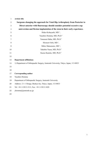
Osteoarthritis of the KneesUsually affects both knees (oftenasymmetrically)SIGNS Walking aids Varus deformity of the knees(medial OA) TKR Scars (midline) Arthroscopy scars
Osteoarthritis of the Knees Knees swollen Pain on flexion and extension Limited range of movement Effusion Crepitus Generalised laxity in all ligaments
Present your findings:“This lady has a swollen left knee which isminimally tender on palpation and movement.She has an old midline scar on the right knee.She is able to mobilise using one stick andappears systemically well. My top differential isan osteoarthritic knee.I would like to examine the hip and ankle andobtain AP and lateral radiographs of the knee”
Top questions: (1)What are your differentials? Osteoarthritis Rheumatoid arthritis Gout / Pseudogout“In the acute setting, I would also consider ” Septic joint Fracture
Top questions: (2)What are the causes of osteoarthritis?Primary osteoarthritis(wear and tear) Risk factors:AgeFemale SexJoint surface injuryAnything resulting inabnormal load bearing onthe joint e.g. Obesity,occupation etcSecondary osteoarthritis Septic jointRAFracturesConnective TissueDisordersMetabolic disorders
Top questions: (3)What signs would you see on an xray? Loss of Joint SpaceOsteophytesSubchondral SclerosisSubchondral CystsRemember the acronym LOSS
Orthopaedic Stations1)2)3)4)Shoulder pathologyHand pathologyKnee pathologyHip pathology
Key Hip PathologiesCommon Total Hip Replacement Previous neck of femurfracture OsteoarthritisAcute conditions: Fracture Periprosthetic Fracture Prosthesis dislocation
Hip arthroplastyPatient Demographics:– Elderly– Reduced Mobility– High BMI– OA of other joints
Hip arthroplastySIGNS Walking aids (opposite side) / antalgic gait Scars: antero-lateral, posterior Abductor weakness (Sound Side Sags) Reduced range of motion (particularly internal andexternal rotation)
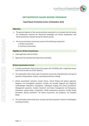
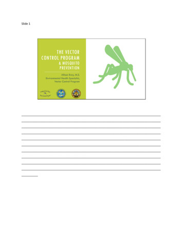
Abductor Weakness Positive Trendelenburg sign Weakened from OA Weakened from Antero-Lateralapproach (Gluteus medius andminimus removed andreattached)
Present your findings:“This elderly lady has bilateral surgical scars onboth hips. She is able to mobilise using a stick buthas marked abductor weakness bilaterally. Sheis likely to have had bilateral hip replacementsdue to severe osteoarthritis.I would like to examine the knees and the lumbarspine and review AP and lateral radiographs ofthe pelvis and hips”
Top questions: (1)Which prosthesis are used and when? Elective – total hip replacement only Intracapsular fractures– THR: if mobile and active– Hemiarthroplasty: if comorbidities80 000 hip fractures per year in UK10% 1-month and 30% 1-year mortalityVERY IMPORTANT FOR EVERYONE
Top questions: (2)How else may hip fractures be treated?– Dynamic hip screw– Cannulated hip screws– Intramedullary nailing
Written by: Dr David FergusonPrevious contributions: Dr Sara Khoyratty & Dr Will ChaundyEdited by: Dr Celine LakraHosted by: OSCE-AidFor tips on Orthopaedics OSCE stations:click here to visit our Orthopaedics page
Present your findings: "This lady has a swollen left knee which is minimally tender on palpation and movement. She has an old midline scar on the right knee. She is able to mobilise using one stick and appears systemically well. My top differential is an osteoarthritic knee.