Transcription
Color profile: Generic CMYK printer profileComposite Default screenPlant Molecular Biology Reporter 18: 143a–143t 2000 2000 International Society for Plant Molecular Biology. Printed in Canada.Publish by AbstractThe Use of the Luciferase Reporter System for inPlanta Gene Expression StudiesWESSEL VAN LEEUWEN1,*, MARC J.M. HAGENDOORN1, TOMRUTTINK2, REMCO VAN POECKE3, LINUS H.W. VAN DER PLAS1 andALEXANDER R. VAN DER KROL11Laboratory of Plant Physiology, Wageningen University, Arboretumlaan 4, 6703 BDWageningen, The Netherlands; 2Laboratory of Molecular Biology, WageningenUniversity, Dreijenlaan 3, 6703 HA Wageningen, The Netherlands; 3Laboratory ofEntomology, Wageningen University, Binnenhaven 7, 6709 PD Wageningen, TheNetherlandsAbstract. The properties of the firefly luciferase (LUC) make it a very goodnon-destructive reporter to quantify and image transgene promoter activity in plants. Theshort half-life of the LUC mRNA and protein, and the very limited regeneration of theLUC protein after reacting with luciferin, enables monitoring of changes in gene activitywith a high time resolution. However, the ease at which luciferase activity is measured inplanta, using a light sensitive camera system (2D-luminometer), contrasts sharply with thecomplications that arise from interpreting the results. A variegated pattern of luciferase activity, that is often observed in in planta measurements, might either be caused by differences in influx, availability of the substrates (luciferin, oxygen, ATP) or by localdifferences in reporter gene activity. Here we tested the possible contribution of differences in the availability of each substrate to the variegated in planta luciferase activity, andwe show when in planta luciferase activity is measured under substrate equilibrium conditions and can be related to the promoter activity of the reporter gene. Furthermore, wedemonstrate the effects of protein stability, apparent half-life of luciferase activity, regeneration of luciferase and pH on the in vivo and in vitro luciferase measurements. The combined results give the prerequisites for the correct utilisation of the luciferase reportersystem, especially for in vivo gene expression studies in plant research.Key words: Cauliflower Mosaic Virus 35S promoter, luciferase reporter system, Petuniahybrida, reporter gene, transgene expression, variegationAbbreviations: FW, fresh weight; LUC, luciferase; rlu, relative light units.LuciferaseIntroductionforgene expression studiesVan Leeuwen et al.The luciferase gene from the North American firefly Photinus pyralis hasemerged as a popular choice for in vitro and in vivo reporting of transcriptionalactivity in eukaryotic cells. Since the cloning of the cDNA encoding the enzyme*Author for correspondence. e-mail: Wessel.vanLeeuwen@Algem.PF.WAU.NL; fax: 31 317 484740; ph: 31 317 482822.J:\PMBR\Pmbr18\02\R00-022.vpThursday, September 07, 2000 1:49:47 PM
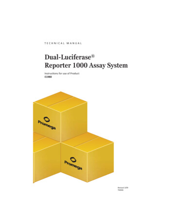
Color profile: Generic CMYK printer profileComposite Default screen143bVan Leeuwen et al.1.LUC luciferin Mg ATP (LUC luciferin-AMP) MgPPI2.(LUC luciferin-AMP) O2 (LUC oxyluciferin* AMP) CO 23.(LUC oxyluciferin* AMP) (LUC oxyluciferin AMP) hv4.(LUC oxyluciferin* AMP) LUC oxyluciferin AMPFigure 1. The luciferase reaction (Aflalo, 1991). Brackets and bullets indicate the complexes formed.Step 1 is a fast equilibrium reaction. Step 2 is the oxidative decarboxylation, in which the oxyluciferin is excited. Step 3 is the fast photon emission at 562 nm. Step 4 is the very slow release of productfrom the active site of the LUC protein.luciferase (LUC) by DeWet et al. in 1985, the luciferase gene has been expressedin tobacco and carrot plants (Ow et al., 1986), mammalian cells (DeWet et al.,1987) and zebrafish (Mayerhofer et al., 1995) and Drosophila (Brandes et al.,1996). In firefly, the LUC protein is targeted to the peroxisomes and theC-terminal peroxisome import signal was shown to function in plants as well. Forenhanced expression in mammalian cells and plants, the luciferase coding sequence was modified and the peroxisomal import sequence (luc , Promega) wasremoved (Sherf and Wood, 1994).Luciferase catalyses the oxidative decarboxylation of the substrate (firefly)luciferin (Figure 1). The reaction causes the release of a photon at 562 nm in 90%of the catalytic cycles with the substrates luciferin, Mg2 -ATP and oxygen(DeLuca et al., 1974; Aflalo, 1991). The luciferase enzyme is only slowly regenerated after reacting with the substrate, because the end product of the reaction,oxyluciferin, is only slowly released from the Luciferase ! Oxyluciferin -complex(Figure 1, step 4, Denburg et al., 1969). In vitro in the presence of high ATP concentrations, Coenzyme A enhances the light production through removal ofoxyluciferin from luciferase resulting in a nearly constant production of light(Ford et al., 1995). We will discuss whether the enhanced regeneration of luciferase by the presence of Coenzyme A occurs in vivo.This slow regeneration in combination with the short half-life of luciferase(Nguyen et al., 1989; Thompson et al., 1991), implies that in the presence of allsubstrates, each luciferase molecule can only react once and emit one photon. Inthe presence of all substrates the LUC protein will therefore not accumulate invivo. Luciferase as a reporter gene thus represents gene expression as the flux ofprotein molecules (LUC) made in the cell ( LUC/sec), while more stable reporter genes only show the accumulation of protein molecules as an indication ofgene expression (total amount of reporter protein in the cell at any given timepoint). Therefore, luciferase can be used as a non-invasive reporter in plants to accurately mark changes in transgene expression.After the plant tissue has been provided with luciferin, the only substratethat is not naturally present in the plant cell, in planta luciferase activity can bemonitored with a 2D-luminometer. However, in order to relate the changes in luciferase activity to changes in transgene expression, the availability of each of thesubstrates (luciferin, oxygen and ATP) should remain constant during the periodJ:\PMBR\Pmbr18\02\R00-022.vpThursday, September 07, 2000 1:49:50 PM
Color profile: Generic CMYK printer profileComposite Default screenLuciferase for gene expression studies143cover which the luciferase activity is monitored. In order to relate differences in luciferase activity within a tissue to local differences in transgene expression, theavailability of each of the substrates should also be similar in different parts ofthe tissue.When we used the luciferase reporter system to measure gene expression invivo in Petunia leaves, we noticed a high degree of variation in light emissionwithin each leaf (variegation) as was observed before by Schneider et al. (1990)and Quandt et al. (1992). In this article we studied the possible contribution of thedifferent substrates to differences and changes in in planta luciferase activity. Wediscuss what precautions have to be taken when the luciferase reporter system isused in plant research and under which circumstances the observed light production directly reflects luciferase gene expression.Materials and MethodsLuc reporter gene constructsAgrobacterium tumefaciens (A. tum. strain ABI) was transformed with the binaryvector pMON721 containing either a CaMV 35S promoter - luc construct(pGM46) or a CaMV m35S promoter - luc construct (pGM107). The CaMV promoter used in our constructs consists of the -343 to 8 sequence (Gardner et al.,1981; Benfey et al., 1989). The modified CaMV 35S (m35S) promoter, containsthe -90 to 8 fragment of the CaMV 35S promoter, with four copies of the B3 domain and four copies of an optimised AS-1 binding site placed upstream (van derKrol et al., 1993), thereby increasing potential binding of B-ZIP transcription factors. The luc gene that is used in the pGM46 construct is the original luciferasecoding sequence cloned by deWet et al. (1985, 1987). For the pGM107 constructa modified firefly luciferase gene (luc , Promega) was used (Sherf and Wood,1994). In the luc gene the peroxisomal translocation sequence is removed, aswell as several restriction sites. Codon usage is improved for mammalian cellsand consensus glycosylation sites, and consensus sequences for transcription factor binding sites were eliminated (Sherf and Wood, 1994). It was shown that luc had a 10-100 times higher expression than luc in mammalian cells (Groskreutz etal., 1995). In tobacco no significant effect on expression was found, but in maizeand wheat a 20- and 55-fold increase in activity was obtained respectively (Lonsdale et al., 1998). In the pGM46 and pGM107 constructs an N-terminal SV40 Nuclear Localisation Signal (van der Krol and Chua, 1991) was present in front ofthe luc coding sequence, which had no apparent effect on its activity.Plant materialPetunia hybrida (Vilm.) plants (cv. V26) were transformed by A. tum. clones containing either pGM46 or pGM107, and transformed shoots were, after rooting,transferred to soil and grown in growth chambers with a 16 h light period (30 Wm-2, 22 C, and 70% RH) and an 8 h dark period (20 C, and 65% RH). For the experiments shown here the F1 progeny plants of a back-cross with wild-type V26were used.J:\PMBR\Pmbr18\02\R00-022.vpThursday, September 07, 2000 1:49:50 PM
Color profile: Generic CMYK printer profileComposite Default screen143dVan Leeuwen et al.Petunia cell suspensions were made by using seedlings of the back-crossfrom the 35S-luc Petunia plants. Seedlings were grown in 250 mL Erlenmeyerflasks on a rotary shaker at 100 rpm in 60 mL MS medium (Murashige andSkoog, 1962) supplemented with sucrose (30 g/L) and 2,4-D (1 mg/L). The suspension was sub-cultured every 10-12 d (10 mL culture with 50 mL fresh medium). After several weeks the cell suspension was sieved ( 120 µm). Thesub-culturing resulted in a homogenous cell suspension after several months.Tobacco plants containing a 35S-luc construct were kindly provided by Dr.Nap, Wageningen University and Research centre –Plant Research International.In vivo luciferase activity measurement with the 2D-luminometerPetunia or tobacco luc reporter plants were sprayed with a luciferin solution(1 mM firefly D-luciferin, sodium-salt, Duchefa, 0.01% Tween 80) by using anair-brush dispenser to obtain a fine mist, 24 h, 16 h and 2 h before measurement.Cell suspensions derived from the Petunia luc reporter plants were treated with0.5 mM luciferin 2 h before measurement. Luciferase activity was imaged with a2D-luminometer, consisting of an intensified CCD camera (C2400-77,Hamamatsu Photonics, Japan), or with a liquid nitrogen cooled slow-scan CCDcamera (512-TKB, Princeton, Trenton, NJ, USA). Photon emission by luc-expressing plants (or suspensions) was quantified by computer (Argus-50 ImageProcessor, Hamamatsu Photonics, Japan). Luciferase activity is shown in relativelight units per pixel (rlu/pixel). Integration intervals varied from 2 to 30 min. Images of luciferase activity are depicted with false colour scales (blue indicatinglow activity, red indicating high activity) or grey scales (dark grey indicating lowactivity, white indicating high activity).LUC protein extractionLeaf parts up to 100 mg, frozen in liquid nitrogen were ground and suspended in100 FL luciferase extraction buffer (25 mM Tris H2PO4, 2 mM EDTA, 10% Glycerol, 1% Triton X-100, 2 mM DTT, pH 7.8). Cell fragments were removed by4-10 min centrifugation at 16,000 g (14,000 rpm, Eppendorf centrifuge 5414C)and the supernatant was frozen in liquid nitrogen and stored at -80 C, until use inan in vitro luciferase activity assay.In vitro luciferase flash-assayFor measurement of luciferase activity the frozen luciferase extract was thawed onice and a 5 FL aliquot was pipetted in a 96-wells microtiter plate and measured ina Labsystem Luminoskan DS luminometer by addition of 100 FL flash-assaybuffer (20 mM Tricine, 2.67 mM MgSO4, 0.1 mM EDTA, 2 mM DTT, 470 FMD-Luciferin, 5 mM ATP (pH 7.8)). Two seconds of light production of the initialflash (caused by the rapid single use of the LUC protein after which a complex isformed with oxyluciferin) was quantified and shown as relative light units (rlu) asmeasured in two seconds. A dilution of luciferase (Boehringer) in extractionbuffer was used for calibration (0.1 U/mL – 200 U/mL).J:\PMBR\Pmbr18\02\R00-022.vpThursday, September 07, 2000 1:49:51 PM
Color profile: Generic CMYK printer profileComposite Default screenLuciferase for gene expression studies143eIn vitro luciferase assay with Coenzyme AThe luciferase extract can also be measured with a luciferase assay buffer containing Coenzyme A (CoA), which will prolong the light production (Ford et al.,1995). The steady state light production can then be quantified for 5 s after a 10 sinterval directly after addition of the CoA-assay buffer (20 mM Tricine, 2.67 mMMgSO4, 0.1 mM EDTA, 33.3 mM DTT, 270 FM CoA, 470 FM D-Luciferin,530 FM ATP (pH 7.8), Luehrsen and Walbot, 1993) and is shown as relative lightunits (rlu) averaged over 2 s (for easier comparison with flash-assay).For the CoA experiments, shown in Table 2 the following buffers used wereall containing 20 mM Tricine, 2.67 mM MgSO4, 0.1 mM EDTA with a final pHof 7.8 and one of the following:a) Buffer: without extra additions;b) Flash2: ( flash-assay buffer) containing 2 mM DTT, 470 FM D-Luciferin,5 mM ATP;c) CoAdil. (dilution): buffer with 270 FM CoA;d) CoA33: ( CoA-assay buffer) containing 33.3 mM DTT, 270 FM CoA,470 FM D-Luciferin, 530 FM ATP;e) CoA2: buffer with 2 mM DTT, 270 FM CoA, 470 FM Luciferin, 530 FMATP.Oxygen determination in Petunia cell suspensionOxygen levels were measured with a Clark oxygen monitor at 25 C in a stirredPetunia cell suspension during the in vivo luciferase activity measurement withthe 2D luminometer.Determination of stomatal aperture by silicone rubber imprintsSilicone rubber imprints were made from a Petunia leaf surface to determine thestomatal aperture of the leaf. Two parts of silicon rubber (Xantopren light body,thin flowing silicone precision impression material; ADA specification No.19,type II, low viscosity, Bayer Dental) were mixed with one part accelerator andsubsequently mixed thoroughly for 30 s, without introduction of air bubbles in themixture. An imprint of the leaf should be made within 2 min, after which an additional 2 min are required for polymerisation. By making two silicone-rubber imprints on the same location, a cleaner imprint can be acquired. Approximately 3 gpolystyrene (Mw 100,000, BDH Chem. Ltd.) was dissolved in 12 mL toluol(Merck) at a temperature of 45 C. The polystyrene solution was applied to thesurface of the rubber imprint as thinly and evenly as possible, with a brush. Thepolystyrene film was carefully removed by gentle bending of the rubber replicaafter 3 min and laid upside down on a glass slide. The replica was covered with acover glass, which was subsequently fixed by tape. Stomatal aperture in the preparation could now be examined through a microscope.J:\PMBR\Pmbr18\02\R00-022.vpThursday, September 07, 2000 1:49:51 PM
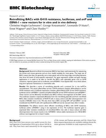
Color profile: Generic CMYK printer profileComposite Default screen143fVan Leeuwen et al.Figure 2. A 35S-luc Petunia leaf showing variegated patterns of light emission (luciferase activitymeasured for 5 min). Scale on the right indicates the colour scale used to represent the luciferase activity. The size of the leaf is approximately 13 by 18 mm.Results and DiscussionImaging of in planta luciferase activity in transgenic Petunia leaves expressingthe 35S- or m35S-luciferase reporter gene shows different patterns of luciferaseactivity that can vary up to 16-fold within a leaf (variegation, Figure 2). In orderto distinguish between variegated luciferase reporter gene expression and a variegated distribution of one or more of the substrates (luciferin, Mg2 -ATP or oxygen), we tested the possible contribution of differences in the availability of eachof the substrates to the variegated in planta luciferase activity. As ATP and oxygen are present within plant cells, only the substrate luciferin needs to be appliedfrom the outside. We first investigated whether local differences in the penetrationof luciferin into plant cells may be the cause of the variegated luciferase activitypattern in leaves.Effects of luciferin on in planta luciferase measurementsLuciferin (in aqueous solution) can be applied either by repeated spraying on theplant or by uptake through the roots and vascular tissue. In order to optimise thedistribution of luciferin over the leaf surface we used for spraying an air-brushdispenser to create a fine mist of luciferin and we used 0.01% Tween 80 as a surface-active agent (especially necessary when applied to hairy plant structures, likeleaf surfaces or roots).The solution of the luciferin as described by Millar et al. (1992) contains0.01% Triton X-100 as a surfactant. However, Triton X-100 may cause necrosis ofthe leaf after prolonged application. We tested the possible necrotic effect of prolonged application of different surfactants on leaves. When Petunia leaves wereput in a petri-dish, on a solution with different concentrations of either Tween 20,J:\PMBR\Pmbr18\02\R00-022.vpThursday, September 07, 2000 1:49:54 PM
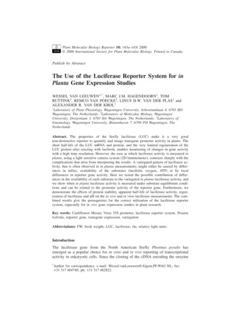
Color profile: Generic CMYK printer profileComposite Default screenLuciferase for gene expression studies143gFigure 3. Luciferase activity of a 35S-luc Petunia branch as measured in 15 min (rlu/pixel) plottedagainst time (h). The measurement is continuously repeated for 40 h. The branch is put in water att 0 h and sprayed at t 0 h, t 7 h, t 20 h and t 30 h with a 1 mM luciferin 0.01%Tween-80 solution.Another branch from the same 35S-luc Petunia plant is put in 1 mM luciferin at t 0 h. The luciferaseactivity reaches the same equilibrium after 20 to 30 h as is shown in the inserted panel A (22 h) andpanel B (36 h). In panels A and B, the left branch is put in 1 mM luciferin at t 0 h, the right branchis the branch described above (put in water at t 0 h and sprayed with luciferin). Scale on the right inpanels A and B indicates the colour scale used to represent the luciferase activity. After 55 h bothbranches are sprayed with a 1 mM luciferin 0.01% Tween-80 solution and measured for 10 h. Theblack line represents the luciferase activity in the branch put on water at t 0 h, the dashed line represents the luciferase activity in the branch put on 1 mM luciferin at t 0 h.Tween 80, Non-Idet P40 or Triton X-100, severe necrosis was observed with Triton X-100 (1%) after a few hours or, with 0.01%, after a few days. The severityof necrosis was with Triton X-100 Non-Idet P40 Tween 20 Tween 80. Prolonged application of a 0.01% solution of Tween 80 had no visible necrotic effecton leaves (equal to water, data not shown). Therefore, in all experiments in whichplants were sprayed with a luciferin solution, 0.01% Tween 80 was used as asurfactant.Luciferin readily penetrates most plant tissues when applied by spraying. Inplanta luciferase activity can be imaged within seconds after spraying when theluciferase reporter gene is expressed in epidermal cells. With vascular uptake,luciferin is transported through the plant within minutes. We compared the effectof luciferin application, by repeated spraying with 1 mM luciferin and throughvascular uptake of a 1 mM luciferin solution, on in planta luciferase activity intwo branches from the same 35S-luciferase Petunia plant. In Figure 3 the quantified luciferase activity of the sprayed branch is shown in time. Spraying of the Petunia branch with 1 mM luciferin at t 0, 7, 20 and 30 h, results in an increase ofluciferase activity at 0 and 7 h, but has almost no effect at 20 and 30 h. The panels inserted in Figure 3 show the two branches at t 22 h (panel A) and t 36 h(panel B). With both types of luciferin application, a similar variegated pattern ofin planta luciferase activity emerges. When after 55 h both branches are sprayedwith 1 mM luciferin, both branches show a comparable and only small increase inluciferase activity, indicating that for both ways of luciferin application anJ:\PMBR\Pmbr18\02\R00-022.vpThursday, September 07, 2000 1:50:00 PM
Color profile: Generic CMYK printer profileComposite Default screen143hVan Leeuwen et al.equilibrium is reached in luciferin influx (Figure 3). The slow overall decrease ofluciferase activity after 30 h might reflect a decrease in luciferase gene expression, due to the prolonged absence of light during measurement under theluminometer. These results indicate that a continuous application of luciferin isnot required, once equilibrium between luciferase activity and luciferin inflow isreached. Intermittent spraying of luciferin twice a day is sufficient to keep the luciferase activity at the same level as continuous application through the vascularfeeding. The substrate luciferin itself is very stable in plant cells, because luciferase-expressing plants that previously have been sprayed with luciferin can stillshow luciferase activity after 7-10 d without further addition of luciferin.Spraying the plants requires less luciferin than application of luciferin bywatering, which induces patterns caused by vascular luciferin uptake when theplants are imaged too soon (Schneider et al., 1990; Quandt et al., 1992). However,some plant structures will not take up luciferin, either when applied from the outside or through the vascular system. For instance, the locules of stamen or developing seeds initiate a dehydration program at a certain stage of their development,which will block an influx of water and consequently an influx of luciferin. A luciferase reporter gene that is expressed in these tissues will only show in plantaluciferase activity when luciferin is applied at an early stage of development,when the structure is still in contact with the vascular system of the entire plant,or when the mature tissue is damaged to facilitate substrate penetration.The luciferase substrate luciferin may have an adverse effect on plant cellswhen used at high concentrations ( 10 mM). Repeated spraying of plants with a1 mM solution (e.g. daily for several weeks) does not markedly inhibit Petunia ortobacco plant growth or reproduction. Sensitive cell systems like tobacco suspension cells or protoplasts can survive in luciferin concentrations of up to 80 FM,but concentrations 400 FM luciferin were found to kill the tobacco suspensioncells (Ow et al., 1986). A comparable toxic effect on somatic carrot embryo development at 400 FM luciferin was also found by Toonen et al. (1997). In Petunia cell suspensions, we found no toxic effect (within the 10 d subculture) whenwe used 500 FM luciferin, which was enough to bring the luciferase reaction inthe cell suspensions to an equilibrium (raising the concentration to 1.0 mM or1.5 mM luciferin had no effect on the level of light produced by the cells, data notshown).In conclusion, different ways of luciferin application have no effect on thevariegated pattern of luciferase activity in plants. In plants pre-sprayed 3x withluciferin, additional re-spraying does not significantly influence the level and pattern of luciferase activity. We therefore conclude that in our experimental set-upthe observed differences in luciferase activity (Figure 2) were not caused by differences in luciferin availability.Effects of oxygen on in planta luciferase measurementsThe luciferase reaction is dependent on oxygen. When the oxygen availabilitywithin a leaf is decreased (e.g. by submergence in water) light production as a result of luciferase activity drops to zero within 15-20 min and is immediately restored after re-exposure to air (data not shown). By measurement of luciferaseJ:\PMBR\Pmbr18\02\R00-022.vpThursday, September 07, 2000 1:50:00 PM
Color profile: Generic CMYK printer profileComposite Default screenLuciferase for gene expression studies143iFigure 4. Panel A. Oxygen dependence of luciferase mediated light production in a 35S-luc Petuniacell suspension. Plotted are relative light units (rlu) per pixel versus oxygen concentration (FM). Thedashed line shows the maximum level of oxygen when the suspension is oxygenated with air. PanelB. Four cross sections of a variegated m35S-luc Petunia leaf. The main vein (left) and a lateral veinare visible. The scale bar represents 1 mm. Panel C. Variegated luciferase activity in the cross sections shown in panel B. Scale on the right indicates the colour scale used to represent the luciferaseactivity.activity in a Petunia cell suspension at different levels of oxygenation, the dependence on oxygen of the luciferase reaction is illustrated (Figure 4A).We investigated whether possible local variations in oxygen concentrationwithin a leaf contribute to the observed variegated patterns of luciferase activity.Cross sections were made of an excised Petunia leaf with different levels of luciferase activity within the leaf. In these cross sections (Figure 4B) the luciferaseactivity remains variegated (Figure 4C), indicating that variable oxygen availability within the leaf is not the cause for the variegated luciferase activity pattern.Local oxygen levels may however vary depending on the opening ofstomata and photosynthetic activity of the leaf. It has been shown before thatstomatal aperture may vary within a leaf (Laisk et al., 1980, 1983). A variegatedstomatal aperture may actually cause or contribute to the variegated luciferase activity that is observed in 35S-luc transgenic leaves (Figure 2). We therefore testedwhether the stomatal aperture can influence the luciferase activity in 35S-luc Petunia plants. Three genetically identical plants with comparable luciferase activityJ:\PMBR\Pmbr18\02\R00-022.vpThursday, September 07, 2000 1:50:04 PM
Color profile: Generic CMYK printer profileComposite Default screen143jVan Leeuwen et al.Table 1. The effects of stomatal aperture on the level of luciferase activity in 35S-luc Petunia plants*.Treatmentluciferase activity(rlu/pixel)Aperture stomata(ratio open/closed (n))LightDarkABA3.53.33.56.7 (115)0.26 (122)0.21 (104)*Three genetically identical plants with comparable luciferase activitywere kept under greenhouse conditions (light), kept in the dark (dark) ortreated with 10 µ M ABA (ABA) for 24 h. The average luciferase activityin the first expanded leaf after 24 h treatment is shown (rlu/pixel). The aperture of the stomata in these leaves is subsequently measured under themicroscope (shown as ratio open/closed). The number of stomata used tocalculate this ratio is shown between brackets (n).were either kept under greenhouse conditions for 24 h, kept in the dark for 24 h,or treated with 10 FM ABA (sprayed three times in 24 h). One leaf of each plantwas then measured in close-up with the luminometer and stomatal aperture wasdetermined in this leaf by microscopic analysis (Table 1). The analysis shows thatboth the dark treatment and the ABA treatment resulted in a similar ratio of opento closed stomata, which was 3-4 % of that in control leaves. However, the luciferase activity in these leaves was not significantly affected by stomatal aperture,indicating that variegated stomatal aperture within a leaf does not contribute tothe variegated luciferase activity in leaves.We therefore conclude from these experiments that the variegated luciferaseactivity in Petunia leaves is not caused by local differences in oxygen availabilitybetween the cells within the leaf.Availability of ATP in in planta luciferase measurementsBecause of the direct relation between ATP and the photon production of the luciferase reaction, and the high sensitivity at which photons can be detected, the luciferase reaction is often used to quantify ATP in plant extracts (Malik andThimann, 1980). It can be concluded from these experiments by Malik andThimann (1980) that the cellular steady state concentration of ATP in plant leafcells under normal physiological conditions is in the range of 100-200 pmol/mgfresh weight (FW). We determined the ATP level that is required for the reactionof a high amount of luciferase that can be present in a plant with high expressionof a luciferase reporter gene. We first used an in vitro luciferase flash assay inwhich different ATP dilutions were added to a fixed amount of LUC protein(20,000 U). Light production was detectable above 10 fmol ATP added (Figure 5).The resulting light production from a fixed amount of ATP can now be comparedwith the light production we normally obtain in leaf extracts per mg FW (0.2-20rlu/mg FW, grey area Figure 5). Apparently, the amount of luciferase present inour Petunia leaves requires 20 fmol ATP/mg FW to a maximum of 2 pmolATP/mg FW. This is 2 to 4 orders of magnitude below the ATP concentration inleaves (Malik and Thimann, 1980).J:\PMBR\Pmbr18\02\R00-022.vpThursday, September 07, 2000 1:50:04 PM
Color profile: Generic CMYK printer profileComposite Default screenLuciferase for gene expression studies143kFigure 5. ATP dependence of the luciferase reaction as determined in an in vitro luciferase flash assay. Plotted are the relative light units (rlu) versus the ATP levels added (pmol). Points are means ofduplicate determinations. The linear regression line has an R2 of 0.999. The grey area shows therange of rlu we generally obtain in luciferase extracts from transgenic leaves and the correspondingpicomoles of ATP required.Assuming that 10% of the fresh weight consists of cytoplasm, 100 pmolATP/mg FW would correspond to a concentration of approximately 1 mM ATP,which is far above the Km of luciferase for ATP (50 FM) (Lembert and Idahl,1995). The variegated luciferase activity that we observe in 35S-luc Petunialeaves (Figure 2) utilises at maximum only 1-2% of the available ATP pool (provided that no regeneration of luciferase activity takes place). We conclude fromthese experiments that the luciferase activity proba
Publish by Abstract The Use of the Luciferase Reporter System for in Planta Gene Expression Studies WESSEL VAN LEEUWEN1,*, MARC J.M. HAGENDOORN1,TOM RUTTINK2, REMCO VAN POECKE3, LINUS H.W. VAN DER PLAS1 and ALEXANDER R. VAN DER KROL1 1Laboratory of Plant Physiology, Wageningen University, Arboretumlaan 4, 6703 BD Wageningen, The Netherlands; 2Laboratory of Molecular Biology, Wageningen