Transcription
MetabolomicsDOI 10.1007/s11306-014-0622-5ORIGINAL ARTICLELuciferase does not alter metabolism in cancer cellsCaroline H. Johnson Timothy S. FisherLinh T. Hoang Brunhilde H. Felding Gary Siuzdak Peter J. O’Brien Received: 22 November 2013 / Accepted: 14 January 2014Ó Springer Science Business Media New York 2014Abstract Luciferase transfected cell lines are usedextensively for cancer models, revealing valuable biological information about disease mechanisms. However, thesegenetically encoded reporters, while useful for monitoringtumor response in cancer models, can impact cell metabolism. Indeed firefly luciferase and fatty acyl-CoA synthetases differ by a single amino acid, raising thepossibility that luciferase activity might alter metabolismand introduce experimental artifacts. Therefore knowledgeof the metabolic response to luciferase transfection is ofsignificant importance, especially given the thousands ofresearch studies using luciferase as an in vivo bioluminescence imaging reporter. Untargeted metabolomicsexperiments were performed to examine three differentElectronic supplementary material The online version of thisarticle (doi:10.1007/s11306-014-0622-5) contains supplementarymaterial, which is available to authorized users.C. H. Johnson L. T. Hoang G. Siuzdak (&)Departments of Chemistry, Molecular, and ComputationalBiology, and the Scripps Center for Metabolomics and MassSpectrometry, The Scripps Research Institute,La Jolla, CA, USAe-mail: siuzdak@scripps.eduT. S. Fisher P. J. O’Brien (&)Pfizer Worldwide Research and Development, La JollaLaboratories, 10724 Science Center Drive, San Diego, CA, USAe-mail: peter.obrien2@pfizer.comB. H. FeldingDepartments of Molecular and Experimental Medicineand Chemical Physiology, The Scripps Research Institute,La Jolla, CA, USAtypes of lymphoblastic leukemia cell lines (Ramos, Rajiand SUP-T1) commonly used in cancer research, each wereanalyzed with and without vector transduction. The Rajimodel was also tested under perturbed starvation conditions to examine potential luciferase-mediated stressresponses. The results showed that no significant metabolicdifferences were observed between parental and luciferasetransduced cells for each cell line, and that luciferaseoverexpression does not alter cell metabolism under basalor perturbed conditions.Keywords Luciferase Metabolomics Bioluminescenceimaging Reporter geneAbbreviationsACNAcetonitrileBLIBioluminescence imagingdPBSDulbecco’s phosphate buffered salineESIElectrospray ionizationEVEmpty vectorFAFormic acidFACSFatty Acyl CoA synthetaseFLucPhotinus pyralis luciferaseHILICHydrophilic interaction liquidchromatographyHPLCHigh performance liquid chromatographyIPAIsopropanolLC/MSLiquid Chromatography/Mass SpectrometryMeOHMethanolMSMass SpectrometryNH4AcAmmonium acetateNH4OHAmmonium hydroxideQ-TOF/MS Quadrupole Time-Of-Flight MassSpectrometerRPLCReversed-phase liquid chromatography123
C. H. Johnson et al.1 IntroductionThe clinical relevance of cancer models depends in largepart on their similarity to primary tumors, and whethertreatment responses can be discerned from other sources ofvariation (van Staveren et al. 2009). Tumor cell linesexpressing reporter genes are widely employed as cancermodels, and their use has revealed valuable biologicalinformation about disease mechanisms and treatmentresponses. Bioluminescence imaging (BLI) for example isused to monitor the abundance, localization, and functionof tumor cells over time in vivo, most often via engineeredexpression of insect luciferase enzymes. However, thesetypes of genetic manipulations or variations in cultureconditions can alter cell physiology and may affect cellmetabolism.Luciferases are oxidative enzymes best known for theirlight-producing, ATP- and oxygen-dependent metabolismof luciferin substrates (Greer and Szalay 2002). Fireflyluciferases (FLuc) and fatty acyl-CoA synthetases (FACS)are structurally and functionally related (McElroy et al.1967; Gulick 2009); both enzyme activities can be localized to peroxisomes (Keller et al. 1987; Watkins and Ellis1822), and both can catalyze the synthesis of dinucleosidepolyphosphates and acyl-CoA derivatives (Guranowskiet al. 1990; Oba et al. 2003). Indeed, a single amino acidchange converts FLuc to a FACS (Oba et al. 2009).Numerous small molecule luciferase inhibitors have beenidentified (Thorne et al. 2012) and luciferase activity canbe modulated by xenobiotics and endogenous conditionsin vivo (Keyaerts et al. 2012; Brutkiewicz et al. 2007; Simet al. 2011; Czupryna and Tsourkas 2011). These findingsand the increasing use of BLI-optimized FLuc raise thepossibility that highly expressed, long-lived luciferasesmight alter cellular metabolism and introduce experimentalartifacts.Recent studies have examined luciferase bioluminescence effects on tumor models and come to contradictoryconclusions. We (Zhang et al. 2012) and others (Milsomet al. 2013) have observed that under certain circumstancesluciferase-expression is associated with altered cell growthin vivo. Furthermore, Brutkiewicz and colleagues (Brutkiewicz et al. 2007) noted that tumor growth retardationoccurred after serial imaging of ovarian tumor cellsexpressing high levels of luciferase, an affect the authorsattributed to the bioluminescence reaction itself. In contrast,Tiffen et al. (Tiffen et al. 2010) examined the growth characteristics of breast tumor and melanoma clones expressingdifferent levels of luciferase activity, and found that neitherluciferase expression nor biophotonic activity causeddetectable cytotoxicity. Similarly conflicting results wereobtained on examination of luciferase bioluminescence inthe presence of photosensitizing agents. Luciferase activity123was reported to be sufficient to drive photodynamic cytotoxicity in NIH 3T3 cells (Theodossiou et al. 2003), but thisfinding was not generalizable to other cells (Schipper et al.2006).Increasingly, researchers are using unbiased profilingmethods to assess experimental cell models. Untargetedmetabolomics permits comprehensive analyses of all thesmall molecules within a tissue or cell population, whichcumulatively reflect the activity of all cellular processes(Patti et al. 2013). Recent advances in liquid chromatography/mass spectrometry (LC/MS)-based metabolomicshave enabled more complete recovery and identification ofmetabolites (Patti et al. 2013; Zhu et al. 2013; Ivanisevicet al. 2013; Tautenhahn et al. 2012; Patti et al. 2012),making it an ideal technology for identifying metabolicperturbations caused by genetic or environmental stresses.In the present study, we use untargeted metabolomics toexamine the impacts of genetic manipulations on lymphoma lines used in BLI. We show that even seeminglyminor nutrient stress induces significant metabolicresponses in the lines tested. In contrast, the same lineswere metabolically stable in sub-confluent cultures, evenafter luciferase vector transduction and antibiotic selection.The implications of these findings for BLI models, and thegeneral utility of this approach for unbiased characterization of experimental cell manipulations are discussed.2 Experimental procedures2.1 Cell culture and biological reagentsRamos (CRL-1596), Raji (CCL-86) and SUP-T1 (CRL1942) cell lines were obtained from American Type Culture Collection (ATCC, Manassas, VA, USA). Unlessotherwise noted, cell culture conditions are as described(Ivanisevic et al. 2013), and all plasticware, buffers, andmedia components were from single manufacturing lots.Fetal bovine serum (FBS) was purchased from GEHealthcare Bio-Sciences Corp. (PAA #A15-204, Piscataway, NJ, USA); Sigma Aldrich (#F4135, St. Louis, MO,USA); and Thermo Fisher (Hyclone #SH30070.03, Waltham, MA, USA). According to the manufacturer, PAAFBS used in these studies consisted of *77 % FBS supplemented with a bulk additive (Hypep 1510, consisting of31.5 g/L Bovine Serum Albumin (BSA) 30.25 g/L SoyPeptone, 3.8 g/L Sodium Chloride, and 1 g/L glucose. Soypeptones are used to enhance protein production inbioreactors.LucSh, is a codon-optimized Photinus pyralis luciferase(FLuc) fused to the Streptoalloteichus hindustanus bleomycin resistance protein ble domain (Gatignol et al. 1988)in place of the carboxy-terminal 13 amino acids of FLuc
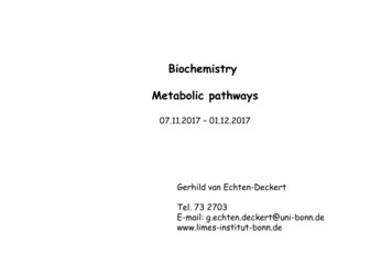
Luciferase does not alter metabolism(Invivogen technical service). LucSh was sub-cloned fromthe pMOD-LucSh vector (InvivoGen; San Diego, CA,USA) into the retroviral transfer vector pMSCV (Clontech;Mountain View, CA, USA). Amphotrophic pseudotypedretrovirus was prepared by co-transfection of 293T cells(ATCC) with LucSh or empty retroviral transfer vectorplus pCL10A1, a retroviral packaging vector (Imgenex,San Diego). Viral supernatants were used to transduce Raji,Ramos, and Sup-T1 cells in the presence of 8 lg/mLpolybrene. Transduced cells were grown in either 2 mg/mLpuromycin or 200 mg/mL ZeocinTM to select drug resistantcell pools. LucSh expression was confirmed using theBright GloTM luciferase assay (Promega, Madison, WI,USA), and by flow cytometry using a goat anti-luciferasepolyclonal antibody (#G745A, Promega, 1:200 dilution)and an Alexa-fluor 488 conjugated donkey anti-goat IgGdetection antibody (#A11055, Invitrogen, Carlsbad, CA,USA).Lymphoma metabolomic profiling was performed asdescribed (Ivanisevic et al. 2013) with the followingmodifications. 16–24 h prior to isolation and extraction,cell viability was assessed, and cultures with greater than93 % viability were re-suspended to a final dilution of*500,000 cells/mL in fresh growth medium containingantibiotics as indicated. 1 h prior to isolation, cell viabilitywas again measured, and cultures with greater than 93 %viability were adjusted to 1 9 106 cells/mL in fresh medium containing FBS and antibiotics as indicated, thenincubated at 37 C in a 5 % CO2 incubator until isolation.Twenty-five million cells were processed per experimentalreplicate, split into five equal aliquots, then analyzed byLC/MS in 5 technical replicates.2.2 Serum withdrawal stressSub-confluent cultures of Raji cells transduced withpMSCV-empty vector (puromycin selection; one passagepost-transduction) or pMSCV-LucSh (FLuc-Sh ble fusiongene; passage 2 post-transduction) were maintained ingrowth medium supplemented with antibiotics as described.Aliquots of 25 million cells were removed and centrifugedat 4009g at room temperature for 2–4 min. 24 mL of eachsupernatant was immediately removed by aspiration, andcell pellets were re-suspended by adding 24 mL of eitherRPMI 1640 (starvation medium) or growth medium consisting of RPMI 1640 supplemented with 10 % FBS, andincubated for 4 h at 37 C in a 5 % CO2 incubator.Adherent cells were dislodged from flasks by gentle pipetting to enable cell counting and cell viability assessmentusing the ViacountTM assay as described (Ivanisevic et al.2013). Cells were processed and frozen for extraction andmetabolite profiling as described above.2.3 Sample preparation for LC/MS and untargetedmetabolomic analysesCell extractions and analysis were performed as described(Ivanisevic et al. 2013). For normalization, the cells werecounted using the Guava ViacountÒ assay Millipore,Billerica, MA, USA) and placed into 5 mL aliquots of 10million cells per replicate before extraction. This ensureduniform cell numbers in each replicate.3 Results and discussion3.1 Metabolomic comparisons of three lymphoma linesgrown under optimized conditionsThese studies sought to determine whether expression of aBLI-optimized FLuc reporter gene affected lymphomametabolism in a manner that might alter tumor treatmentresponses. Experiments were performed in vitro usingpools of transduced cells to minimize clone-specific artifacts. To isolate the effects of transductant selection andvector expression from environmental effects, the metabolism of parental and vector-transduced cells were firstexamined in nutrient-replete, sub-confluent cultures. Previously optimized conditions were used, including carefulcontrol of cell culture reagents and plasticware; cell densityand media freshness; metabolite extraction and data analysis; and strict adherence to cell viability criteria at thetime of sample collection (Ivanisevic et al. 2013).Raji, Ramos, and SUP-T1 lymphoblastic cells weregrown under identical conditions, harvested, and stored at-80 C before extraction. Samples were analyzed by LC/MS and the data were exported into XCMS Online softwarefor pair-wise analysis (Tautenhahn et al. 2012). The datawas filtered for noise, statistical significance (\ 0.001), andthe magnitude of dysregulation (fold change from comparator C 2). Cloud plots (Patti et al. 2013), were generatedto display dysregulated features after inter-line metabolomic comparisons (Figs. 1 and Table 1). Data sets for allanalyses are available to view on XCMS Online under thePublic Shares tab. The analysis shows total ion chromatogram plots, cloud plots, multidimensional scaling plots andprincipal component analysis plots. Metabolite profilesobserved were consistent within each line (see below) and,as expected, significant metabolomic differences were seen(46–272 dysregulated features) between parental lines.3.2 Metabolomic effect of growth medium serumadditivesLymphoma metabolism was next examined in media prepared with different lots of FBS. Specifically, Ramos cells123
C. H. Johnson et al.Table 1 Pairwise comparisons of lymphoma cells with luciferaseanalyzed by XCMS OnlineCell comparisonTotalnumberof featuresNumber ofdysregulatedfeaturesRaji parental vs. Raji luciferase (zeo)4,6572 (NS)Raji parental vs. Raji luciferase (puro)5,8621 (NS)Raji Empty vector vs. Raji luciferase(puro)4,1061 (NS)SUP-T1 parental vs. SUP-T1 luciferase(zeo)4,7260 (NS)SUP-T1 parental vs. SUP-T1 luciferase(puro)4,7011 (NS)Ramos parental vs. Ramos luciferase(zeo)7,7916 (NS)Ramos parental vs. Ramos luciferase(puro)5,5476 (NS)Raji parental vs. Raji Empty vector4,7360 (NS)Raji parental vs. Ramos parentalRaji parental vs. SUP-T1 parental9,7329,3327346SUP-T1 parental vs. Ramos parental7,154118NS Not statistically significant where the number of dysregulatedfeatures is less than 0.01 %Fig. 1 Cloud plots (Patti et al. 2013) of dysregulated features of Rajiparental versus Raji luciferase (zeo), and Raji parental compared toRamos parental cells. Cloud plots, which display the dysregulated(differentially regulated) features, provide a bubble which representsa metabolic feature. Features upregulated can be seen the upperportion of the plot, while features downregulated can be seen in thelower portion of the plot. The larger and darker the bubblescorrespond to larger fold change and smaller the p-values, respectively. The data was filtered for noise, p value B 0.001 and foldchange C 2were grown for 2 days in manufacturing grade mediacontaining FBS and soy peptone, washed, and split intothree separate cultures using either fresh, identical media,or media supplemented with one of two different lots ofFBS. After 16 h of culture, between 8 and 20 metabolitedifferences were seen among these cultures, indicating thatboth media freshness and FBS lots have significant effectson Ramos cell metabolism.3.3 Metabolomic comparisons of vector-transducedlymphoma lines grown in different antibioticselection mediaSince genetic modification of cells typically requires antibiotic selection, we asked whether vector-driven resistanceto a protein synthesis inhibitor (puromycin) or a DNAdamaging agent (ZeocinTM) alters lymphoma metabolism.123Specifically, cells were transduced with vectors encoding apuromycin N-acetyl transferase antibiotic resistance genealone or in combination with LucSh, firefly luciferase fusedto Sh ble; a bleomycin-binding protein that confers additional resistance to ZeocinTM. LucSh transduced cells hadcomparable luciferase protein expression (Figure S1) andin vitro bioluminescence (data not shown) under eitherantibiotic selection regime. Figures 1 and 2 compare Raji,Ramos and Sup-T1 parental metabolism to those ofderivative lines expressing LucSh under ZeocinTM selection (LucSh-zeo). Table 1 shows the number of featuresobserved for each experiment, and the number of featuressignificantly dysregulated when p B 0.001. No significantdifferences were seen between parental and LucSh-zeo celllines. For example, parental Ramos cells compared toRamos LucSh-zeo cells revealed 7,791 total aligned features, of which only six were dysregulated. At p B 0.001,these six were most likely to occur by chance or wereattributable to noise. ZeocinTM detoxification can beincomplete in resistant cells, leading to ongoing DNAdamage (Oliva-Trastoy et al. 2005). Our results show thatthese cells are metabolically insensitive to ZeocinTMexposure, suggesting that either DNA damage is minimalor cells metabolically adapt to the effects of this antibiotic.It is also worth noting that ZeocinTM fragments are toolarge to be detected using these methods, and are notexpected to appear among dysregulated metabolites.
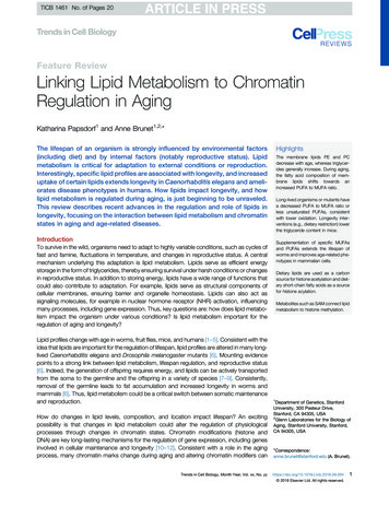
Luciferase does not alter metabolismTable 2 Pairwise comparisons of Raji cells under nutritional stressanalyzed by XCMS RPLCRaji emptyvectorstarved vs.Raji lucpuro starved1,0470 (NS)4,3050 (NS)Raji emptyvector fedvs. Raji lucpuro fed1,1830 (NS)4,5920 (NS)Raji luc-purostarved vs.Raji lucpuro fedRaji emptyvectorstarved vs.Raji emptyvector fed1,277374,867271,147214,8199NS Not statistically significantcells during tumor engraftment in vivo, but does not addresspotential luciferase impacts on treatment responses.Fig. 2 Cloud plots of dysregulated features p \ 0.001, foldchange [ 2, intensity [ 10,000 of SUP-T1 parental cells comparedto SUP-T1 luciferase (zeo), and SUP-T1 parental compared to RajiparentalWe also examined the effect of LucSh expression withpuromycin selection on Raji cells. Again, only six featureswere significantly different between parental Raji cells andtheir LucSh-transduced derivative line, which would mostlikely have occurred due to chance (the total number ofaligned features was 5,862 and p B 0.001) whereas onlyone metabolite was different between empty vector andRaji cells. Notably, that single difference was identified asN-acetyl puromycin, a product of the detoxifying antibioticresistance enzyme puromycin N-acetyl-transferase; thismetabolite was identified in the METLIN database, andconfirmed by accurate mass measurement and tandem MS.The absence of puromycin itself in the empty vectorsamples demonstrates the efficiency of cell washing procedures, and suggests that the metabolites being measuredare intracellular products of puromycin metabolism.These results demonstrate that engineered lymphoma linescan be inured to BLI reporter gene expression and are metabolically indistinguishable from their parental lines grownunder identical conditions. This finding increases confidencethat cultured BLI reporter cells behave similarly to parental3.4 Luciferase expression does not alter stressed cellmetabolismRaji cells transduced with an empty puromycin resistancevector or the same vector expressing LucSh, were grownfor 4 h in fresh medium or under serum starvation stress,then extracted for RPLC/MS and HILIC/MS metaboliteprofiling. Previous experiments had shown that serumwithdrawal caused increased lactate dehydrogenaserelease from Raji cells, after 120 min. Cells weretherefore starved for 4 h, tested for viability in theViacountÒ assay, and extracted for metabolite profiling.By 4 h, the starved cells were noticeably more adherentthan cells growing in complete medium, and dye permeability was increased by about 10 % in both emptyvector and luciferase lines (data not shown). Meta-analysis of HILIC/MS and RPLC/MS data showed that Rajicell starvation responses were pronounced, but largelyunaffected by luciferase expression when compared tothose of empty vector controls (p B 0.001, Table 2).HILIC results, for example, show that starvation leads tothe dysregulation of 37 and 21 metabolites in LucShand empty vector-transduced cells, respectively whencompared to fed cells. In contrast, no significant differences were seen when comparing these cell lines withina given nutrient condition.123
C. H. Johnson et al.4 ConclusionLuciferase-expressing cell lines have been used in thousandsof cancer studies, including BLI studies where luciferase ishighly overexpressed, highlighting the question of whetherthis manipulation creates metabolic perturbations. Untargeted metabolomics was used to examine three differenttypes of lymphoblastic leukemia cell lines, Ramos, Raji andSUP-T1 to determine if they underwent significant metabolic change in response to luciferase overexpression. Whilemetabolic differences were seen between parental Ramos,Raji and SUP-T1 cancer cell lines, no significant differenceswere observed when comparing the respective parental andluciferase-expressing cells, revealing that luciferase did notsignificantly perturb cellular metabolism. Furthermore, neither an empty expression vector nor the LucSh constructused here affected lymphoma metabolism under basal orperturbed conditions, suggesting luciferase reporter geneexpression itself is unlikely to alter cellular metabolism orinject ambiguity into interpretations of treatment responses.It is possible that luciferase effects on cellular metabolism remain undetected here because they take place duringor immediately after vector transduction and antibioticselection, and that our results instead reflect a steady-stateadaptation of lymphoma cells to our luciferase expressionsystem. The remarkable metabolic reproducibility ofparental and transduced lymphoma cell lines argues againstthis, and further highlights the utility of our approach forexamining additional aspects of luciferase biology andcellular stress responses. For example, treatment withluciferase inhibitor drugs or inducible genetic knockdownof luciferase in cells could be pursued to determine whetherdifferences in luciferase activity alters cell metabolismover a different time course than that examined in thesestudies. It also remains possible that other enzymaticreporters might alter cellular metabolism, such as wild-typeluciferases localized to peroxisomes [(Gould et al. 1987),(Ellis et al. 2010)], but overall our metabolomic analysis ofRaji, SUP-T1 and Ramos cancer cell lines has revealed thatthis BLI-optimized luciferase construct does not confer asignificant metabolic signature to the cells under basal andperturbed conditions.5 Availability of supporting dataThe metabolomics data sets are available on XCMS Onlineunder the public shares tab (https://xcmsonline.scripps.edu/).Acknowledgments The authors thank Cathy Zhang and Max Hallin(Pfizer Oncology Research) for insightful discussions. These studieswere funded by Pfizer and National Institutes of Health grants5R01CA170737-02 (GS and BHF), P30 MH062261-13 (GS),1R21CA170492-01 (GS) and W81XWH-13-1-0402 (GS).123Conflict of interestsThe authors declare no Conflict of interests.ReferencesBrutkiewicz, S., Mendonca, M., Stantz, K., Comerford, K., Bigsby,R., Hutchins, G., et al. (2007). The expression level of luciferasewithin tumour cells can alter tumour growth upon in vivobioluminescence imaging. Luminescence, 22, 221–228.Czupryna, J., & Tsourkas, A. (2011). Firefly luciferase and RLuc8exhibit differential sensitivity to oxidative stress in apoptoticcells. PLoS ONE, 6, e20073.Ellis, J. M., Frahm, J. L., & Li, L. O. (2010). Acyl-coenzyme asynthetases in metabolic control. Current Opinion Lipidol, 21,212–217.Gatignol, A., Durand, H., & Tiraby, G. (1988). Bleomycin resistanceconferred by a drug-binding protein. FEBS Lett, 230, 171–175.Gould, S. G., Keller, G. A., & Subramani, S. (1987). Identification ofa peroxisomal targeting signal at the carboxy terminus of fireflyluciferase. J Cell Biol, 105, 2921–2923.Greer, L. F, 3rd, & Szalay, A. A. (2002). Imaging of light emissionfrom the expression of luciferases in living cells and organisms:A review. Luminescence, 17, 43–74.Gulick, A. M. (2009). Conformational dynamics in the Acyl-CoAsynthetases, adenylation domains of non-ribosomal peptidesynthetases, and firefly luciferase. ACS Chem Biol, 4, 811–827.Guranowski, A., Sillero, M. A. G., & Sillero, A. (1990). Fireflyluciferase synthesizes P1, P 4-bis (50 -adenosyl) tetraphosphate(Ap4A) and other dinucleoside polyphosphates. FEBS Lett, 271,215–218.Ivanisevic, J., Zhu, Z., Plate, L., Tautenhahn, R., Chen, S., O’Brien, P.J., et al. (2013). Toward ‘Omic’ scale metabolite profiling: a dualseparation—mass spectrometry approach for coverage of lipidsand central carbon metabolism. Anal Chem, 85, 6876–6884.Keller, G. A., Gould, S., Deluca, M., & Subramani, S. (1987). Fireflyluciferase is targeted to peroxisomes in mammalian cells. ProcNatl Acad Sci USA, 84, 3264–3268.Keyaerts, M., Remory, I., Caveliers, V., Breckpot, K., Bos, T. J.,Poelaert, J., et al. (2012). Inhibition of firefly luciferase bygeneral anesthetics: effect on in vitro and in vivo bioluminescence imaging. PLoS ONE, 7, e30061.McElroy, W. D., DeLuca, M., & Travis, J. (1967). Molecularuniformity in biological catalyses. The enzymes concerned withfirefly luciferin, amino acid, and fatty acid utilization arecompared. Science, 157, 150–160.Milsom, C. C., Lee, C. R., Hackl, C., Man, S., & Kerbel, R. S. (2013).Differential post-surgical metastasis and survival in SCID, NODSCID and NOD-SCID-IL-2Rgamma(null) mice with parental andsubline variants of human breast cancer: implications for hostdefense mechanisms regulating metastasis. PLoS ONE, 8, e71270.Oba, Y., Iida, K., & Inouye, S. (2009). Functional conversion of fattyacyl-CoA synthetase to firefly luciferase by site-directed mutagenesis: a key substitution responsible for luminescence activity.FEBS Lett, 583, 2004–2008.Oba, Y., Ojika, M., & Inouye, S. (2003). Firefly luciferase is abifunctional enzyme: ATP-dependent monooxygenase and along chain fatty acyl-CoA synthetase. FEBS Lett, 540, 251–254.Oliva-Trastoy, M., Defais, M., & Larminat, F. (2005). Resistance tothe antibiotic Zeocin by stable expression of the Sh ble gene doesnot fully suppress Zeocin-induced DNA cleavage in human cells.Mutagenesis, 20, 111–114.Patti, G. J., Tautenhahn, R., Rinehart, D., Cho, K., Shriver, L. P.,Manchester, M., et al. (2013). A view from above: cloud plots tovisualize global metabolomic data. Anal Chem, 85, 798–804.
Luciferase does not alter metabolismPatti, G. J., Tautenhahn, R., & Siuzdak, G. (2012). Meta-analysis ofuntargeted metabolomic data from multiple profiling experiments. Nat Prot, 7, 508–516.Schipper, M. L., Patel, M. R., & Gambhir, S. S. (2006). Evaluation offirefly luciferase bioluminescence mediated photodynamic toxicity in cancer cells. Mol Imaging Biol, 8, 218–225.Sim, H., Bibee, K., Wickline, S., & Sept, D. (2011). Pharmacokineticmodeling of tumor bioluminescence implicates efflux, and notinflux, as the bigger hurdle in cancer drug therapy. Cancer Res,71, 686–692.Tautenhahn, R., Patti, G. J., Rinehart, D., & Siuzdak, G. (2012).XCMS Online: a web-based platform to process untargetedmetabolomic data. Anal Chem, 84, 5035–5039.Theodossiou, T., Hothersall, J. S., Woods, E. A., Okkenhaug, K.,Jacobson, J., & MacRobert, A. J. (2003). Firefly luciferinactivated rose bengal: in vitro photodynamic therapy byintracellular chemiluminescence in transgenic NIH 3T3 cells.Cancer Res, 63, 1818–1821.Thorne, N., Shen, M., Lea, W. A., Simeonov, A., Lovell, S., Auld, D.S., et al. (2012). Firefly luciferase in chemical biology: acompendium of inhibitors, mechanistic evaluation of chemotypes,and suggested use as a reporter. Chem Bol, 19, 1060–1072.Tiffen, J. C., Bailey, C. G., Ng, C., Rasko, J. E., & Holst, J. (2010).Luciferase expression and bioluminescence does not affecttumor cell growth in vitro or in vivo. MolCancer, 9, 299.van Staveren, W. C., Solis, D. Y., Hebrant, A., Detours, V., Dumont,J. E., & Maenhaut, C. (2009). Human cancer cell lines:Experimental models for cancer cells in situ? For cancer stemcells? Biochim et Biophys Acta, 1795, 92–103.Watkins, P. A., & Ellis, J. M. (1822). Peroxisomal acyl-CoAsynthetases. Biochim et Biophys Acta, 2012, 1411–1420.Zhang, C. C., Yan, Z., Li, W., Kuszpit, K., Painter, C. L., Zhang, Q.,et al. (2012). [(18)F]FLT-PET imaging does not always ‘‘lightup’’ proliferating tumor cells. Clin Cancer Res, 18, 1303–1312.Zhu, Z. J., Schultz, A. W., Wang, J., Johnson, C. H., Yannone, S. M.,Patti, G. J., et al. (2013). Liquid chromatography quadrupoletime-of-flight mass spectrometry characterization of metabolitesguided by the METLIN database. Nat Prot, 8, 451–460.123
Luciferase does not alter metabolism in cancer cells . 10724 Science Center Drive, San Diego, CA, USA e-mail: peter.obrien2@pfizer.com B. H. Felding Departments of Molecular and Experimental Medicine and Chemical Physiology, The Scripps Research Institute, La Jolla, CA, USA 123 Metabolomics DOI 10.1007/s11306-014-0622-5. 1 Introduction