Transcription
Yang et al. Fisheries and Aquatic (2020) 23:24RESEARCH ARTICLEOpen AccessIshige okamurae reduces blood glucoselevels in high-fat diet mice and improvesglucose metabolism in the skeletal muscleand pancreasHye-Won Yang1†, Myeongjoo Son2,3†, Junwon Choi2,3, Seyeon Oh3, You-Jin Jeon1,4, Kyunghee Byun2,3* andBo Mi Ryu1,4*AbstractBrown alga (Ishige okamurae; IO) dietary supplements have been reported to possess anti-diabetic properties.However, the effects of IO supplements have not been evaluated on glucose metabolism in the pancreas andskeletal muscle. C57BL/6 N male mice (age, 7 weeks) were arranged in five groups: a chow diet with 0.9% saline(NFD/saline group), high-fat diet (HFD) with 0.9% saline (HFD/saline group). high-fat diet with 25 mg/kg IO extract(HFD/25/IOE). high-fat diet with 50 mg/kg IO extract (HFD/50/IOE), and high-fat diet with 75 mg/kg IO extract (HFD/75/IOE). After 4 weeks, the plasma, pancreas, and skeletal muscle samples were collected for biochemical analyses.IOE significantly ameliorated glucose tolerance impairment and fasting and 2 h blood glucose level in HFD mice.IOE also stimulated the protein expressions of the glucose transporters (GLUTs) including GLUT2 and GLUT4 andthose of their related transcription factors in the pancreases and skeletal muscles of HFD mice, enhanced glucosemetabolism, and regulated blood glucose level. Our results suggest Ishige okamurae extract may reduce bloodglucose levels by improving glucose metabolism in the pancreas and skeletal muscle in HFD-induced diabetes.Keywords: Ishige okamurae, Diabetes, High-fat diet mice, Skeletal muscle, PancreasBackgroundType 2 diabetes is the complex metabolic disorders characterized by an abnormal glucose metabolism arisingfrom pancreatic β-cell dysfunction and glucose intolerance (Hamza et al. 2010, Guilherme et al. 2008, Bellet al. 2001). Glucose metabolism is an essential target totreat type 2 diabetes (Rines et al. 2016, Tsuneki et al.2004). Controlling blood glucose levels requires the establishment of balance between glucose utilization,* Correspondence: khbyun1@gachon.ac.kr; bmryu@jejunu.ac.kr†Hye-Won Yang and Myeongjoo Son contributed equally to this work.2Department of Anatomy & Cell Biology, Gachon University College ofMedicine, Incheon 21565, Republic of Korea1Department of Marine Life Science, School of Marine Biomedical Sciences,Jeju National University, 1 Ara 1-dong, Jejudaehak-ro, Jeju 63243, Republic ofKoreaFull list of author information is available at the end of the articleglucose production, and glucose transport (Giuglianoet al. 2008). Previous studies have emphasized adults ina state of glucose intolerance and obesity of children andadolescents are at increased risk of disrupted glucosemetabolism (Wiegand et al. 2005). Furthermore, it hasbeen established glucose intolerance and β-cell dysfunction are characteristics of the pathogenesis of impairedglucose metabolism. Hyperglycemia and glucose intolerance are suppressed by improved glucose transport,which is regulated by the GLUT transporter family (e.g.,GLUT2 and GLUT4), and members of this family are essential for the controlling blood glucose level (Nishiumiet al. 2010, Yamashita et al. 2012). Herman and Kahn reported glucose transport occurs in the pancreatic β-cells,skeletal muscle, adipose tissue, and brain and that it is The Author(s). 2020 Open Access This article is licensed under a Creative Commons Attribution 4.0 International License,which permits use, sharing, adaptation, distribution and reproduction in any medium or format, as long as you giveappropriate credit to the original author(s) and the source, provide a link to the Creative Commons licence, and indicate ifchanges were made. The images or other third party material in this article are included in the article's Creative Commonslicence, unless indicated otherwise in a credit line to the material. If material is not included in the article's Creative Commonslicence and your intended use is not permitted by statutory regulation or exceeds the permitted use, you will need to obtainpermission directly from the copyright holder. To view a copy of this licence, visit http://creativecommons.org/licenses/by/4.0/.
Yang et al. Fisheries and Aquatic Sciences(2020) 23:24essentially required for blood glucose homeostasis (Herman et al. 2006).The glucose transporter 2 (GLUT2) in pancreatic βcells is demanded for glucose-stimulated glucose tolerance and insulin secretion, which are both reduced indiabetic mouse models (Bonny et al. 1997). On the otherhand, glucose transporter 4 (GLUT4) in the skeletalmuscle is a key glucose transporter and plays an essential role in the extracellular glucose transport intoinsulin-sensitive cells (Chang et al. 2004, Shan et al.2011).Seaweeds are valuable sources containing diverse bioactive substances with potentials as nutraceutical andpharmaceutical agents (Lee et al. 2010, Kiuru et al.2014), and recent studies have reported seaweeds andtheir active substances can improve glucose metabolismby stimulating glucose transport in diabetes (Muruganet al. 2015, Sharifuddin et al. 2015). Ishige okamurae(IO), as an edible brown seaweed, has been shown tocontain biologically active substances like diphlorethohydroxycarmalol (DPHC), ishophloroglucin A, and fucoxanthin and associated secondary metabolites (Sanjeewaet al. 2017). Furthermore, it was shown in a previousstudy that an ethanolic IO extract (IOE) regulated bloodglucose level by increasing the level of glucosemetabolizing enzyme in the liver and promoting insulinresistance in db/db mice which is a leptin receptordeficient model (Min et al. 2011). However, the effect ofIOE has not been investigated on the glucose metabolism in the pancreas and muscle of high-fat diet (HFD)fed mice (a well-established model of glucose intolerance) (Honors et al. 2012, Riant et al. 2009). Accordingly, we evaluated the effects of an IOE on the proteinlevels of glucose GLUT2 and GLUT4 and on those oflinked transcription factors of the pancreas and skeletalmuscle and on blood glucose levels in HFD mice.Materials and methodsExtraction of Ishige okamuraeThe 50% of ethanol extract of Ishige okamurae (IOE)was supplied by Shinwoo Co. Ltd. (Lot No. SW9E29SA,Republic of Korea) as previously described by Yang et al.(2019). Depending on a previous method (Ryu et al.2018), the IOE used to this study was standardized byassuming diphlorethohydroxycarmalol (DPHC, 2.37%)via HPLC analysis. The HPLC chromatogram of DPHCis shown in Figure A1. The chromatographic analyseswere carried out by an Alliance 2695 Separations Module equipped with a 2998 PDA detector (all from Waters, Milford, MA, USA) and an Agilent poroshell 120EC-C18 column (4.6 100 mm, 4 μm). And, the mobilephase contained (A) 0.1% formic acid in water and (B)ACN with 0.1% formic acid. The eluting conditions ofHPLC were as follows: 20–40% B for 25 min, followingPage 2 of 9that re-equilibration time of the column for 10 mins.The rate of flow was kept at 0.3 mL/min and the injection volume was 10 μL. The content of DPHC was validated at Korea Health Supplement Institute (KHSI;Seongnam-si, Gyeonggi-do, Republic of Korea). Thepresence of DPHC was inspected by quadrupole timeof-flight lipid chromatography-mass spectrometry (QTOF LC-MS/MS) using an electrospray ionization (ESI)source (maXis-HD, Bruker Daltonics, Breman, Germany)at the Korea Basic Science Institute (KBSI; Ochang,South Korea).High-fat diet-fed mouseC57BL/6N male mouse (age, 7 weeks) was purchasedfrom Orient Bio Inc. (Republic of Korea) and housed individually in stainless steel cages at 24 C and 45–50%RH with a 12-h light-dark cycle. Before the experimentalperiod, all mice were fed a control chow diet for 1 week.All mice were randomly allocated to five groups (n 3for each group): mice were fed a chow diet for 8 weekswith 0.9% saline (wt/wt on chow diet) for the last 4weeks (the NFD/saline group); mice were fed a 45%high-fat diet (HFD) (Research Diet Inc., USA) for 8weeks with 0.9% saline (wt/wt on HFD) for the last 4weeks (the HFD/saline group); and mice were fed the45% HFD for 8 weeks with 25 mg/kg (HFD/25 IOE), 50mg/kg (HFD/50 IOE), or 75 mg/kg (HFD/75/IOE) ofIOE during the latter 4 weeks. Mice were sacrificed onexperimental day 57 in accordance with guidelinessigned by the Animal Care and Use Committee ofGachon University (IACUC; LCDI-2018-0112).Glucose tolerance testing (IPGTT)The intraperitoneal injection of glucose (2 g/kg of bodyweight) in mice that had been fasted for 8 h and bloodwas obtained from a tail vein 30, 60, 90, or 120 minlater, as previously described (Konrad et al. 2002). Serumglucose level was measured using a blood glucose meter(AccuChek Advantage; Roche Diagnostics Corp., USA).ImmunohistochemistryParaffin blocks of pancreatic tissue were sectioned at5 μm, laid on coating slides, dried at 40 C for 12 h, anddeparaffinized using xylene. And then, the deparaffinizedtissue sections were incubated in 0.3% hydrogen peroxide (Sigma-Aldrich, USA) for 30 min, washed 3 timeswith PBS (phosphate-buffered saline), blocked with normal animal serum, incubated with anti-PCNA antibody(Abcam, USA), and rinsed with PBS 3 times. The tissuesections were incubated with biotinylated secondaryantibodies in the ABC kit (Vector Laboratories, USA),washed 3 times with PBS, treated with DAB (3, 3diaminobenzidine) substrate to 0.03% hydrogen peroxidefor 10 min, mounted using a xylene-based DPX
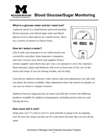
Yang et al. Fisheries and Aquatic Sciences(2020) 23:24mounting solution (Sigma-Aldrich, USA), and photographed under the microscope (Olympus Optical Co.,Japan).ImmunofluorescenceThe pancreas and skeletal muscle (soleus and gastrocnemius muscle) paraffin block section (5 μm) was deparaffinized, treated with animal serums for blockingantibody binding, and incubated with anti-GLUT2 andanti-GLUT4 antibodies (Santa Cruz Biotechnology, Inc.,USA) at 4 C for 1 day. And then, the tissue sectionswere washed with PBS, incubated with fluorescence conjugated secondary antibody at room temperature for 1 h,stained with DAPI (4′ 6-diamidino-2-phenylindole;Sigma-Aldrich, USA) solution for 1 min, rinsed with PBS3 times, and mounted using Vectorshield solution (Vector Laboratories). Fluorescences were detected usingconfocal microscopy (LSM 700; Carl Zeiss Ltd.,Germany).Quantitative real-time polymerase chain reactionTotal RNAs of mouse’s pancreases and skeletal muscles(soleus and gastrocnemius) were lyzed using the RNAisoPlus kit (TAKARA, Japan). Briefly, cell pellets were suspended using 1000 μl RNAiso Plus by pipetting, and lysates were mixed with 100 μl chloroform (Amresco LLC,USA) and centrifuged at 12,000 g at 4 C for 15 mins.Obtained supernatants were mixed with 250 μl isopropanol and centrifuged, and the RNA pellets so obtainedwere either rinsed with 70% ethyl alcohol and centrifuged at 7500 g at room temperature for 5 min or dissolved into 30 μl of diethyl pyrocarbonate (DEPC)treated water. The levels of RNA were quantified by aNanodrop 2000 (Thermo Fisher Scientific, Inc., USA). Inorder to conduct quantitative real-time polymerase chainreaction (qRT-PCR), complementary DNA (cDNA) synthesis was performed using a cDNA synthesis kit(TAKARA). Subsequently, cDNAs, SYBR green (TAKARA), and adaptive primers were mixed and amplified inPCR (Bio-Rad, USA) to profile gene expression. Validated genes are listed in Table A1.Hematoxylin and eosin stainingParaffin block slides of pancreas tissues were deparaffinized, stained with Mayer’s hematoxylin solution(DAKO, England) for 30 s and eosin Y solution(Sigma-Aldrich, USA) for 10 s, rinsed 3 times withdistilled water, and mounted using xylene-based DPXmounting solution. H&E stained tissue slide imageswere captured by light microscopy and pancreaticislet sizes were calculated using the Image J software(NIH, USA).Page 3 of 9Data analysisThe significances of intergroup differences were identified using the Kruskal-Wallis test. And, post-hoc comparisons were determined using Mann-Whitney U test.The analysis was conducted using SPSS software, and results are expressed to means SDs. Statistical analysiswas admitted by p values of 0.05. An asterisk indicatessignificantly different from the NFD/saline group and anumber sign significantly different from the HFD/salinegroup.ResultsEffect of IOE on glucose tolerance in HFD miceTo determine whether IOE reduces elevated blood glucose level in HFD mice, we investigated glucose tolerance in HFD mice. We supplemented HFD mice feedwith IOE (25, 50, or 75 mg/kg, the HFD/IOE groups) for4 weeks after mice had been fed HFD for 4 weeks. Intraperitoneal glucose tolerance test (2 g/kg, IPGTT) wasconducted at 30, 60, 90, or 120 min after injection ofglucose to investigate glucose tolerance. After 8 weeks,the HFD group gained more body weight compared withthe NFD group. However, the HFD/IO (25, 50, and 75mg/kg) group showed a slightly decreased in bodyweight, but no significance compared with the HFDgroup. Body weight is shown in Figure 2A. As shown inFig. 1a, HFD/saline controls (fed an HFD diet for 4weeks and followed by saline for another 4 weeks) hadlower glucose tolerance with higher blood glucose levelsat each time after glucose injection than the NFD controls (fed a chow diet for 8 weeks). However, HFD/IOEgroups (fed a HFD diet for 8 weeks and IOE at 25, 50, or75 mg/kg for the latter 4 weeks) showed significant reductions in glucose levels at 120 min after glucose injection than were observed in the HFD group. AUC (areaunder the curve) levels of IPGTT were expressed to estimate the level of glucose tolerance impairment in eachgroup. In line with the results shown in Fig. 1, the AUCof IPGTT in the HFD group was remarkably greater (p 0.001) than in the NFD group (Fig. 1b). HFD/IOE (25,50, or 75 mg/kg) groups showed dose-dependent decreases in the AUC of IPGTT. The maintenance of fasting and 2 h glucose levels is a major therapeutic goal indiabetes (Ratner 2001). In Fig. 1c and d, both fasting and2 h glucose levels in the HFD group were significantlyhigher than in the NFD group. However, fasting and 2 hglucose levels in the HFD/IOE 50- and 75-mg/kg groupswere significantly lower than in the HFD group. Theseresults demonstrate that IOE can ameliorate glucose tolerance in HFD mice. Furthermore, we examined the improvement of IOE on glucose metabolism in thepancreas and muscle tissue of HFD mice in controllingblood glucose levels.
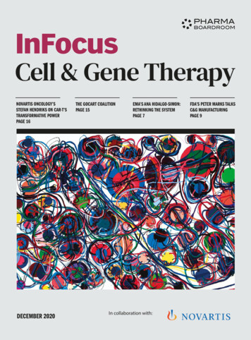
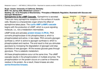
Yang et al. Fisheries and Aquatic Sciences(2020) 23:24Page 4 of 9Fig. 1 Effect of IO on serum glucose levels in high-fat diet (HFD)-fed mice model. The mice receiving 45% HFD for 8 weeks exhibit amelioratedglucose tolerance; however, IOE oral administration decreased glucose tolerance. a, b Intraperitoneal glucose tolerance test (IPGTT) was performed andanalyzed area under the curve (AUC) from the IPGTT in all mice groups. c Fasting and d 2 h glucose levels were in all mouse groups. Data arepresented as mean S.D. **p 0.01 or ***p 0.001 vs NFD/saline group; #p 0.05, ##p 0.01, or ***p 0.001 vs HFD/saline group. NS, not significantEffect of IOE on β-cell function in the pancreatic islets ofHFD miceIn order to investigate the effect of IOE on the morphologies and areas of the pancreatic islets of HFD mice, we conducted hematoxylin and eosin (H&E) staining andimmunohistochemistry (Matveyenko et al. 2009, Sun et al.2016). The proliferating cell nuclear antigen (PCNA, a proliferation marker) was used to detect the proliferation of βcells (Bringhenti et al. 2013). In order to identify the effectof IOE on the proliferation of β-cells in pancreatic islets, wemeasured PCNA intensities (Fig. 2a, b). PCNA intensitieson β-cells in pancreatic islets of the HFD group were remarkably greater than in the NFD group, whereas PCNAintensities were dose-dependently decreased in the HFD/IOE groups as compared to those of the HFD group. H&Estaining pancreatic islets were larger in HFD than in NFDmice (Fig. 2c, d), and that this increase in size was significantly and dose-dependently diminished in the HFD/IOEgroups (25, 50, or 75 mg/kg). These data indicated that IOEcan improve β-cell function in the pancreatic islet of HFDmice.Effect of IOE on pancreas dysfunction in HFD miceHFD-induced diabetes is related to failure of β-cell function (Matveyenko et al. 2009, Cerf. 2006), and GLUT2 isessential for β-cell glucose sensing, which leads toglucose-induced insulin secretion (Cerf. 2006, Folli et al.2011). In order to examine the effect of IOE on GLUT2stimulation in pancreases of HFD mice, we evaluate theintensity of GLUT2 by immunofluorescence (Fig. 3a, b).The immunofluorescence intensity of GLUT2 in HFDmice was remarkably lower than in NFD mice (p 0.05),and this reduction in immunofluorescence was slightlyreduced in the HFD/IOE 20- and 50-mg/kg groups, butsignificantly reduced in the 75-mg/kg group.In addition, the GLUT2 transcription is controlled bypancreatic duodenal homeobox 1 (PDX1) and Hexokinase isoforms 1 (HK1) and 2 (HK2), which are also included in glucose sensing and glucose metabolism. Weevaluated the effect of IOE on the mRNA levels ofPDX1, HK1, and HK2 in pancreas tissues of HFD miceusing qRT-PCR. mRNA levels of PDX1, HK1, and HK2were remarkably decreased in HFD controls comparedwith NFD controls, which showed levels of GLUT2related transcription factors are impaired in HFD mice(Fig. 3c). However, these falls in PDX1 and HK1 mRNAlevels were significantly prevented in the HFD/IOE 50and 75-mg/kg groups. Also, HK2 mRNA levels were significantly increased in the three HFD/IOE groups compared to those of HFD controls. These results showed
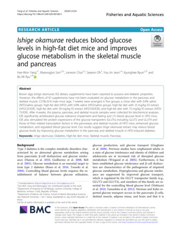
Yang et al. Fisheries and Aquatic Sciences(2020) 23:24Page 5 of 9Fig. 2 Effects of IO on the morphologies and areas of pancreatic islets in high-fat diet (HFD)-fed mice model. a Proliferation marker of cell(proliferating cell nuclear antigen, PCNA) was evaluated by immunohistochemistry, and b quantitative graph shows the PCNA expression level onpancreatic islets from representative images. c Hematoxylin and eosin (H&E)-stained pancreatic islets (round shape and pale color) and d the sizeof pancreatic islets were measured by the Image J software from representative images (scale bar 100 μm). Data are presented as mean S.D.**p 0.01 vs NFD/saline group; #p 0.05, ##p 0.01, or ###p 0.001 vs HFD/saline groupFig. 3 Effects of IO on GLUT2 expression increase of pancreas tissues in high-fat diet (HFD)-fed mice model. a Confocal images show GLUT2(green) expression and nuclei (blue; DAPI) were evaluated of pancreas tissues of all mice groups. b Quantitative graph shows the level of GLUT2expression from representative images using the Zen 2012 software. c The mRNA levels of Pdx1, Hk1, and Hk2 were evaluated by qRT-PCR (scalebar 100 μm). Data are presented as mean S.D. *p 0.05 or **p 0.01 vs NFD/saline group; #p 0.05 or ##p 0.01 vs HFD/saline group. NS,not significant
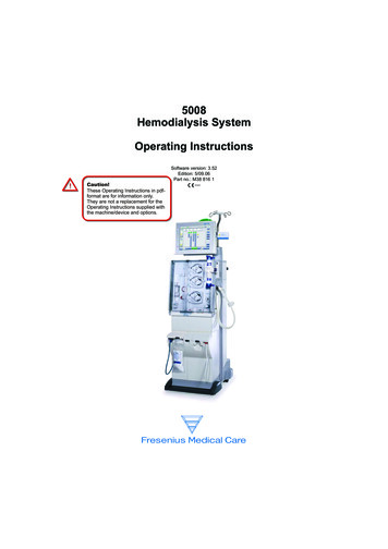
Yang et al. Fisheries and Aquatic Sciences(2020) 23:24that the increased GLUT2 and its related transcriptionfactors PDX1, HK1, and HK2 by IOE can improve glucose metabolism in the pancreas.Effect of IOE on glucose metabolism in the skeletalmuscle tissue of HFD miceGLUT4 levels and the levels of its related transcriptionfactors were evaluated in the skeletal muscle of HFDmouse for glucose metabolism, which is the primary siteof insulin-mediated GLUT4 translocation (Shepherdet al. 1998). The GLUT4 translocation into the membrane of the skeletal muscle in HFD mice was significantly decreased compared with NFD mice (p 0.01;Fig. 4a, b), and GLUT4 translocation was significantlygreater in the HFD/IOE groups than in HFD groups (p 0.05). These observations suggest IOE can improveglucose metabolism in HFD mice by increasing translocation of GLUT4 into the membrane of the skeletalmuscle.Glucose metabolism in the skeletal muscle is achievedby activation of HK2, phosphoinositide-3-kinase (PI3K)/AKT, and AMP-activated protein kinase (AMPK)pathways (Fukumoto et al. 1988, Tsuchiya et al. 2010).We measured the mRNA level of AKT, AMPK, andHK2 in the skeletal muscle tissue of HFD mice by qRTPCR to assess the related level of GLUT4 and glucosemetabolism by IOE. Consistent with our GLUT4Page 6 of 9translocation results, qRT-PCR showed the mRNA levelof AKT, AMAPK, and HK2 in the skeletal muscle wereremarkably lower in HFD than in NFD controls (Fig.3c), but that the mRNA levels of AKT and AMPK in thethree HFD/IOE groups were significantly increased compared to those of the HFD controls (p 0.01), and HK2mRNA levels in HFD/IOE 50- and 75-mg/kg groupswere significantly higher than in HFD controls (p 0.01). These results demonstrate that IOE enhanced thetranscription of GLUT4 and those of its related transcription factors AKT, AMPK, and HK2 in our mouseHFD model.DiscussionGiven that the underlying molecular mechanisms of type2 diabetes with insulin resistance are accompanied by βcell dysfunction, which is probable that the cross-talkbetween the pancreatic cells and skeletal muscle playsan important part. Thus, identification of a novel therapeutic agent that can control the glucose metabolism byenhancing the function of cells is key to restore theblood glucose homeostasis in type 2 diabetes patients.Of note is that IOE has been announced to have various biological activities related to diabetes. For example,it has been reported to inhibit α-glucosidase, improveglucose tolerance, upregulate levels of hepatic glucosemetabolites, and reduce insulin resistance in db/db miceFig. 4 Effects of IO on GLUT4 expression increase of skeletal muscle tissues in high-fat diet (HFD)-fed mice model. a Confocal images showGLUT4 (green) expression and nuclei (blue; DAPI) were evaluated of skeletal muscle tissue of all mice groups. b Quantitative graph shows thelevel of GLUT4 expression from representative images using Zen 2012 software (scale bar 100 um). c The mRNA levels of Akt, Ampk, and Hk2were evaluated by qRT-PCR. Data are presented as mean S.D. **p 0.01 vs NFD/saline group; #p 0.05 or ##p 0.01 vs HFD/saline group. NS,not significant
Yang et al. Fisheries and Aquatic Sciences(2020) 23:24(Min et al. 2011, Ryu et al. 2018, Heo et al. 2009). However, the effect of IOE on functions of glucose transportin the pancreases and skeletal muscle tissues is yet to beassessed in HFD mice that have been fed with an imbalanced diet to induce mild hyperglycemia, a conditionthat may resemble the pre-diabetic state in humans.Therefore, we firstly confirmed that the decreased glucose transporter function in the pancreases and skeletalmuscle tissues of HFD mice fed for 4 weeks was significantly ameliorated alongside improved glucose metabolism upon IOE administration for another 4 weeks.The severe glucose intolerance in HFD mice with decreased insulin sensitivity may decelerate β-cell functionin pancreatic islets, leading to excessive β-cell proliferation and increased pancreatic islet size (Matveyenkoet al. 2009, Carey et al. 1996, Butler et al. 2003). Hullet al. have shown that rodents fed with an HFD to induce insulin resistance develop increased pancreatic isletsize due to hyperplasia rather than hypertrophy of β-cells(Hull et al. 2005). We herein observed that the deceleratedβ-cell function in the pancreatic islet of HFD mice was noticeably improved by IOE administration, probably because of the restoration of the GLUT2 level. The majorglucose transporter GLUT2 in hypertrophied pancreaticislets is associated with glucose sensing to stimulate insulin secretion. The mechanism of the improvement of thepancreas function by IOE through GLUT2 upregulationin the islets is linked with the regulation of PDX1, HK1,and HK2 mRNA levels, enabling acquisition of glucoseresponsive insulin secretion (Frantz et al. 2013). PDX1mutant β-cells have been shown to activate the isletspecific GLUT2 and amyloid polypeptide, and these twofactors have been shown to be low in diabetic mice andpatients (Nicolino et al. 2010, Suzuki et al. 2003, Waeberet al. 1996). HK1 and HK2 play key roles in glucose sensing by catalyzing glycolysis through which glucose is phosphorylated to produce glycos-6-phosphate (Hashimotoet al. 2005). Epstein et al. have reported that the elevatedhexokinase activity of both HK1 and HK2 in β-cells canreduce the blood glucose level in diabetes patients (Epstein et al. 1992).The skeletal muscle of HFD mice is known to be defective in the membrane translocation of GLUT4 alongside impaired glucose metabolism and reduced insulinstimulated glucose transport (Fukumoto et al. 1988,Tremblay et al. 2001, Savage et al. 2007). IncreasedGLUT4 translocation by IOE probably facilitates glucoseuptake of the skeletal muscle in HFD mice, activatingthe PI3K/AKT and AMPK pathways, thereby regulatingthe glucose metabolism, including glycogen synthesis,gluconeogenesis, and glucose transport as well as the energy expenditure (Schultze et al. 2012). Consistent withthese findings, we found that the mRNA level of HK2,which promotes glucose transport and utilization, wasPage 7 of 9also upregulated in HFD mice fed with IOE (Fukumotoet al. 1988).In this study, our findings are summarized as follows:(1) IOE supplementation ameliorated blood glucose intolerance, (2) improved glucose metabolism in the pancreas, and (3) enhanced glucose transporters in theskeletal muscle. Additionally, our results suggest that theeuglycemic effects of IOE in HFD mice are because ofthe stimulation of the glucose metabolism.ConclusionThis study shows IOE (an ethanolic extract of Ishige okamurae) had euglycemic effects in our HFD-induced micemodel of diabetes and that these effected were associatedwith improved glucose metabolism in the pancreas andskeletal muscle and lower blood glucose levels. We suggest that IOE is considered a potential starting point fordeveloping the therapeutic or functional foods targetingthe alleviation of diabetes.Supplementary informationSupplementary information accompanies this paper at nal file 1: Figure A1. HPLC chromatogram of DPHC from IOEAdditional file 2: Table A1. List of qRT-PCR primer sequencesAdditional file 3: Figure A2. Effect of IO on body weight in high fatdiet (HFD)-fed mice model. Data are presented as mean S.D (n 3 foreach group). *** p 0.01 vs NFD/saline group. NS, not significant.AbbreviationsAMPK: AMP-activated protein kinase; cDNA: Complementary DNA;DEPC: Diethyl pyrocarbonate; DPHC: Diphlorethohydroxycarmalol;ESI: Electrospray ionization; GLUT2: Glucose transporter 2; GLUT4: Glucosetransporter 4; H&E: Hematoxylin and eosin; HFD: High-fat diet;HK1: Hexokinase isoforms 1; HK2: Hexokinase isoforms 2; IO: Ishige okamurae;IOE: Ethanolic IO extract; IPGTT: Intraperioneal glucose tolerance testing;PCNA: Proliferating cell nuclear antigen; PDX1: Pancreatic duodenalhomeobox 1; PI3-K: Phosphoinositide-3-kinase; qRT-PCR: Quantitative realtime polymerase chain reaction; Q-TOF LC-MS/MS: Quadrupole time-of-flightlipid chromatography-mass spectrometryAcknowledgementsThis research was a part of the project titled “Development of functionalfood products with natural materials derived from marine resources (no.20170285),” funded by the Ministry of Oceans and Fisheries, Korea. The IOEin this study was kindly provided by Dong-Min Chung in Shinwoo Co. Ltd.,Republic of Korea.Authors’ contributionsH.-W.Y.: Provided the substance and draft preparation. M.S. and S.O.: Dataassembly and interpretation. S.O. and J.C.: Formal analysis and datacollection. Y.-J.J. and K.B.: Study conceptualization and methodology. K.B. andB.R.: Supervision and provision of funding acquisition. The authors read andapproved the final manuscript.Availability of data and materialsThe data used to support the findings of this study are included within themanuscript.
Yang et al. Fisheries and Aquatic Sciences(2020) 23:24Ethics approval and consent to participateAnimal experiments were performed according to the guidelines signed bythe Animal Care and Use Committee of Gachon University (IACUC; LCDI2018-0112).Competing interestsThe authors declare that they have no competing interests.Author details1Department of Marine Life Science, School of Marine Biomedical Sciences,Jeju National University, 1 Ara 1-dong, Jejudaehak-ro, Jeju 63243, Republic ofKorea. 2Department of Anatomy & Cell Biology, Gachon University College ofMedicine, Incheon 21565, Republic of Korea. 3Functional Cellular NetworksLaboratory, Lee Gil Ya Cancer and Diabetes Institute, Gachon University,Incheon 21999, Republic of Korea. 4Marine Science Institute, Jeju NationalUniversity, Jeju 63333, Republic of Korea.Received: 21 May 2020 Accepted: 24 July 2020ReferencesBell GI, Polonsky KS. Diabetes mellitus and genetically programmed defects in βcell function. Nature. 2001;414:788.Bonny C, Roduit R, Gremlich S, Nicod P, Thorens B, Waeber G. The loss of GLUT2expression in the pancreatic β-cells of diabetic db/db mice is associated withan impaired DNA-binding activity of islet-specific trans-acting factors. MolCell Endocrinol. 1997;135:59–65.Bringhenti I, Moraes-Teixeira JA, Cunha MR, Ornellas F, Mandarim-de-Lacerda CA,Aguila MB. Maternal obesity during the preconception and early life periodsalters pancreatic development in early and adult life in male mouseoffspring. PLOS ONE. 2013;8:e55711.Butler AE, Janson J, Bonner-Weir S, Ritzel R, Rizza RA, Butler PC. β-cell deficit andincreased β-cell apoptosis in humans with type 2 diabetes. Diabetes. 2003;52:102–10.Carey DG, Jenkins AB, Campbell LV, Freund J, Chisholm DJ. Abdominal fat andinsulin resistance in normal and overweight women: direct measurementsreveal a strong relationship in subjects at both low and high risk of NIDDM.Diabetes. 1996;45:633–8.Cerf ME. High fat diet modulation of glucose sensing in the beta-cell. Med SciMonit. 2006;13:RA12–7.Chang L, Chiang S-H, Saltiel AR. Insulin signaling and the regulation of glu
However, the effects of IO supplements have not been evaluated on glucose metabolism in the pancreas and skeletal muscle. C57BL/6N male mice (age, 7weeks) were arranged in five groups: a chow diet with 0.9% saline (NFD/saline group), high-fat diet (HFD) with 0.9% saline (HFD/saline group). high-fat diet with 25mg/kg IO extract