Transcription
Gebeyew et al. Journal of Animal Science and 733-z(2022) 13:85RESEARCHOpen AccessLipid metabolism and m6A RNAmethylation are altered in lambssupplemented rumen-protectedmethionine and lysine in a low-protein dietKefyalew Gebeyew1,2, Chao Yang1,2, Hui Mi1,2, Yan Cheng1,2, Tianxi Zhang1,2, Fan Hu1,2, Qiongxian Yan1,2,Zhixiong He1,2,3, Shaoxun Tang1,2* and Zhiliang Tan1,2,3AbstractBackground: Methionine or lysine has been reported to influence DNA methylation and fat metabolism, but theircombined effects in N6-methyl-adenosine (m6A) RNA methylation remain unclarified. The combined effects ofrumen-protected methionine and lysine (RML) in a low-protein (LP) diet on lipid metabolism, m6A RNA methylation,and fatty acid (FA) profiles in the liver and muscle of lambs were investigated. Sixty-three male lambs were dividedinto three treatment groups, three pens per group and seven lambs per pen. The lambs were fed a 14.5% crudeprotein (CP) diet (adequate protein [NP]), 12.5% CP diet (LP), and a LP diet plus RML (LP RML) for 60 d.Results: The results showed that the addition of RML in a LP diet tended to lower the concentrations of plasmaleptin (P 0.07), triglyceride (P 0.05), and non-esterified FA (P 0.08). Feeding a LP diet increased the enzymeactivity or mRNA expression of lipogenic enzymes and decreased lipolytic enzymes compared with the NP diet.This effect was reversed by supplementation of RML with a LP diet. The inclusion of RML in a LP diet affected thepolyunsaturated fatty acids (PUFA), n-3 PUFA, and n-6 PUFA in the liver but not in the muscle, which might belinked with altered expression of FA desaturase-1 (FADS1) and acetyl-CoA carboxylase (ACC). A LP dietsupplemented with RML increased (P 0.05) total m6A levels in the liver and muscle and were accompanied bydecreased expression of fat mass and obesity-associated protein (FTO) and alkB homologue 5 (ALKBH5). The mRNAexpressions of methyltransferase-like 3 (METTL3) and methyltransferase-like 14 (METTL14) in the LP RML diet groupwere lower than those in the other two groups. Supplementation of RML with a LP diet affected only liver YTHdomain family (YTHDF2) proteins (P 0.05) and muscle YTHDF3 (P 0.09), which can be explained by limited m6Abinding proteins that were mediated in mRNA fate.* Correspondence: sxtang@isa.ac.cn1CAS Key Laboratory for Agro-Ecological Processes in Subtropical Region,National Engineering Laboratory for Pollution Control and Waste Utilizationin Livestock and Poultry Production, Hunan Provincial Key Laboratory ofAnimal Nutritional Physiology and Metabolic Process, Institute of SubtropicalAgriculture, The Chinese Academy of Sciences, Changsha 410125, Hunan,ChinaFull list of author information is available at the end of the article The Author(s). 2022 Open Access This article is licensed under a Creative Commons Attribution 4.0 International License,which permits use, sharing, adaptation, distribution and reproduction in any medium or format, as long as you giveappropriate credit to the original author(s) and the source, provide a link to the Creative Commons licence, and indicate ifchanges were made. The images or other third party material in this article are included in the article's Creative Commonslicence, unless indicated otherwise in a credit line to the material. If material is not included in the article's Creative Commonslicence and your intended use is not permitted by statutory regulation or exceeds the permitted use, you will need to obtainpermission directly from the copyright holder. To view a copy of this licence, visit http://creativecommons.org/licenses/by/4.0/.The Creative Commons Public Domain Dedication waiver ) applies to thedata made available in this article, unless otherwise stated in a credit line to the data.
Gebeyew et al. Journal of Animal Science and Biotechnology(2022) 13:85Page 2 of 17Conclusions: Our findings showed that the inclusion of RML in a LP diet could alter fat deposition throughmodulations of lipogenesis and lipolysis in the liver and muscle. These changes in fat metabolism may beassociated with the modification of m6A RNA methylation.Keywords: Lambs, Lipid metabolism, Low-protein, Lysine, Methionine, m6A RNA methylationIntroductionIncreased price of dietary protein sources and environmental concerns leading to feeding trends towards reduced dietary protein levels to meat-producing animals,including bulls and pigs [1, 2]. Reducing the levels ofdietary protein below the NRC recommendation is themost successful nutritional approach in lowering feedcost and nitrogen emissions [3]. However, feeding a lowprotein (LP) diet has been shown to affect the carcasscharacteristics by reducing the carcass lean content andincreasing fat accretion depending on the levels of dietary protein reduction and its amino acids composition[2, 4]. It also increased fat deposition by increasing thequantity of energy accessible for fat mass and restrictingprotein synthesis [5, 6]. One possible explanation couldbe a deficiency of one or more essential amino acid(EAA). Adding dietary EAA to the LP diet may balancethe fat accretion and improve protein synthesis. In thisregard, dietary methionine (Met) and lysine (Lys), toplimiting EAA in growing lambs, have been reported topromote protein synthesis and modulate lipid metabolism [7, 8].A diet containing deficient or adequate Met levels affected lipid turnover by altering the expression of genesrelated to fat metabolism in the liver and other tissues[8, 9]. The inclusion of low Met levels in the LP diet haslittle influence on fat accumulation, but those in a highprotein diet reduced fat accretion [5], suggesting thatthe effects of Met in the LP diet are influenced by otherEAAs in the diet. Meanwhile, increasing the levels ofdietary Lys has been reported to reduce lipid accretionand improve muscle protein synthesis through regulating key genes involved in various pathways [7]. This effect was connected with improved biosynthesis of Lcarnitine, which is reported to decrease carcass fat content and liver total lipids [10, 11]. Conversely, feedingLys deficient diet increased intramuscular fat content inlongissimus dorsi muscle [12]. The findings stated in theabove studies point out that dose-dependent influencesof dietary Met or Lys in lipid metabolism. However, thecombined effects of Met and Lys on the mechanisms ofcontrolling fat removal and accretion and de novo fattyacid synthesis in liver and muscle tissues of lambs underLP diet conditions are not clarified so far. In addition,considering the essence of evaluating N6-methyladenosine (m6A) methylation in the liver and muscleand the vital role of liver tissue in lipoprotein and lipidmetabolism, the present study focused on the liver andmuscle tissues even though the main site of de novo FAssynthesis in sheep is adipose tissue.Methionine and betaine serve as donors of methylgroups that are involved in the DNA and m6A RNAmethylation process. In this content, supplementation ofdietary Met affected the DNA methylation process by altering the SAM concentration [13]. Likewise, supplementation of dietary betaine affected the process of m6ARNA methylation [14, 15]. These researches suggested amodulatory role of methyl donors in epigenetic modification. The m6A is considered as the most commonform of intrinsic messenger RNA modification ineukaryotic cells [16]. It is a dynamic and reversibleprocess, and installation of a methyl group to m6A iscatalyzed by methyltransferases like 3 (METTL3), methyltransferases like 14 (METTL14), and Wilms’ tumor 1associating protein (WTAP) [17]. Fat mass and obesityassociated (FTO) and alkB homologue 5 (ALKBH5) havebeen identified as demethylase [18, 19], which promotethe dumping of a methyl group from m6A via differentmechanisms. Subsequently, the YTH domain family(YTHF1–3) proteins have recognized the modification ofm6A to influence mRNA maturation, splicing stability,decay, translation, and export [16]. These modificationsof RNA could affect several biological functions, such aslipid metabolism and energy homeostasis [20]. Moreover, alters in the m6A RNA methylation and lipid metabolism in various tissues may be inter-relatedmechanisms in response to supplementation of Met andLys. Thus, we hypothesized that the inclusion of rumenprotected methionine and lysine (RML) in the LP dietmay influence lipid metabolism through modulations ofm6A RNA methylation in growing lambs. Therefore, thestudy aimed to investigate m6A RNA methylation, lipidmetabolism, and fatty acid profiles in the liver and longissimus dorsi muscle of lambs fed a LP diet supplemented with RML.Materials and methodsAnimal Care and Use Committee of the Institute of Subtropical Agriculture were checked and approved all theexperimental protocols used in this trial (202020). Animals were handled in accordance with the protocol approved by the committee. The feeding trial wasperformed in Hulunbuir city, Inner Mongolia Autonomous Region, China.
(2022) 13:85Gebeyew et al. Journal of Animal Science and BiotechnologyAnimals, diets, and experimental designThis experimental procedure was in conjunction withour earlier research [21]. Briefly, the rumen-protected lysine (RPL, LysiPEARL) and methionine (RPM, MetiPEARL) products were obtained from Kemin IndustriesInc. (Iowa, USA). The RPL and RPM products are contained L-Lys monohydrochloride at 475 g/kg and DLMet at 500 g/kg, respectively. The rumen passage ratesof Lys and Met were about 85% and 66%, respectively,[22]. At four months of age and the initial body weight(BW) of 24.82 2.03 kg, sixty-three fat tail male Hulunbuir lambs were randomly allocated into three dietarygroups with three pens per group and seven lambs perpen. The lambs were fed a 14.5% crude protein (CP) diet(positive control, adequate-protein diet [NP]); a 12.5%CP diet (negative control; LP); and a LP diet with RML(LP RML). The quantity of RPM (0.253 g/kg DM) andTable 2 Amino acids and fatty acid composition of theexperimental dietsItemTreatmentaNPLP20.0026.0LP RMLIngredient, % of DM basisCorn26.00Barley20.8720.8720.87Wheat bran8.728.728.72Soybean meal12.56.56.5Cottonseed meal3.03.03.0Sodium bicarbonate0.70.70.7Salt0.50.50.5Magnesium oxide0.50.50.5Calcium carbonate0.20.20.2Beet molasses1.01.01.0Wheat en protected methionine, g/kg of DM––0.253Rumen protected lysine, g/kg of 4612.46NDF34.133.230.5ADF15.214.113.3Metabolize energy, MJ/kg9.559.55GE, MJ/kg17.0517.04NPLPLP C18:1n-9c17.0115.8115.54Fatty acids composition, %C18:2n-6c45.9045.9746.01C18:3n-37.497.317.32aNP adequate protein diet, LP low protein diet, LP RML low protein diet withrumen-protected methionine and lysineChemical composition, % of DMaTreatmentaAmino acid composition, %Table 1 Ingredients and chemical composition (% DM basis) ofthe treatment dietsItemPage 3 of 179.5517.01NP adequate protein diet, LP low protein diet, LP RML low protein diet withrumen-protected methionine and lysinebSupplied per kilogram of dietary DM: 221.31 mg of Fe, 16.23 mg of Cu, 71.31mg of Zn, 71.31 mg of Mn, 1.23 mg of I, 0.54 mg of Co, 1.70 mg of Se, 16,250IU of vitamin A, 5000 IU of vitamin D, and 40 IU of vitamin ERPL (0.50 g/kg DM) supplemented to the lambs were selected following our previous results [23, 24].The total experimental period was 75 d, including 15 dof acclimatization and 60 d of actual periods. The compositions and nutrient levels of the experimental diets areshown in Tables 1 and 2. Determination of dry matter(DM), ash, and crude protein (CP) of the feed were carriedout using the standard protocol of the Association of Official Analytical Chemists (AOAC) [25]. Acid-detergentfiber (ADF) and neutral detergent fiber (NDF) were measured following the methods of Van Soest et al. [26]. Theisothermal automatic calorimeter (5E-AC 8018, ChangshaKaiyuan Instruments Co. Ltd., Changsha, China) was usedto assay the gross energy (GE) content of feed. Extractionof total amino acids (AA) from the experimental feed wascarried out according to the standard procedure descriedby the AOAC [27]. The quantification of individual aminoacids from the extract was performed using HPLC
Gebeyew et al. Journal of Animal Science and Biotechnology(2022) 13:85(Agilent 1100; Agilent Technologies) following the standard protocol of Henderson et al. [28].Sample collectionBlood was sampled on d 60 (before morning feeding)from the jugular vein of lambs into 5-mL heparinizedtubes (Changsha Yiqun Medical Equipment Co., Ltd.,Hunan, China) to analyze plasma hormone and lipidrelated metabolites. The blood plasma was separated bycentrifugation at 3000 g for 15 min at roomtemperature and frozen at 80 C until analysis. Sevenor six lambs per treatment with BW close to the groupweight were chosen for slaughter. After evisceration, theweight of the carcass was recorded within 30 min ofslaughter. The liver samples were taken quickly, sealedin foil paper, and stored at 80 C for later analysis.Then, the longissimus dorsi muscle (muscle) was sampled at the 12/13th rib of each carcass, sealed in foilpaper, and stored at 80 C until analysis. The dressing percentage was computed as described by Wanget al. [4].Analysis of plasma metabolite and hormonesThe concentrations of plasma triglycerides (TAG), highdensity lipoprotein-cholesterol (HDL-C), total cholesterol (TC), and low-density lipoprotein-cholesterol(LDL-C) were analyzed using a Cobas C311 analyzer(Roche Di-agnostics, Rotkreuz, Switzerland) based onthe procedure described by Wang et al. [4]. The nonesterified fatty acid (NEFA), leptin (LEP), adiponectin(ADPN), and insulin (INSU) concentrations wereassayed using the enzyme-linked immunosorbent assay(ELISA) kits specific to sheep following the protocolgiven by the manufacturer’s manual (Fankel Co., Ltd.,Shanghai, China) as described by Gebeyew et al. [29].The results are presented as pg/mL. The sensitivity ofthe kits was 0.1 pg/mL.Measurement of tissue enzyme activity, concentration ofS-adenosylhomocysteine and L-carnitineLiver and muscle samples were weighed (100 mg) andhomogenized (1:9 w/v) with ice-cold normal saline buffer. Then, the upper liquids were separated by centrifugation at 3000 g for 15 min at 4 C to detect enzymeactivity. Another 100 mg of liver and muscle sampleswere weighed and homogenized (1:9 w/v) using a mixture of n-butanol, methanol, and deionized water at theratio of 5:25:70 (v:v:v). Then, the supernatants wereretained by centrifugation at 5000 g for 10 min at 4 Cto detect the L-carnitine. The activities of fibroblastgrowth factor 21 (FGF21), hormone-sensitive lipase(HSL), acetyl-CoA carboxylase (ACC), malic enzyme(ME), lipoprotein lipase (LPL), carnitine palmitoyltransferase I B (CPT1B), fatty acid-binding protein 4 (FABP4),Page 4 of 17fatty acid desaturase 1 (FADS1), and stearoyl-CoA desaturase (SCD) were determined in the liver and muscletissues using the ELISA kits specific to sheep followingthe recommended procedure (Jiangsu Meimian industrial Co., Ltd., Yancheng, China). The results were presented as pg/mg protein or ng/mg protein. The intraassay and inter-assay coefficient of variation (CV) of allELISA kits used to detect FGF21, HSL, ACC, LPL,CPT1B, FABP4, FADS1, SCD, and ME were 10% and 12%, respectively. The protein concentrations in the liverand muscle tissues were quantified using the BCA protein assay kit (Beyotime Biotech Inc., Shanghai, China).The levels of S-adenosylhomocysteine (SAH) and Lcarnitine were measured using the ELISA kits specific tosheep based on the manufacturers’ protocol (FankeweiCo., Ltd., Shanghai, China). The results are presented asng/g or pg/g [30]. The intra-assay and inter-assay CV ofELISA kits used to SAH and L-Carnitine were 8%and 10%, respectively.Fatty acid profilingThe liver and muscle tissue samples were freeze-dried.Then, the total lipids from lyophilized samples were extracted using the chloroform-methanol (2:1, v/v) following the methods described elsewhere [31]. Fatty acidmethyl esters (FAME) of the tissues and experimentaldiets were prepared using a solution of KOH-methanolas reported by Tan et al. [32] . The FAME was identifiedby an Agilent 7890A gas chromatographer (GC) systemequipped with a flame ionization detector (Agilent Technologies Inc., Santa Clara, CA, USA) as previously described by Wang et al. [4]. The concentration of eachfatty acids was quantified by comparing the retentiontimes of the peak area with those authentic standards(Sigma Chemical Co., St. Louis, MO, USA), and presented as a percentage of total fatty acids. The partialsums of saturated fatty acids (SFA), monounsaturatedfatty acids (MUFA), polyunsaturated fatty acids (PUFA),n-3 PUFA, and n-6 PUFA were computed.RNA extraction and gene expression profilingCytoplasmic RNA was isolated from liver and muscletissues of lambs using the cytoplasmic and nuclear RNApurification kit following the kit procedures (NorgenBiotek Corp., Thorold, ON, Canada). The concentrationand purity of the extracted RNA were checked using aNanoDrop 2000 spectrophotometer (Thermo Fisher Scientific, Waltham, MA, USA). The integrity of total RNAwas verified by agarose-formaldehyde (1%) gel electrophoresis. Aliquots of RNA samples were reverse transcribed into complementary DNA (cDNA) using Evo MMLV Reverse Transcription Kit (AG11707, AccurateBiotechnology Co., Ltd., Changsha, Hunan, China)
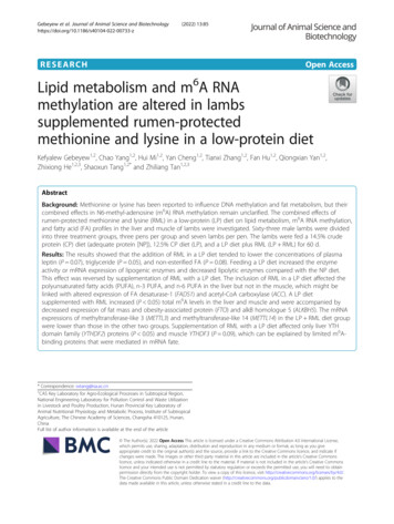
Gebeyew et al. Journal of Animal Science and Biotechnology(2022) 13:85following the manufacturer’s guidelines. Then, the synthesized cDNA was placed at 20 C until analysis.The lipid metabolism-related genes investigated in thisstudy were ACC, SCD, HSL, LPL, CPT1B, FABP4,FGF21, adipose triglyceride lipase (ATGL), fatty acidsynthase (FASN), sterol regulatory element-binding protein 1 (SREBF1), peroxisome proliferator-activatedreceptor-gamma (PPARγ), and peroxisome proliferatoractivated receptor-alpha (PPARα) (Additional file 1:Table S1). As internal reference genes, expression yde-3-phosphate dehydrogenase (GAPDH)were analyzed. The stability of the reference genes wereassessed using BestKeeper, Genorm, and NormFindertools [33]. The RPS9 and GAPDH were identified as themost stable reference gene across experimental treatment. Therefore, the two reference genes were used tonormalize the data. The amplification efficiency of allprimers used in this study was computed using serialcDNA dilution curves. All primers showed efficiency between 90% and 110%, and correlation coefficients wereclose to 1.0. All gene-specific primers sequences used inthe present study were synthesized by Sangon BiotechCo., Ltd. (Shanghai, China). The real-time PCR was performed using SYBR Green Premix Pro Taq HS qPCRKit (AG11707, Accurate Biology Co., Ltd., Changsha,China) on a fluorescence LightCycler 480 II system(Roche, Basel, Switzerland) following the manufacturer’sdirection. The details of the reaction program were described elsewhere [34]. The relative mRNA expressionsof the target genes were calculated using the 2-ΔΔCtmethod [35].Quantification of m6A contentThe m6A RNA methylation quantification kit (EpigentekGroup Inc., Farmingdale, NY, USA) was used to detectthe total content of m6A in liver and muscle tissues according to the protocol given by the manufacturer’smanual. The total content of m6A was computed usingthe formula: m6A % {[(ODSample – ODNC)/S] [(ODPC – ODNC)]} 100%, and S, NC, and PC denotethe total quantity of input RNA, negative control andpositive control, respectively, as described by Heng et al.[17].Statistical analysisStatistical analyses were executed using origin pro 2020band the SPSS version 23.0 (SPSS Inc., Chicago, IL, USA).First, Shapiro–Wilk and Levene’s tests were employed toconfirm the normality and homoscedasticity of data, respectively. Then, Tukey’s post hoc tests were used to determine the differences between treatment means,significant difference was regards as P 0.05 and trendswere recognized 0.05 P 0.1. The data are presented asPage 5 of 17Table 3 Effect of a low-protein diet supplemented with rumenprotected methionine and lysine on live body weight, hotcarcass weight, and dressing entsNPLPLP RMLLBW, kg36.2236.44ADG, kg/d0.1950.187ADFI, kg/d1.458G:F0.134HCW, kg17.0417.5818.220.3590.102DP, %47.5048.4949.441.6410.737Data are presented as means with their pooled SEMNP adequate protein diet, LP low-protein diet, LP RML low-protein dietsupplemented with rumen-protected methionine and lysine, LBW Live bodyweight, kg, ADG Average daily gain, kg/d, ADFI Average daily feed intake, kg/d;G:F: ADG/ADFI, HCW hot carcass weight, kg, DP dressing percentage, %the means standard errors of the mean (SEMs). Thecorrelations between genes-related to lipid metabolismand methylation were performed using Spearman’s rankcorrelation coefficient in R package and the network wasconstructed on the Omics-studio (LC-Bio TechnologyCo., Ltd., Hangzhou, China).ResultsCarcass parameter, lipid-related metabolites, andhormonesThe live body weight, average daily gain and feed intake,G:F ratio, dressing percentage, and hot carcass weightwere similar (P 0.05) among the three dietary groups(Table 3). The concentrations of plasma TAG and NEFAtended to be increased in the LP diet group than theother two groups (P 0.1, Fig. 1). The concentration ofplasma LEP (P 0.07) in the LP RML diet grouptended to be lower than that in the LP diet group. Incomparison to the other two groups, the concentrationsof L-carnitine in the liver and muscle were enhanced(P 0.05) in the RML supplemented group (Fig. 2).Fatty acids profiles in liver and muscle tissuesThe compositions of FA in the liver and muscle tissuesare presented in Tables 4 and 5. In liver, the C16:0 concentration was greater (P 0.05) in the LP diet groupthan that in the NP diet group, but comparable withthe RML supplemented group. The concentration ofC16:1 was greater (P 0.05) in the LP and LP RMLdiet groups than that in the NP diet group. In comparison to the NP diet group, the concentration of C18:2n6c was reduced in the LP diet group but equivalent tothe LP RML diet group. The LP diet group had ahigher (P 0.05) concentration of C18:1n-9 t than thosein the other two groups. In comparison to the NP dietgroup, the LP and LP RML diet groups had lower(P 0.05) concentrations of C18:3n-3, C20:3n-6, n-3
Gebeyew et al. Journal of Animal Science and Biotechnology(2022) 13:85Page 6 of 17Fig. 1 Effect of a low-protein diet supplemented with rumen-protected methionine and lysine on lipid-related metabolites and hormone levels.Values are expressed as means SEM indicated by vertical bars. Different letters indicate a significant difference among three dietary treatments(P 0.05). NP, adequate protein diet; LP, low-protein diet; LP RML, low-protein diet supplemented with rumen-protected methionine and lysineand n-6 PUFA. The LP diet group had greater (P 0.05) SFA and lower (P 0.05) PUFA and n-6 PUFAthan that in the NP diet group but were equivalent tothe RML supplemented group. However, neither a LPdiet nor LP RML affected (P 0.05) the partial sumsof MUFA in the liver. In muscle, the concentrations ofC15:0 and C18:3n-3 in the LP and LP RML dietgroups were reduced (P 0.05) than that of the NP dietgroup. In addition, the n-3 PUFA concentration tendedto be reduced (P 0.07) in the LP and LP RML dietgroups. Conversely, the LP diet increased (P 0.05) theconcentrations of C18:1n-9 t and C20:4n-6 than that ofthe other two diets. The partial sums of SFA, PUFA,and n-6 PUFA in the muscle were unaffected (P 0.05)by dietary groups.Enzyme activity in the liver and muscle tissuesAs depicted in Table 6, adding RML in a LP diet depressed(P 0.05) ACC, ME, and SCD enzyme activities in the livercompared with the LP diet. However, those enzyme activities were unaffected in muscle (P 0.05) by the supplementation of RML with a LP diet except for SCD (Table 7).In liver and muscle, the CPT1B enzyme activity in the LP RML diet group was higher (P 0.05) than that in the othertwo diet groups. In comparison with the LP diet group, theLP RML diet group increased (P 0.05) the activity ofHSL only in the liver. The LP diet increased (P 0.05) theenzyme activities of FABP4 and LPL in the liver and musclecompared with the NP and LP RML diets. Meanwhile,the LP diet increased (P 0.05) the FADS1 and FGF-21 enzyme activities only in the liver. However, no variationsFig. 2 Effect of a low-protein diet supplemented with rumen-protected methionine and lysine on L-carnitine levels in liver and muscle. On eachside of the gray line is a kernel density estimation to illustrate the distribution of shape of the data in a group. Wider and skinnier sections of theviolin plot represent a higher and lower probability. The white dot represents the mean and the thick red bar in the center represents theinterquartile range. * significantly different means (P 0.05). NP, adequate protein diet (n 7); LP, low-protein diet (n 6); LP RML, low-proteindiet supplemented with rumen-protected methionine and lysine (n 7)
Gebeyew et al. Journal of Animal Science and Biotechnology(2022) 13:85Table 4 Effect of a low-protein diet supplemented with rumenprotected methionine and lysine on the fatty acids compositionof liver in growing lambs, % of total fatty acidsFatty acids,%TreatmentNPLPLP 20.128Page 7 of 17Table 5 Effect of a LP diet supplemented with rumenprotected methionine and lysine on the fatty acids compositionof muscle in growing lambs, % of total fatty .0140.890PvalueFatty acids,%TreatmentNPLPLP 3290.3160.0150.062a0.678bbC15:00.9810.7150.037 035C18:1n-9 t0.636b0.750a0.501b0.0450.006C18:1n-9 23 .5870.297ab0.6060.3620.0510.0430.6990.533Fatty acid partial sumsFatty acid partial .4000.2590.3530.721 n-6 PUFA46.5506.1456.5950.2440.4660.3080.007 n-3 FA243.6943.2843.630.551PUFA311.98a10.20b10.97ab n-6 PUFA49.14a7.73b n-3 PUFA52.83a2.47bDifferent letters across a row indicate a significant difference among the threedietary treatments (P 0.05). Data are presented as means with theirpooled SEMNP adequate protein diet (n 7), LP low-protein diet (n 6), LP RML lowprotein diet supplemented with rumen-protected methionine andlysine (n 7)1SFA (saturatedfatty acid) C12:0 C14:0 C15:0 C16:0 C17:0 C18:0 C20:0 C24:02MUFA (monounsaturated fatty acid) C14:1 C16:1 C18:1n-9 t C18:1n-9c3PUFA (polyunsaturatedfatty acid) C18:2n-6c C18:3n-3 C20:3n-6 C20:4n-6 C22:6n-34n-3 PUFA C18:3n-3 C22:6n-35n-6 PUFA C18:2n-6c C20:3n-6 C20:4n-6were found between dietary groups for FADS1, FGF21, andHSL enzyme activities in the muscle (P 0.05).Lipid metabolism related genes in the liver and muscletissuesIn the liver, the mRNA expressions of ACC, SCD, LPL,FABP4, and SREBF1 were greater (P 0.05) in the LPdiet group than those in the other two groups (Fig. 3).Meanwhile, the expression of FADS1 (P 0.05) wasdown-regulated in the LP RML diet group comparedwith the other two groups (Fig. 3A). Conversely, the expression of CPT1B tended to be enhanced (P 0.06) inDifferent letters across a row indicate a significant difference among the threedietary treatments (P 0.05). Data are presented as means with their pooledSEM. NP, adequate protein diet (n 7); LP, low-protein diet (n 6); LP RML,low-protein diet supplemented with rumen-protected methionine andlysine (n 7)1SFA (saturatedfatty acid) C12:0 C14:0 C15:0 C16:0 C17:0 C18:0 C20:0 C24:02MUFA (monounsaturated fatty acid) C14:1 C16:1 C18:1n-9 t C18:1n-9c3PUFA (polyunsaturatedfatty acid) C18:2n-6c C18:3n-3 C20:3n-6 C20:4n-6 C22:6n-34n-3 PUFA C18:3n-3 C22:6n-35n-6 PUFA C18:2n-6c C20:3n-6 C20:4n-6the LP RML diet group than those in the other twogroups (Fig. 3B). In comparison to the NP diet group,the mRNA expression of FGF21 was up-regulated (P 0.05) in the LP and LP RML diet groups. The mRNAexpressions of HSL and PPARα were up-regulated (P 0.05) by the supplementation of RML with a LP diet,while mRNA expression of PPARγ in the LP RML dietgroup was similar (P 0.05) with the other two groups(Fig. 3C). However, the mRNA expressions of liver FASNand ATGL
A diet containing deficient or adequate Met levels af-fected lipid turnover by altering the expression of genes-related to fat metabolism in the liver and other tissues [8, 9]. The inclusion of low Met levels in the LP diet has little influence on fat accumulation, but those in a high protein diet reduced fat accretion [5], suggesting that