Transcription
TICB 1461 No. of Pages 20Feature ReviewLinking Lipid Metabolism to ChromatinRegulation in AgingKatharina Papsdorf1 and Anne Brunet1,2,*The lifespan of an organism is strongly influenced by environmental factors(including diet) and by internal factors (notably reproductive status). Lipidmetabolism is critical for adaptation to external conditions or reproduction.Interestingly, specific lipid profiles are associated with longevity, and increaseduptake of certain lipids extends longevity in Caenorhabditis elegans and ameliorates disease phenotypes in humans. How lipids impact longevity, and howlipid metabolism is regulated during aging, is just beginning to be unraveled.This review describes recent advances in the regulation and role of lipids inlongevity, focusing on the interaction between lipid metabolism and chromatinstates in aging and age-related diseases.IntroductionTo survive in the wild, organisms need to adapt to highly variable conditions, such as cycles offast and famine, fluctuations in temperature, and changes in reproductive status. A centralmechanism underlying this adaptation is lipid metabolism. Lipids serve as efficient energystorage in the form of triglycerides, thereby ensuring survival under harsh conditions or changesin reproductive status. In addition to storing energy, lipids have a wide range of functions thatcould also contribute to adaptation. For example, lipids serve as structural components ofcellular membranes, ensuring barrier and organelle homeostasis. Lipids can also act assignaling molecules, for example in nuclear hormone receptor (NHR) activation, influencingmany processes, including gene expression. Thus, key questions are: how does lipid metabolism impact the organism under various conditions? Is lipid metabolism important for theregulation of aging and longevity?Lipid profiles change with age in worms, fruit flies, mice, and humans [1–5]. Consistent with theidea that lipids are important for the regulation of lifespan, lipid profiles are altered in many longlived Caenorhabditis elegans and Drosophila melanogaster mutants [6]. Mounting evidencepoints to a strong link between lipid metabolism, lifespan regulation, and reproductive status[6]. Indeed, the generation of offspring requires energy, and lipids can be actively transportedfrom the soma to the germline and the offspring in a variety of species [7–9]. Consistently,removal of the germline leads to fat accumulation and increased longevity in worms andmammals [6]. Thus, lipid metabolism could be a critical switch between somatic maintenanceand reproduction.How do changes in lipid levels, composition, and location impact lifespan? An excitingpossibility is that changes in lipid metabolism could alter the regulation of physiologicalprocesses through changes in chromatin states. Chromatin modifications (histone andDNA) are key long-lasting mechanisms for the regulation of gene expression, including genesinvolved in cellular maintenance and longevity [10–12]. Consistent with a role in the agingprocess, many chromatin marks change during aging and altering chromatin modifiers canTrends in Cell Biology, Month Year, Vol. xx, No. yyHighlightsThe membrane lipids PE and PCdecrease with age, whereas triglycerides generally increase. During aging,the fatty acid composition of membrane lipids shifts towards anincreased PUFA to MUFA ratio.Long-lived organisms or mutants havea decreased PUFA to MUFA ratio orless unsaturated PUFAs, consistentwith lower oxidation. Longevity interventions (e.g., dietary restriction) lowerthe triglyceride content in mice.Supplementation of specific MUFAsand PUFAs extends the lifespan ofworms and improves age-related phenotypes in mammalian cells.Dietary lipids are used as a carbonsource for histone acetylation and dietary short-chain fatty acids as a sourcefor histone acylation.Metabolites such as SAM connect lipidmetabolism to histone methylation.1Department of Genetics, StanfordUniversity, 300 Pasteur Drive,Stanford, CA 94305, USA2Glenn Laboratories for the Biology ofAging, Stanford University, Stanford,CA 94305, USA*Correspondence:anne.brunet@stanford.edu (A. Brunet).https://doi.org/10.1016/j.tcb.2018.09.004 2018 Elsevier Ltd. All rights reserved.1
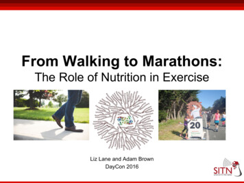
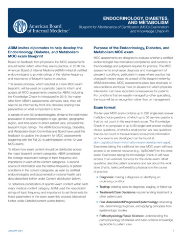
TICB 1461 No. of Pages 20extend lifespan in invertebrate and vertebrate species [10–12]. Chromatin marks are linked tolipid metabolism. Transfer of chromatin marks requires metabolites shared by lipid metabolismpathways, including acetyl-CoA, S-adenosyl methionine (SAM), and even lipids themselves[13]. Reciprocally, chromatin modifications control the expression of genes involved in variousaspects of lipid metabolism, including synthesis, degradation, and storage. Thus, a tantalizingpossibility is that lipid metabolism could interact with chromatin modifications to regulate aging.In this review, we highlight recent work showing the importance of lipid metabolism for theregulation of aging. We focus on the relationship between lipid metabolism and chromatinmodifiers by reviewing recent findings identifying how chromatin modifiers influence lipidcomposition and, vice versa, how lipids affect chromatin marks. We discuss the importanceof the connection between lipid metabolism and chromatin for longevity. Finally, we proposethat targeting lipid metabolism, or even lipids themselves, could be a promising strategy forlifespan-extending interventions.Lipid Metabolism Changes During Aging and Impacts LongevityLipids are crucial for a variety of biological processes, including aging and longevity. Lipids are adiverse class of molecules that comprise lipophilic molecules (e.g., free fatty acids), steroids, andcomplex lipids (e.g., triglycerides and phospholipids) (Figure 1), and a number of methods havebeen developed to assess lipid levels and composition (Box 1). Lipid classes are differentiallyaffected by aging- and longevity-promoting interventions. In this section, we review the recentevidence that implicates lipid levels and composition in longevity in a variety of species.Lipid Levels and Composition Are Altered During Aging and in Long-Lived OrganismsAging is associated with increased fat storage and altered complex lipid profiles [14]. In humansand mice, plasma triglyceride levels and circulating lipid–protein complexes increase with age[14] (Table 1). In mice, the levels of the membrane phospholipids phosphatidylethanolamine(PE), phosphatidylcholine (PC), and sphingomyelin decrease in old liver and brain, perhapsreflecting a change in plasma membrane composition [15,16] (Table 1). One possibility is thatmembrane fluidity is altered by membrane composition changes. Membranes are more fluidwhen they contain a low PC:PE ratio, a high degree of fatty acid unsaturation, a low concentration of oxidized lipids, or a low cholesterol content [17]. During aging, membrane fluiditydecreases in the brain, liver, and heart of rats [17]. Long-lived organisms (e.g., naked mole-rats)exhibit a specific phospholipid/fatty acid saturation profile [18], which may help maintainmembrane fluidity. Furthermore, longevity-promoting interventions such as dietary restriction(DR) protect from the age-dependent decline in membrane fluidity in mice [19]. Thus, increasedmembrane fluidity may be a key component of longer lifespan.The notion that membrane composition is central to longevity is supported by studies suggesting that membrane lipids, notably sphingolipids, are biomarkers of human aging [20]. Serumprofiling of long-lived humans revealed an increase in specific sphingolipids [20]. In addition,several plasma sphingolipids and their metabolites are also increased in long-lived naked molerats compared with wild type mice [21]. Sphingolipids can be converted into sphingosine-1phosphate (S1P) and ceramides, two bioactive lipids with opposing biological effects (Figure 1)[22]. Whereas S1P promotes cell proliferation and survival, ceramides promote apoptosis[22–24]. The balance between S1P and ceramides, termed the S1P/ceramide axis, modulatesthe aging process [22]. Age-related diseases, including Alzheimer’s disease and diabetes, areassociated with low levels of S1P [25,26]. Consistently, ceramides accumulate in old wormsand humans [27,28] (Table 1). In addition, a ceramide-rich diet shortens lifespan in C. elegans[28]. Conversely, worm mutants that lack the enzyme that synthesizes ceramides are long-lived2Trends in Cell Biology, Month Year, Vol. xx, No. yy
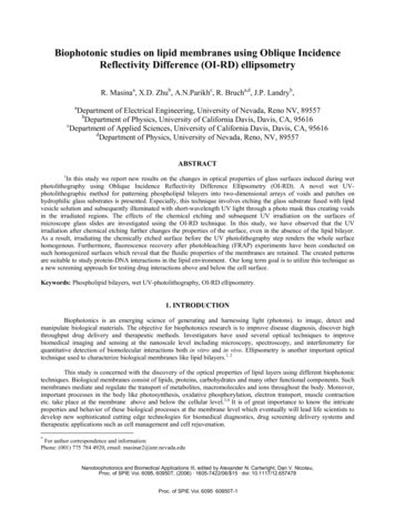
TICB 1461 No. of Pages 20FaƩy acid rasesMUFAsPhospholipid synthesisTriglyceride atePSTriacylglycerolPCPIS1P/ceramide glyceride degradaƟonLipophagyLipid dropletsMembranesLysosomeLysosomal lipases(LIPL1-5/LIPF)Lipid droplet lipase(ATGL-1/ATGL)Membrane lipid degradaƟonFaƩyacidsVery long chain/branchedfaƩy acidsLysosomeFaƩy acid degradaƟonMitochondriaPeroxisomesβ-oxidaƟon of faƩy acidsα/β-oxidaƟon of faƩy acidsFigure 1. Lipid Synthesis and Degradation Pathways. Fatty acids, including MUFAs and PUFAs, are synthesized and subsequently incorporated intotriglycerides and phospholipids. Triglycerides are synthesized and stored in lipid droplets. Triglycerides can be degraded by two different mechanisms. Lysosomescan fuse with lipid droplets, and lysosomal lipases (LIPL1-5/LIPF) in turn can hydrolyze triglycerides during lipophagy, thereby releasing free fatty acids. Alternatively,lipases located directly at the lipid droplet interface such as the adipose tissue lipases (ATGL-1/ATGL1) hydrolyze triglycerides. Free fatty acids are degraded inmitochondria for energy generation. Peroxisomes degrade very long or branched fatty acids, which can be subsequently shuffled to mitochondria for furtherdegradation. Membrane lipid synthesis pathways that are mentioned within the review are shown. Abbreviations: MUFAs, monounsaturated fatty acids; PC,phosphatidylcholine; PE, phosphatidylethanolamine; PI, phosphatidylinositol; PS, phosphatidylserine; PUFAs, polyunsaturated fatty acids; S1P, sphingosine-1phosphate; SFAs, saturated fatty acids.[29]. Thus, a shift in membrane composition during aging could modulate lifespan via the S1P/ceramide axis.Aging is accompanied not only by changes in lipid levels, but also by changes in the fatty acidcomposition of complex lipids, notably fatty acid desaturation [30]. The level of saturation of theTrends in Cell Biology, Month Year, Vol. xx, No. yy3
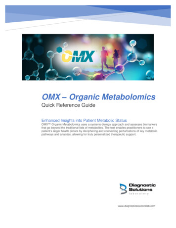
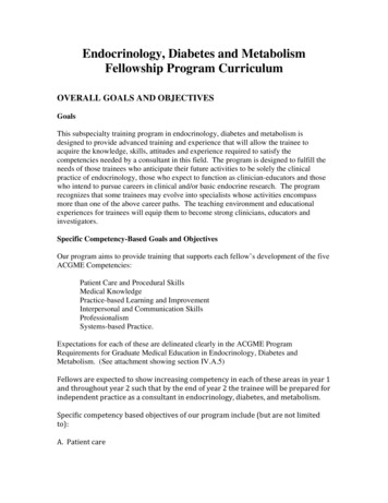
TICB 1461 No. of Pages 20Table 1. Lipid Changes Detected During AgingLipid classOrganismTissueChange with ageRefsCardiolipinRatBrain mitochondria,heart mitochondriaDecrease[158]CeramideC. elegans, humanWhole wormIncrease[27,28]Free fatty acidsHumanPlasmaIncrease[161]Fatty acidsC. elegansWhole wormDown after reproduction[3]PhosphatidylcholineMouse, ratLiver, brainDecrease[15,16]PhosphatidylcholineC. elegansWhole wormDecrease[3]PhosphatidylethanolamineMouse, ratLiver, brain, lamineC. elegansWhole wormDecrease[3]SphingomyelinMouseLiver, brainDecrease[15,16]SphingomyelinC. elegansWhole wormIncrease in late life[3]TriglycerideHumanPlasma, liverIncrease[161]TriglycerideRatLiver, plasmaIncrease[162]TriglycerideMouseLiver, mitochondriabrainIncrease[5,15,16]lipid chain confers specific chemical properties to lipids, including solubility and fluidity, whichcould in turn affect the status of membranes. Saturation also alters lipid susceptibility tooxidative damage. For example, polyunsaturated fatty acids (PUFAs), which contain multipledouble bonds in their carbon chains, are more susceptible to oxidation than monounsaturatedfatty acids (MUFAs) [31]. Oxidized lipids are particularly detrimental to cellular function because(i) they catalyze free radical chain reactions which can damage both lipids and proteins, (ii) theydiffuse over large distances in membranes, (iii) they can have long-lasting effects, and (iv) theyalter membrane properties, eventually leading to loss of organelle integrity [32]. A metric tocharacterize the susceptibility to oxidation is the peroxidizability index (PI), which correlates withthe number of unsaturated bonds in lipids [30]. During aging, the PI of membrane phospholipidsincreases, with higher PUFA to MUFA ratio in old flies and rat liver [30,33]. Conversely, the PI ofphospholipids is lower in the heart of long-lived wild-derived mouse strains than in that ofcontrol mouse strains [34]. Long-lived naked mole-rats also have low levels of phospholipidswith highly unsaturated PUFAs (e.g., docosahexaenoic acid) compared with mice [18]. Thiscorrelation between low PI and longevity seems to extend to many species. Indeed, a low PI inphospholipids is a predictor of longevity across 11 mammalian species [35]. Furthermore,offspring of long-lived individuals have high MUFA to PUFA ratios in erythrocyte membranelipids compared with controls [36]. Hence, elevated products of lipid peroxidation may lead tocumulative cell and tissue damage, and possibly accelerate aging.Intriguingly, changes in lipid profile during aging are tissue specific. Mitochondrial lipids of oldmouse brains show a decrease in PUFAs, a change normally associated with longevity [37]. Bycontrast, mitochondrial lipids of old mouse muscles exhibit increased triglycerides anddecreased PEs [37] (Table 1), changes generally associated with aging [37]. These differentialchanges in organelle lipid profiles in various tissues during aging could reflect diverse tissuespecific functions and energy requirements or compensatory protective mechanisms.4Trends in Cell Biology, Month Year, Vol. xx, No. yy
TICB 1461 No. of Pages 20Consistently, lipid species that are predictors of long lifespan, including specific triglycerides,differ across various tissues [38]. More investigation is needed to elucidate the dynamic lipidprofiles of different cell types and organelles during aging.DR, Which Delays Aging, Remodels Lipid ProfilesDR is a well-known strategy to delay aging and slow the onset of age-related phenotypes[39–42]. DR can reverse age-associated lipid changes in mice [5,43]. In mouse liver, thephospholipid PE decreases during aging, but this decrease is blunted when old animals aresubjected to DR [43]. In addition, DR increases S1P levels and decreases ceramide levels,thereby ameliorating age-dependent changes in these lipids [44]. Finally, the accumulation oftriglycerides during aging in mouse liver is blunted by DR [5,43]. Interventions that mimicaspects of DR, such as resveratrol, also decrease triglyceride content in mouse plasma [45] andzebrafish plasma [46]. The effect of DR on triglycerides is conserved across species. DRdecreases triglyceride levels in C. elegans (Figure 2) and longevity induced by DR in Drosophilarequires an intact machinery for triglyceride synthesis and breakdown [47–49]. It will beimportant to determine whether overall triglyceride levels or triglyceride composition play afunctional role in lifespan extension by DR, and if other aspects of lipid metabolism are alsoinvolved in lifespan extension by DR.Known Longevity Signaling Pathways Affect Lipid MetabolismSignaling pathways that regulate longevity, such as the insulin pathway and the mTORautophagy pathway, also impact lipid metabolism. These longevity pathways mostly act byinfluencing the activity of transcription factors that in turn modulate enzymes involved in lipidmetabolism.Insulin Signaling PathwayDeficiency in the insulin receptor DAF-2 extends lifespan in C. elegans by activating thetranscription factor DAF-16/FOXO [50–52]. DAF-16/FOXO transcriptionally upregulates lipidmetabolic enzymes, such as the fatty acid desaturase FAT-7 and the lysosomal lipase LIPL-4,that subsequently promote MUFA synthesis and lipid degradation processes [53,54]. Consistent with these transcriptional changes, lipid profiling of daf-2 mutant worms shows increasedMUFA/PUFA ratios and elevated triglycerides [55–58] (Figure 2). In fact, long-lived insulinpathway mutants exhibit triglycerides whose side chains are enriched in MUFAs [56]. Inaddition, inhibiting this switch to MUFAs reduced the longevity of daf-2 mutant worms [53].Thus, high concentrations of cellular triglycerides may not always be detrimental to longevity,for example when they are enriched in MUFAs or when they occur in tissues rather than blood.The transcriptional response that is responsible for the switch to MUFAs upon insulin signalingdeficiency can be blunted by other types of lipids. Indeed, high cholesterol concentrationabrogates lifespan extension induced by deficiency in the insulin pathway [59]. An unansweredquestion is whether high cholesterol concentrations prevent the switch to MUFAs and howtriglyceride composition mediates the beneficial effect of insulin signaling on lifespan.The concomitant regulation of lifespan and lipid metabolism by the insulin pathway is conservedin other species. Long-lived insulin pathway deficiency in Drosophila (e.g., InR and chico) alsoincreases triglyceride storage [60,61], possibly by affecting FOXO activity [62]. The insulinsignaling pathway also influences longevity and lipid metabolism in mammals [60]. Insulinlowers plasma fatty acid levels by promoting triglyceride uptake from the blood into the adiposetissue and increases triglyceride synthesis [63]. In mice, the heterozygote mutation of Igf1rextends lifespan [64] and a mutation of the insulin receptor in the adipose tissue is sufficient toextend lifespan [65]. However, in contrast to invertebrates, mice harboring a mutation of theTrends in Cell Biology, Month Year, Vol. xx, No. yy5
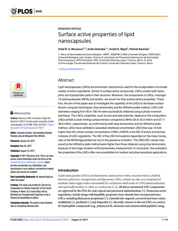
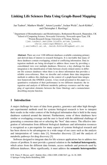
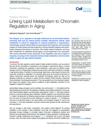
TICB 1461 No. of Pages 20Dietary restricƟonDietary restricƟonInsulin/IGF signalingAutophagyChromaƟnDietary restricƟoneat-2 mutantsdaf-2/InR mutantsmTORXH3K4me3 transferasedeficiencyDNA methylationTranscripƟon P1Inhibition of fattyacid elongases(elo-1/Elovl5)Mitochondri- Fatty acidal β-oxidation transport(acs-2/(lbp-8Acsf2)Fabp)Activation ofMDT-15/MED15AcƟvaƟon of lipases(lipl-2/3/4/5/Lipf)lipophagyFaƩy hortened chain faƩyacidsC. elegansMouseGermline signalingglp-1 mutantsTranscripƟon factorsacƟvatedNHR-49/ DAF-16/ TCER-1/TCERG1FOXOTranscripƟonalchangesFatty n(acs-2/Acsf2)FaƩy aciddesaturase(fat-7/Scd1)FaƩy l-3/Lipf)drialFaƩy acid enuatedlipid accumulaƟonLipidchangesIncreasedMUFAsand Lipa)C. elegansFigure 2. Lipid Metabolism Is Targeted by Several Longevity Pathways. Longevity pathways that target lipid metabolism in Caenorhabditis elegans andmouse. Activated transcription factors/activators are circled. Upper panel: longevity pathways other than germline depletion. Dietary restriction in mouse leads to globalDNA methylation changes that inhibit SREBP1. DNA hypermethylation is found on the bodies of genes that are important for fatty acid elongation. Dietary-restrictedmice show a decrease in triglyceride level and a shift towards shorter chain fatty acids. In C. elegans, dietary restriction by eat-2 mutation results in activation of thenuclear hormone receptors NHR-49/PPAR and NHR-62/HNF4. The eat-2 mutants show decreased triglyceride content. Depletion of insulin signaling via daf-2/InRmutation activates DAF-16/FOXO and the coactivator MDT-15/MED15. This leads to an increase in triglycerides and a higher MUFA to PUFA ratio. Activation ofautophagy by mTOR depletion activates multiple transcription factors (HLH-30/TFEB, SKN-1/NRF, DAF-16/FOXO, and PHA-4/FOXA). Depletion of the COMPASSH3K4me3 modifiers ash-2/set-2 in the germline activates the transcription factors/activators SBP-1/SREBP and MDT-15/MED15 in the worm soma. H3K4me3modifier deficient worms have increased triglycerides and a higher MUFA content. Lower panel: longevity signaling upon germline depletion induced by glp-1 mutationin C. elegans. Multiple transcription factors and regulators are activated in germline-deficient animals (NHR-49/PPAR, MDT-15/MED15, NHR-80/HNF4, DAF-12/LXRa,DAF-16/FOXO, TCER-1/TCERG1, SKN-1/NRF, HLH-30/TFEB, and PHA-4/FOXA,). Germline-deficient animals show higher triglyceride content, an increase in(Figure legend continued on the bottom of the next page.)6Trends in Cell Biology, Month Year, Vol. xx, No. yy
TICB 1461 No. of Pages 20insulin receptor in the adipose tissue have reduced whole body triglycerides [65]. Thus, highlevels of triglycerides may not be important for insulin-mediated longevity in certain contexts. Itremains to be determined if triglyceride content in specific tissues is altered or if triglyceride fattyacid composition changes in insulin pathway mutants in mammals.mTOR-Autophagy PathwayAnother longevity pathway that links lipid metabolism to lifespan is the mTOR-autophagypathway. Inhibiting mTOR extends lifespan and induces autophagy [66]. mTOR regulatesoverall fat storage [66–68], and autophagy is required to maintain lipid homeostasis in manyorganisms, including C. elegans [69,70]. The mTOR-autophagy pathway influences longevity inlarge part by modulating transcription factors that in turn regulate lipid metabolism enzymes.For example, autophagy-mediated longevity requires the transcription factor HLH-30/TFEB,which contributes to lipid homeostasis by upregulating the expression of lysosomal lipases (lipl2/3/4/5) [71] (Figure 2). Depletion of mTOR via RNAi increases the expression and activity of thelipase LIPL-4 in C. elegans [72]. Overexpression of LIPL-4 extends lifespan and promotesautophagy by activating the transcription factor PHA-4/FOXA [72].Autophagy-mediated lifespan extension not only involves lipases but also lipid binding proteinscalled vitellogenins [73]. Vitellogenins bind complex lipids and transport them from the intestineto the gonad. Thus, vitellogenins may change intracellular lipid availability, thereby affectinglongevity. While vitellogenins do not have exact orthologs in mammals, they share functionalsimilarity with mammalian large lipid transfer modules such as apolipoprotein B [e.g., lowdensity lipoprotein (LDL)] [74]. Similar to worms, high levels of circulating LDL are detrimental tohealth and are associated with increased incidence of age-related diseases such as cardiovascular diseases [75]. In addition, LDL levels are also regulated by autophagy [75]. It would beinteresting to test if increasing the autophagic flux results in beneficial mammalian lipoproteinabundance and if this improves age-related phenotypes.Chromatin Modifiers Impact Lifespan via MUFA MetabolismChromatin modifiers impact lifespan in worms by regulating fatty acid desaturation. Depletion ofH3K4me3 modifiers of the COMPASS complex (ash-2, set-2, wdr-5) in the germline of fertileanimals extends lifespan [76]. Deficiency of H3K4me3 modifiers in the germline leads to theactivation of the coactivator MDT-15 and the transcription factor SBP-1 in the intestine [77].These transcriptional regulators in turn upregulate fatty acid desaturase fat-7 expression[77–79]. Knockdown of fat-7 abrogates the longevity effect of H3K4me3 modifier deficiency,and FAT-7 overexpression is sufficient to extend lifespan [77]. Mass spectrometry-basedstudies on these long-lived worms showed a switch towards an enrichment of MUFAs, notablyoleic acid, palmitoleic acid, and cis-vaccenic acid [77]. The mammalian FAT-7 ortholog, SCD1,also regulates fat storage and fatty acid profiles [80], and SCD1 overexpression attenuatesreactive oxygen species levels in obese mice [81]. It will be interesting to test if fatty aciddesaturases also extend lifespan in vertebrates.Germline Longevity Signaling Regulates Lipid Metabolism via a Network of TranscriptionFactorsA key dissection of the importance of lipid metabolism for longevity has been done in germlinedeficient animals. Germline deficiency, via laser ablation or by genetic mutations that preventgerm cell proliferation, extends lifespan in worms and flies [82,83]. Concurrently, germlineunsaturated fatty acids, and an increase in the MUFA derivative oleoylethanolamide. Abbreviations: MUFAs, monounsaturated fatty acids; NHR, nuclear hormone receptor; PUFAs, polyunsaturated fatty acids.Trends in Cell Biology, Month Year, Vol. xx, No. yy7
TICB 1461 No. of Pages 20removal leads to a drastic remodeling of lipid fat metabolism, not only in C. elegans but also inmammals [6]. Multiple transcription factors mediate lifespan extension in germline-deficient C.elegans, including the nuclear receptors NHR-49/PPAR [84], NHR-80/HNF4 [85], and DAF-12/LXRa [86], the transcription factors SKN-1/NRF [87] and HLH-30/TFEB [70,88], the Forkheadtranscription factors PHA-4/FOXA [72] and DAF-16/FOXO [82], as well as the transcriptionelongation regulator TCER-1/TCERG1 [89] (Figure 2).NHRs are required for the longevity of germline-deficient C. elegans and directly regulate theexpression of several lipid metabolism genes (Figure 2). For example, NHR-49 upregulatesgenes involved in mitochondrial b-oxidation of lipids [84,90,91]. NHR-49, together with thecoactivator MDT-15, also increases the expression of fatty acid desaturases such as fat-5 andfat-7 that promote the synthesis of various MUFAs, including palmitoleic and oleic acid [78]. Thetranscription factor NHR-80 also upregulates fatty acid desaturase genes involved in MUFAsynthesis [85]. The central role of nuclear receptors in lipid metabolism in nematodes might beconserved in mammals. For example, the nuclear receptor PPARa, an NHR-49 ortholog,upregulates the fatty acid desaturase SCD1 [92]. In mice, PPARd signaling improves intestinalstem cell activity by inducing fatty acid degradation during aging [93], and a small moleculeagonist for PPARd mimics this effect [93]. However, whether PPAR activators also promotelongevity in mammals remains to be further studied.Another example of a nuclear receptor pathway targeting lipid metabolism in long-lived germline-deficient C. elegans is the DAF-12 steroid signaling pathway [86] (Figure 2). In contrast toNHR-49, which is activated by fatty acid signals [94,95], DAF-12 is activated by cholesterolderivatives such as dafachronic acid [86]. One prominent function of DAF-12, and cholesterol,is the regulation of correct reproductive development in C. elegans [86]. Worms cannotsynthesize cholesterol and need to uptake it from their food. Still, cholesterol is abundant inthe gonad because lipid binding proteins (vitellogenins) transport the cholesterol uptaken fromfood into oocytes [96]. DAF-12 and the cholesterol-derivative dafachronic acid modulate lipidmetabolism [97]. For example, one of the downstream targets of DAF-12 is the fatty acylreductase fard-1/far1, which is essential for longevity in germline-deficient C. elegans [97](Figure 2). In addition, DAF-12 promotes the fusion of lipid droplets via unknown lipophilichormones [98]. It remains to be analyzed if this process is involved in germline-deficiencymediated longevity.The regulation of longevity and fat metabolism by the DAF-12 steroid signaling pathway may beconserved in mammals. Indeed, the potential DAF-12 mammalian ortholog, Liver X Receptoralpha (LXRa), is also activated by cholesterol derivatives [99]. A single nucleotide polymorphismin the gene encoding LXRa was found to correlate with human longevity and, surprisingly, withhigher levels of serum triglycerides [100]. While high serum triglyceride levels normally correlatewith rapid aging, it is possible that here, triglyceride composition or downstream processing isdifferent, which could be essential for longevity. Future work is needed to determine ifcholesterol derivatives influence triglyceride composition and if this is involved in aging.Other types of transcription factors, such as the Forkhead transcription factor DAF-16/FOXO,also mediate longevity in response to germline depletion by targeting lipid metabolism [50].DAF-16/FOXO regulates target genes involved in lipid metabolism, including the lysosomallipase lipl-4 [53,101] (Figure 2). Furthermore, DAF-16/FOXO and the transcription elongationand splicing factor TCER-1/TCERG1 can activate lipogenesis via dgat-2, acs-22, and mboa-2[89]. These transcriptional changes result in increased triglycerides levels and increasedunsaturated fatty acids in germline-deficient worms [89].8Trends in Cell Biology, Month Year, Vol. xx, No. yy
TICB 1461 No. of Pages 20Together, these findings highlight the beneficial effect of lipid accumulation for longevity ingermline-deficient worms. Without a functioning germline, lipids can be repartitioned in somatictissues. An exciting possibility is that lipids retained in the soma, for example triglyceridesenriched in MUFAs [84], could contribute to the maintenance of somatic cells during aging.Lipids could activate specific longevity pathways in the soma or directly lower the risk ofoxidative damage in somatic tissues.Dietary Supplementation of Specific Fatty Acids Extends LifespanManipulating lipid metabolism by supplementation of different dietary fatty acids can extendlifespan in C. elegans. One mechanism by which dietary lipids influence lifespan is by activatinga transcription factor network [87,95]. For example, supplementation of the PUFA v-3 fatty acida-linolenic acid (ALA) extends lifespan in C. elegans through regulation of the NHR-49/PPARaand the SKN-1/NRF transcription factors [95] (Figure 3). ALA directly binds to and activatesNHR-49 [95]. ALA indirectly leads to SKN-1/NRF activation via its peroxidized or hydroxylatedcounterparts (oxylipins) such as 9(
Lipid Metabolism to Chromatin Regulation in Aging Katharina Papsdorf1 and Anne Brunet1,2,* The lifespan of an organism is strongly influenced by environmental factors (including diet) and by internal factors (notably reproductive status). Lipid metabolism is critical for adaptati