Transcription
Göttlicher et al. Biomaterials Research (2017) 21:18DOI 10.1186/s40824-017-0104-8RESEARCH ARTICLEOpen AccessFunctionalization of Ti-40Nb implantmaterial with strontium by reactivesputteringMarkus Göttlicher1, Marcus Rohnke1* , Yannik Moryson1, Jürgen Thomas2, Joachim Sann1, Anja Lode3,Matthias Schumacher3, Romy Schmidt2, Stefan Pilz2, Annett Gebert2, Thomas Gemming2 and Jürgen Janek1AbstractBackground: Surface functionalization of orthopedic implants with pharmaceutically active agents is a modernapproach to enhance osseointegration in systemically altered bone. A local release of strontium, a verified bonebuilding therapeutic agent, at the fracture site would diminish side effects, which could occur otherwise by oraladministration. Strontium surface functionalization of specially designed titanium-niobium (Ti-40Nb) implant alloywould provide an advanced implant system that is mechanically adapted to altered bone with the ability tostimulate bone formation.Methods: Strontium-containing coatings were prepared by reactive sputtering of strontium chloride (SrCl2) in aself-constructed capacitively coupled radio frequency (RF) plasma reactor. Film morphology, structure and compositionwere investigated by scanning electron microscopy (SEM), time of flight secondary ion mass spectrometry (ToF-SIMS)and X-ray photoelectron spectroscopy (XPS). High-resolution transmission electron microscopy (HR-TEM) was used forthe investigation of thickness and growth direction of the product layer. TEM lamellae were prepared using the focusedion beam (FIB) technique. Bioactivity of the surface coatings was tested by cultivation of primary human osteoblasts andsubsequent analysis of cell morphology, viability, proliferation and differentiation. The results are correlated with theamount of strontium that is released from the coating in biomedical buffer solution, quantified by inductively coupledplasma mass spectrometry (ICP-MS).Results: Dense coatings, consisting of SrOxCly, of more than 100 nm thickness and columnar structure, were prepared.TEM images of cross sections clearly show an incoherent but well-structured interface between coating and substratewithout any cracks. Sr2 is released from the SrOxCly coating into physiological solution as proven by ICP-MS analysis.Cell culture studies showed excellent biocompatibility of the functionalized alloy.Conclusions: Ti-40Nb alloy, a potential orthopedic implant material for osteoporosis patients, could be successfullyplasma coated with a dense SrOxCly film. The material performed well in in vitro tests. Nevertheless, the Sr2 release mustbe optimized in future work to meet the requirements of an effective drug delivery system.Keywords: Surface coating, Titanium alloy, Osteoporosis, Strontium release, Biocompatibility, Plasma deposition* Correspondence: Marcus.Rohnke@phys.chemie.uni-giessen.de1Institute of Physical Chemistry and Center of Materials Research,Justus-Liebig-University of Giessen, Heinrich-Buff-Ring 17, 35392, Giessen,GermanyFull list of author information is available at the end of the article The Author(s). 2017 Open Access This article is distributed under the terms of the Creative Commons Attribution 4.0International License (http://creativecommons.org/licenses/by/4.0/), which permits unrestricted use, distribution, andreproduction in any medium, provided you give appropriate credit to the original author(s) and the source, provide a link tothe Creative Commons license, and indicate if changes were made. The Creative Commons Public Domain Dedication o/1.0/) applies to the data made available in this article, unless otherwise stated.
Göttlicher et al. Biomaterials Research (2017) 21:18BackgroundTitanium and its alloys are well established biomaterialsfor the production of load-bearing implants for hard tissue replacement and fracture stabilization. They offerbeneficial mechanical properties and develop a stablepassivating oxide film that ensures excellent biocompatibility and good corrosion resistance [1]. Despite of theirsuccessful use as surgical implants the mechanical andsurface properties still need to be improved to meet thespecific challenges that orthopedics and traumatologyare facing. Key weaknesses of currently used α and α/βtype titanium-based implants are their high stiffnessmismatch to bone [2] and their biocompatibility, whichcan be increased further by surface modification to finally enhance osseointegration [3]. As in the future acontinuous rise of population’s life expectancy is expected, more patients will suffer from age-related orpost-menopausal osteoporosis or its secondary forms.These bone disorders negatively affect mineral density,fragility [4, 5] and regenerative ability of bone [6]. Therefore, it is an important aim to develop advanced metallicimplants that are mechanically adapted to the weakenedbone structure and ensure fast bone integration bymodified surface characteristics [3].Beta-type Ti-40Nb alloy is a very promising implant material for the treatment of bone fractures because it exhibits a low elastic modulus combined with moderatestrength [7]. Besides that, it has been shown that niobiumleads to an outstanding corrosion resistance of the passivating oxide film [8]. While this is a major advantage onthe one hand, it makes standard surface treatment protocols for metallic implants ineffective on the other hand, sothat alternative methods were exploited [9].Strontium cations (Sr2 ) are successfully used for themedical treatment of patients with osteoporosis by oraladministration of strontium ranelate. In vitro and in vivostudies showed that strontium ions are able to stimulatenew bone formation and inhibit excessive bone resorption [10], which is occurring in osteoporotic bone andcaused by a characteristic imbalance between bonebuilding osteoblasts and bone-resorbing osteoclasts [11].The success of strontium as an anti-osteoporotic drugsuffered a setback in 2014, when the European Medicines Agency recommended a restricted use of strontium ranelate due to its adverse effects, which include anincreased risk of serious cardiac disorders [12]. Tominimize this risk, a locally administered release ofstrontium, for example by coating of surgical implants,would be highly beneficial [13]. Numerous studies foundan increased biocompatibility and upregulation of osteogenesis related genes in vitro [14–16] and an effectivelyimproved implant osseointegration in vivo, in bonehealthy [17, 18] and recently in ovariectomized rats [19].The spatial dispersion of the therapeutic agent Sr2 inPage 2 of 11rat bone with time in vivo was recently described byRohnke et al. [20]. It was shown that the Sr2 releaseand transport from a biomaterial into bone is a usefullconcept, which can be mathematically described withthe help of Ficks laws of diffusion.Methods for preparation of strontium-releasing implant coatings involve hydro-thermal synthesis routes[15–17], Sr-substituted hydroxyapatite coating [21, 22],NaOH treatment followed by strontium acetate treatment [14], electrolytic deposition techniques [23, 24]and non-reactive magnetron sputtering [18, 19]. Foroptimum medical performance of such coatings it is assumed, that ionic strontium needs to be released continuously over a period of several weeks while thepharmaceutical effect threshold is in the range of0.10 mM and defined by the serum strontium level ofpostmenopausal osteoporosis patients that were treatedwith strontium ranelate [25]. However, an initial burstrelease of strontium after implantation is also unwantedto reduce the risk of a short-time toxic strontium level[18]. In recent approaches such a regulated strontiumrelease was demonstrated by chelation into folic acid[26] and introduction of topographical structures [27].In the present study, strontium-containing thin filmsare deposited on Ti-40Nb substrates by reactive sputteringof strontium chloride in a radio frequency (RF) oxygendischarge. The chemical composition and morphology offilms were characterized by scanning electron microscopy(SEM), X-ray photoelectron spectroscopy (XPS), secondary ion mass spectrometry (SIMS) and transmission electron microscopy (TEM). By immersion of the coatedsubstrates in Tris-buffered saline (TBS) the strontium release of the thin films was determined using inductivelycoupled plasma mass spectrometry (ICP-MS). Finally, thebiological response of primary human osteoblasts to thesefilms was evaluated in vitro.MethodsTi-40Nb sample preparationA beta-phase Ti-40Nb rod with a diameter of 12 mmwas prepared by arc melting and cold crucible castingfollowed by homogenization treatment, as described indetail in reference [28]. Discs with a thickness of 0.7 mmwere prepared by cutting the rod using an IsoMet 5000linear precision saw (Buehler, Switzerland) equippedwith a SiC cut-off wheel (Struers, Denmark). Ti-40Nbdiscs were ground with bound diamond particles(15 μm) using a Phoenix 4000 grinding and polishingmachine (Buehler). The discs were polished with a 9 μmdiamond suspension (MetaDi, Buehler) and subsequentlyfine polished with a 4:1 colloidal SiO2 (20 nm MasterMet2, Buehler)/H2O2 (35% HYPROX , Evonik,Germany) mixture. Finally, the mirror-polished discswere cleaned in acetone, demineralized water and pure
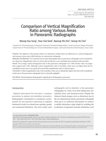
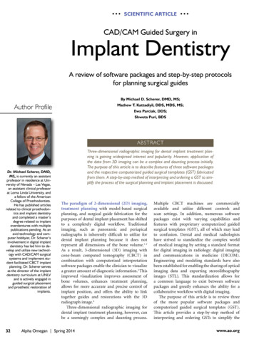
Göttlicher et al. Biomaterials Research (2017) 21:18ethanol using an ultrasonic bath for 10 min in succession. The samples were stored in ethanol until strontiumdeposition was performed.Plasma setupAll deposition experiments were conducted using a selfbuilt plasma setup for excitation of capacitively coupledRF plasma, already introduced in [29] (Fig. 1). Two discshaped electrodes of same size were arranged parallel toeach other inside a spherical vacuum chamber (approx.35 cm in diameter), all made of stainless steel. The distance between the electrodes was 3.5 cm, and they areapprox. 8.0 cm in diameter. The lower electrode washeatable and attached to the grounded vacuum chamberto ensure adjustment of high self-bias values. The topelectrode was powered by an RF generator (PFG RF 300,Trumpf Hüttinger Elektronik, Germany) and matchingnetwork system (PFM 1500 A, Trumpf Hüttinger Elektronik), operating at 13.56 MHz.One special feature of the top electrode is a removablelower side that is coated with a SrCl2 layer (details to thecoating process are described below), therefore acting asstrontium-containing target for the sputtering process.For supply of the process gases (Ar, O2, both 99.999%), asystem of electropneumatic valves (EVI 105 P, PfeifferVacuum, Germany) and mass flow controllers (MKS Instruments, US) was flanged to the vacuum chamber aswell as a manometer (Baratron, MKS Instruments, US)to monitor overall gas pressure (Fig. 2).For evacuation of the vacuum chamber a rotary vanepump (Duo 10, Pfeiffer Vacuum, Germany) was attached. The sample temperature was adjusted using anEurotherm controller (Model 2416, Schneider Electric,Page 3 of 11France) and a type K thermocouple (Thermocoax, France)that was located beneath the lower electrode. Electropneumatic valve control, gas flow and pressure control, RF generator and temperature control were connected to acomputer to automatize the deposition process using LabVIEW (National Instruments, US) software.SrCl2 target preparationA SrCl2 layer was prepared onto the removable part ofthe top electrode (hereinafter referred to as “target electrode”) by dropcasting of a saturated solution of SrCl2(99.9%) and a 1:1 mixture of H2O and pure ethanol. Thetarget electrode was placed on a heating plate, fully covered with the solution and subsequently heated to speedup solvent evaporation.Strontium functionalization by reactive sputteringAfter placing the polished Ti-40Nb samples on thelower, grounded electrode, the plasma chamber wasevacuated to a pressure of p 1 Pa several times forcomplete removal of residual water inside the vacuumchamber including the hygroscopic SrCl2 target. Oncepumped, the reactor was continuously flooded with amixture of 0.25 sccm O2 and 0.68 sccm Ar resulting inan overall gas pressure of 21 Pa. Strontium depositionwas carried out at room temperature with constant RFpower of P 100 W and variable dc bias voltage fort 2000 s. Afterwards this procedure was repeated oncemore. After every deposition process both electrodeswere polished with a polishing paper (grain size 4000)and cleaned with acetone to remove passivating oxidefilms that have been formed due to the oxygen plasmaexposure.Strontium film characterizationFig. 1 Plasma setup; Schematic sketch of the capacitively coupledRF plasma setupSEM images of the strontium film surface were taken witha MERLIN field emission scanning electron microscope(Carl Zeiss Microscopy, Germany) using an accelerationvoltage of 3.0 kV at 4 mm working distance. EDX measurements were carried out with an Oxford Instruments(Abingdon, UK) silicon drift X-Max 50 detector.For TEM investigations of film morphology (thicknesses, roughness), homogeneity and grain structure thincross section lamellae were prepared using the focussedion beam (FIB) technique. A thin slice perpendicular tothe sample surface was cut out in a Zeiss Cross Beam1540 XB (Carl Zeiss Microscopy) with a 30 keV fine focussed gallium ion probe. This lamella was in situ transferred and welded on a special copper grid by use of theelectron beam under SEM control. The final lamellathickness of less than 100 nm was reached by thinningwith a gallium ion probe of lower energy (ca. 5 keV). Toreduce the amorphization and the gallium implantationa protection bar of hydrocarbon and hydrocarbon
Göttlicher et al. Biomaterials Research (2017) 21:18Page 4 of 11Fig. 2 Technical mapping; Technical mapping of the RF plasma equipmentcontaining platinum was deposited onto the sample surface at the cutting position before starting the procedure. For the TEM analyses we used a TEM/STEMmicroscope Tecnai F30 ST (FEI Company, US) equippedwith Ametek/EDAX (US) windowless SDD for energydispersive X-ray spectroscopy (EDXS) and Gatan imaging filter 200 for electron energy loss spectroscopy(EELS). The measurements were done using an acceleration voltage of 300 kV reaching a resolution limit ofbetter than 0.2 nm.The elemental composition of the Sr-coating was analyzed by ToF-SIMS depth profiling using a ToF-SIMS 5100 machine of IONToF company (Germany). Cationdepth profiles were generated by sputtering with 1 keV O 2ions in combination with surface analysis using 25 keV Bi primary ions. The primary ion gun was operated in highcurrent bunched mode with a mass resolution m/Δm 6900 for Ti (m/z 47.95). For anion depth profiling0.5 keV Cs ions were used. Data were evaluated with SurfaceLab 6.7 software (IONToF company). After depth profiling the sputter craters were measured with a confocalmicroscope PLu neox 3D (Sensofar Group) to calibratethe sputter time axis. For calibration of the sputter timeaxis the assumption was made that the sputter yields inthe film and in the bulk material are the same.The surface composition was analyzed by XPS with aPHI Versaprobe 2 instrument (Physical electronics, US)equipped with a monochromatized Al k-alpha sourcewith 1486.6 eV. For depth profiling the instrument isequipped with an Ar ion sputter gun. Chargeneutralization was conducted with 1 eV electrons and 10 eV Ar ions in parallel. The sample was excited witha 200 μm diameter X-ray beam (50 W) and the data wasmeasured with an analyzer pass energy of 23.5 eV and astepsize of 0.2 eV. For depth profiling, we sputtered with1 kV beam energy and a raster size of 2 mm 2 mm for2 min between each measurement.Determination of strontium ion releaseFor analysis of strontium ion release a 0.05 M Tris-HClbuffer with pH 7.6 containing 0.15 M sodium chloridewas prepared by dissolving a TBS BioUltra tablet(Sigma-Aldrich, US) in 500 ml demineralized water. Dueto CO2 uptake from air the pH decreased slightly afterstarting the experiment to physiological conditions. Srfunctionalized Ti-40Nb samples were prepared andimmersed in 5 ml Tris-HCl buffer per sample for oneday. For quantitative analysis of the strontium contentthe buffer solutions were weighted and mixed with concentrated nitric acid at 60 C. The solutions were dilutedwith purified water to achieve a suitable concentrationrange for mass spectrometric analysis. ICP-MS analysiswas conducted with an ELEMENT 2 machine fromThermo Fisher Scientific (Germany). Calibration was
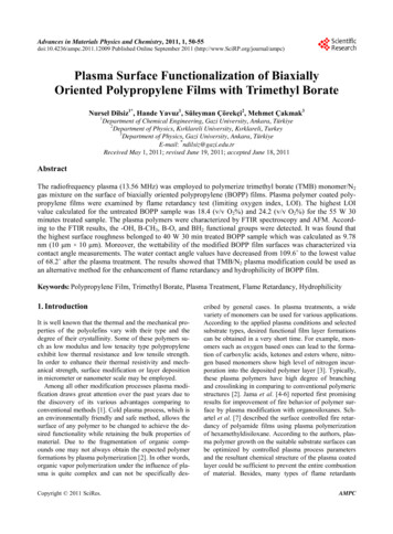
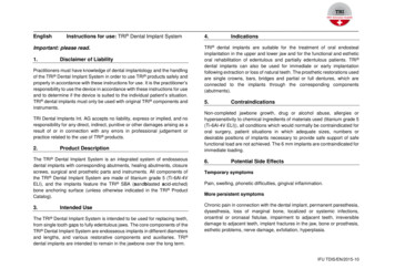
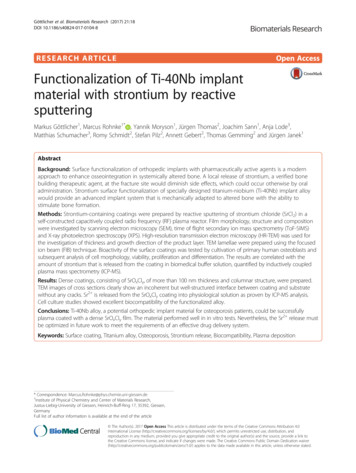
Göttlicher et al. Biomaterials Research (2017) 21:18done by addition of standard Sr2 solutions with 50, 100,500, 1000 and 5000 ppt. The strontium release experiment was reproduced three times.Page 5 of 11AlexaFluor 488 phalloidin and DAPI (both from Invitrogen, USA), respectively. Fluorescence microscopy wasperformed using a Keyence BZ-X700 (Keyence Cooperation, Japan).In vitro biological characterizationBiocompatibility of the Sr-functionalized surface wasstudied in vitro using primary human osteoblasts (hOB).The cells were isolated from spongious bone of humanfemoral heads derived from two osteoarthritic patients(female, age: 56 and 75 years, moderate to servere stateof osteoarthritis) undergoing total hip replacement atthe university hospital‚ Carl Gustav Carus‘ Dresden afterobtaining informed consent. The ethics commission ofTechnische Universität Dresden approved the application of hOB for in vitro experiments (no. EK262092009). The cells were expanded in α-MEM containing 15% fetal calf serum (FCS), 2 mM L-glutamine(L-glu), 100 U/ml penicillin and 100 mg/ml streptomycin (pen/strep) (all from Biochrom, Germany) untilpassage 3. Sr-functionalized samples (Sr-Ti40Nb) and, asreference, unfunctionalized samples (Ti40Nb) wereseeded with 2 104 hOB per disc (d 12 mm,h 0.7 mm) and cultivated in α-MEM containing 9%FCS, L-glu, pen/strep as well as 10 7 M dexamethasone,10 mM β-glycerophosphate and 0.05 mM ascorbic acid2-phosphate (all from Sigma-Aldrich). After 1, 7, 14 and21 days of cultivation, samples were collected for biochemical and microscopic analysis.Cell viability and proliferation were assessed by measurement of the intracellular lactate dehydrogenase(LDH) activity and the DNA content; osteogenic differentiation was evaluated by quantification of alkalinephosphatase (ALP) activity. The samples, frozen aftercell culture, were thawed and incubated for 50 min in1% Triton X-100/PBS on ice; cell lysis was supported by10 min ultrasonication. LDH activity in the lysates wasanalysed using the CytoTox 96 Non-Radioactive Cytotoxicity Assay (Promega, USA), and the DNA contentwas determined using the QuantiFluor Assay (Promega),both according to the manufacturer’s instructions. Themeasured values were correlated with the cell numberusing a calibration series. For quantification of the ALPactivity, the lysates were incubated with p-nitrophenylphosphate (Sigma-Aldrich) in substrate buffer (0.1 Mdiethanolamine, 1% Triton X-100, 1 mM MgCl2, pH 9.8)at 37 C for 30 min, followed by absorbance measurement at 405 nm. The amount of p-nitrophenolate (pNp)produced by ALP in the cell lysate was calculated usinga calibration line. ALP activity was normalized to the cellnumber to express specific ALP activity (μmol pNp/30 min/106 cells). Cell distribution and morphology onthe samples were investigated by fluorescence microscopy: After fixation in 3.7 wt% formaldehyde, actin cytoskeleton and nuclei of the cells were stained byResults and discussionStrontium oxide chloride film characterizationFigure 3 shows a typical SEM image of the film surfaceafter the strontium deposition process. A dense thin filmwith a very smooth surface was produced whereas additional μm-sized particles were present on the surface.An analysis of the particle composition by EDX (notshown) revealed that they contain strontium, fluorineand chlorine. The origin of this surface contamination isdiscussed at the end of this section.Figure 4 shows a cross-sectional TEM image of thelayer structure after strontium deposition. The thin Srcontaining film, from here termed as SrOxCly film, is130 nm thick including an interlayer of about 10 nm directly above the substrate surface. This interlayer is verylikely a passivating Ti-Nb oxide that forms spontaneously after the polishing procedure. The SrOxCly film iscomposed of nanocrystalline grains that are preferablyconfigured in columns with less than 10 nm in widthand oriented in growth direction of the layer. The surface roughness of the deposited film is less than 5 nm. Ahigh resolution image of the interface region betweensubstrate and Sr-film in Fig. 5 shows no epitaxial relations between both phases. Although the TEM image reveals a dense and completely adhering coating layer itcannot be excluded that microcracks or spalling occurs,if too much bending is carried out in a convex way.In Fig. 6 EDX spectra of three different regions insidethe layer structure are depicted. As expected, titaniumand niobium signals emerged in the Ti-40Nb substrateregion. At the thin interlayer a higher amount of oxygenwas found, compared to the substrate region. This confirms the above mentioned assumption that a passivatingoxide film forms an interlayer. A considerable amount offluorine was found, possibly emerging from condensation of gaseous fluorine compounds inside the vacuumchamber before the deposition process. Potential fluorine sources are Teflon and MACOR parts inside thevacuum chamber and volatile compounds from the vacuum pump oil or vacuum grease.Inside the strontium film, mainly strontium, oxygen,chlorine and iron impurities were detected. We concludethat the reaction of SrCl2 to SrO in the oxygen dischargeis incomplete and that iron was also sputtered from thetarget electrode, causing this element composition. Nofluorine signal was found within the SrOxCly film so thatfluorine incorporation during strontium deposition is relatively low. As the detection limit of TEM-EDXS is 1–2 at.%, this does not exclude smaller fractions of fluorine.
Göttlicher et al. Biomaterials Research (2017) 21:18Page 6 of 11Fig. 3 SEM image; Representative scanning electron microscopy image of a coated surface after reactive sputtering of SrCl2 in Ar/O2 plasma(room temperature P 100 W, 0.25 sccm O2, 0.68 sccm Ar, t 2 2000 s)As an additional tool for phase characterization we usedX-ray diffraction. The obtained data is depicted in theAdditional file 1. Unfortunately, the count rate is poor andno well-grounded conclusion is possible. Nevertheless, thesmall reflexes could assigned to a SrOxCly phase.Representative ToF-SIMS anion and cation depth profiles of strontium coated Ti-40Nb are depicted in Fig. 7.As halogens and halogen-containing fragments, such asF , Cl or SrF ions are easily ionized, their signal intensity reaches the saturation limit of the secondary ion detector. Therefore, we used specific halogen-containingsignals (e.g. 37Cl , F 2 ) to determine a reliable depth profile of an element species, especially at interfaces. In caseof the cations, the Sr secondary ion signal was oversaturated. From the two depth profiles, we conclude thatthe deposited strontium film is chemically homogeneousand contains SrO and SrCl2 as well as traces of FexOy.This is in agreement with the reported TEM results.As mentioned before, F and SrF signals in the aniondepth profile are oversaturated (not shown in Fig. 7)from the beginning of the spectra. By monitoring the F 2and F signals, we found that the highest fluorine concentration is located at the interlayer. There are alsofluorine impurities inside the strontium film, but it issafe to assume that their concentration was below1 at.%. Besides the fluorine signals also several metaloxide signals (TiO 2 , NbO 2 , O 2 ) reach a maximum intensity at the interlayer, which is also in agreement with theTEM results. At higher depth, mainly substrate signals(Ti , Nb ) were detected. As observed in [30] the monooxygen fragments TiO and NbO are characteristic signals in pure Ti-40Nb samples. Due to a high oxygensolubility combined with a high oxygen affinity of thepure metals, these fragments were formed during secondary ion emission.XPS measurements were carried out to obtain moreaccurate chemical information of the coating. In Fig. 8 asputter depth profile of the SrOxCly coated Ti-40Nbsample is shown. We measured detail spectra of C1s,O1s, Cl2p, F1 s, Fe2p, Nb3d, Sr3d and Ti2p. The depthFig. 4 TEM image cross section; Cross section TEM brightfield image of the layer stack showing the thicknesses of the Sr-containing film andinterface as well as their crystallinity
Göttlicher et al. Biomaterials Research (2017) 21:18Fig. 5 HRTEM image; High resolution TEM image of the interfacialarea. The film does not show epitaxial relations to the interfaceprofile supports the findings of the ToF-SIMS and TEMmeasurements, even though the profile is not as sharpdue to a larger analysis spot and a less defined sputtercrater. The particles on the surface (Fig. 3) deterioratethe XPS depth resolution. The strontium Sr3d signal isPage 7 of 11present as SrCO3 (only on the surface due to reactionwith CO2 and water from the lab air). SrF2, SrCl2 andSrO, correspond well to the respective signals of C1s,O1s, Cl2p and F1 s. The SrF2 is a contamination fromthe thin film deposition process present as small spotson the surface (as shown from SEM/EDXS). Since theXPS spot size is much wider than the SrF2 spots the signal superimposed with the SrOxCl y film. The film itselfconsists mainly of SrO and SrCl2. Iron is present as another contamination from the deposition process andthus correlates with the strontium signal. The titaniumand niobium signals are not present at the surface, indicating a homogeneous film without cracks or pinholes.During depth profiling both the Nb3d as well as theTi2p signal appear in the fully oxidized state and are reduced to the metal state with ongoing depth profiling.Due to the fitting procedure and background functionused, the titanium amount is underestimated. Thus, thestoichiometry at the end of the sputter process is [Ti]/[Nb] 56/44 instead of the expected atomic ratio ofabout 74/26. The differing atomic ratio for the investigated alloy in XPS spectra is discussed in detail in [31].Strontium ion releasePreliminary studies at different immersion times revealed that strontium is almost completely released intophysiological solution after 1 day. By ICP-MS analysis anFig. 6 TEM images and EDX spectra; (a) TEM brightfield image and EDX spectra acquired from the marked areas (substrate, interface, Sr film). (b)X-ray energy range 0 7000 eV, (c) X-ray energy range 200 800 eV
Göttlicher et al. Biomaterials Research (2017) 21:18Page 8 of 11Fig. 7 TOF SIMS data; ToF-SIMS depth profiles of the SrOxCly coated Ti-40Nb sample. In a) the depth profile obtained from secondary cations isdepicted. In b) the depth profile obtained from secondary anions is depicted. SrOxCly was deposited at room temperature, P 100 W, 0.25 sccmO2, 0.68 sccm Ar, t 5 haverage Sr2 concentration of 0.05 mM in the buffer solution was quantified. This value is close to the pharmaceutical effect threshold of 0.10 mM for osteoblastactivation [10]. In another study Schumacher et al. reported an optimal strontium concentration of 0.01–0.10 mM for stimulation of human-bone-marrow-derived mesenchymal stem cell proliferation and osteogenic differentiation in vitro [32]. However, in contrastto our material, Schumacher et al. tested strontiummodified calcium phosphate bone cements that continuously release 0.03–0.07 mM Sr2 over 20 days. Thereforewe conclude that our coating releases enough strontiumto stimulate osteoblastic activity, but modifications arerequired to generate a continuous release. However, inan in vivo environment we have to deal with more complex interactions, e.g. higher strontium concentrationsaround the implant region because of a lower liquidamount per area that is in contact with the implant.Additionally, adsorbed proteins may block the strontiumFig. 8 XPS data; XPS depth profile of the SrOxCly coated Ti-40Nbsample. SrOxCly was deposited at room temperature, P 100 W, 0.25sccm O2, 0.68 sccm Ar, t 5 hrelease. ToF-SIMS analysis of the Ti-40Nb surface afterthe release experiment revealed only traces of residualstrontium. Obviously, the produced coating dissolvesquickly, generating an initial burst release of strontium.To produce applicable coatings with optimal strontiumrelease kinetics, it is important that the strontiumamount is higher while the coating dissolves with aslower rate and over a longer period. Possible methodsto improve the release characteristics of the coating willbe the deposition of a less water soluble strontium compound (e.g. strontium phosphate [33]) or the depositioninto porous Ti-40Nb implants that might be inserteddirectly into bone defects [34].Biological response of human osteoblasts to SrOxClycoated Ti-40NbFor probing the biological response human osteoblasts(sustained from osteoarthritis patients) were cultivatedon the surface of SrOxCly-coated Ti-40Nb (Sr-Ti40Nbin Fig. 9) and unmodified Ti-40Nb samples (Ti40Nbin Fig. 9). On both surfaces, the cells showed the typicalspreading and cytoskeletal organization 1 day after seedingand their number increased strongly during further cultivation (Fig. 9). Both, measurements of the LDH activity(related to the number of viable cells) and the DNA content (reflecting the total cell number) demonstrated an increase of the cell number over time, which wassignificantly higher on the strontium-modified samples inthe first experiment with hOB from donor 1 (Fig. 10).However, in a second experiment with cells from donor 2,no significant differences were determined between SrTi40Nb and Ti40Nb (data not shown). The activity of theosteoblastic marker ALP increased during the cultivationperiod of 21 days on both surfaces. However, no significant difference between Sr-Ti40Nb and
detail in reference [28]. Discs with a thickness of 0.7 mm were prepared by cutting the rod using an IsoMet 5000 linear precision saw (Buehler, Switzerland) equipped with a SiC cut-off wheel (Struers, Denmark). Ti-40Nb discs were ground with bound diamond particles (15 μm) using a Phoenix 4000 grinding and polishing machine (Buehler).