Transcription
User Guide Sectra OneScreen
User Guide Sectra OneScreenUser Guide Sectra OneScreenAs part of our commitment to providing the highest quality products and services, we would like toencourage feedback on the quality of this documentation.If you think we can improve this document in any way, then please send your suggestions toinfo.imtec@sectra.seDocument TitleRevisionDateUser Guide Sectra OneScreenDOC-JKAN-75WC6Y-14.02016-02-10Copyright 2016, Sectra AB2
User Guide Sectra OneScreenTable of Contents1.1.1Introduction . 4Intended Use . 42.12.22.3Introduction to Sectra OneScreen for Osteoporosis . 5BMD Analysis Process. 5Patient Integrity . 5Additional Integration Options . 53.13.23.33.43.53.6The Sectra OneScreen for Osteoporosis Report . 6Identifiers. 7Patient . 7Examination . 7Reference . 7Reference Graph . 8Exemption to Normal BMD Estimation Guidelines . 84.14.2Preliminary reports . 8Intended use of preliminary reports . 8Limitations of preliminary reports . 82.3.4.5.References . 9Appendix A. Safety and Cautions. 10A.1Cautions – Measuring Change of DXR-BMD . 10Appendix B. Introduction to DXR-BMD . 11B.1Fundamentals of DXR-BMD . 11Appendix C. Sectra OneScreen for Osteoporosis Reference Databases . 13C.1 Generation of a Database . 13C.2 Inclusion Criteria . 13C.3 Exclusion Criteria . 13C.4 Generation of Reference Values for T-scores . 13C.5 Generation of Reference Values for Z-scores . 14Copyright 2016, Sectra AB3
User Guide Sectra OneScreen1.IntroductionThe Sectra DXR-BMD technology enables the Bone Mineral Density (BMD) to be measured from astandard hand radiograph. DXR-BMD measurements are available through the Sectra OneScreen service. The Sectra OneScreen service is based upon Sectra Osteoporosis Package . Sectra OsteoporosisPackage is based on the same digital X-ray radiogrammetry image analysis engine as was previously usedalso by Pronosco X-posure System V2.1.1Intended UseThe intended use of the Sectra OneScreen service is to estimate BMD in the distal forearm and thereby toassess increased risk of osteoporotic fracture according to the World Health Organization (WHO) criteria[1]. The OneScreen report is especially intended to be used for:1) assisting the physician in diagnosing subjects who already have been identified to be at risk forsuffering from osteoporosis, together with other known risk factors.2) comparing the BMD estimate with a reference population comprised of young normals and agematched normals to compute T-scores and Z-scores, respectively.The BMD estimate is obtained from a standard digital X-ray image of the hand by sending the image toSectra OneScreen for analysis.The BMD estimate generated by the Sectra OneScreen service is sometimes referred to as digital X-rayradiogrammetry BMD (DXR-BMD). The general characteristics of the DXR-BMD are: DXR-BMD is highly correlated to forearm DXA-BMD. It is an automated and operator-independent analysis. The precision is high. It is insensitive to relevant variations in image capture conditions. It has been demonstrated to be effective in monitoring the progress of Osteoporosis and responseto treatment. Reference databases are available for relevant ethnicities in a large number of countries. Extensive clinical documentation is available.For a summary of studies and clinical performance, please refer to dxr-online – Clinical and TechnicalInvestigation History [2].Copyright 2016, Sectra AB4
User Guide Sectra OneScreen2. Introduction to Sectra OneScreen forOsteoporosisThis section outlines the Sectra OneScreen analysis process.2.1BMD Analysis ProcessThe following steps are performed during a BMD analysis: An image of the non-dominant hand is acquired. The image is de-identified and sent to Sectra dxr-online. (Optional) A preliminary report is delivered to the referring unit. The image is quality checked and the analysis validated by a by a certified operator at the Sectradxr-online laboratory. The final report with the analysis result is sent to the referring unit by DICOM or HL7integration.2.2Patient IntegrityTo protect patient integrity, patient identifiable data is encrypted locally at the hospital prior to transfer tothe Sectra OneScreen system. Thus Sectra OneScreen operators and system administrators are not able toidentify the person associated with an image.2.3Additional Integration OptionsTo provide an efficient workflow for high volume osteoporosis screening, Sectra OneScreen supports anumber of integration options. Options include report integration with PACS/RIS/HIS via HL7 and/orDICOM, automatic integration of additional fracture risk factors and custom rule based generation ofreports. For details contact your Sectra OneScreen representative.Copyright 2016, Sectra AB5
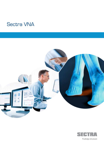
User Guide Sectra OneScreen3.The Sectra OneScreen ReportThe default Sectra OneScreen report format is DICOM Secondary Capture image sent to PACS, asanother series in the same examination as the original hand image. Figure 1 shows the layout of thereport.Figure 1: The Sectra OneScreen report.Copyright 2016, Sectra AB6
User Guide Sectra OneScreen3.13.1.1IdentifiersExamination IDThis is an ID that uniquely identifies the examination in your HIS, RIS or PACS system. Contact yourimaging department for details on which field in your system it corresponds to.3.1.2Referring PhysicianIf the name or code of the referring physician is available from the DICOM image, it is displayed in thisfield.3.1.3Care Provider IDThis is the identifier for your site within the Sectra dxr-online system.3.23.2.1PatientDate of BirthIf the date of birth of the patient is available from the DICOM image, it is displayed in this field.3.2.2SexIf the sex of the patient is available from the DICOM image, it is displayed in this field.3.33.3.1ExaminationDateThis field displays the date the image was captured.3.3.2AgeThis field displays the age of the patient at the day the image was captured.3.3.3BMDThis field displays the BMD of the analyzed image with two decimals.Note: If the measurement will be used to assess BMD change rate over short time intervals or if a higherprecision of the DXR-BMD measurement is required for other reasons, it is recommended to use theDXR-BMD Bone Change examination type instead. For a detailed discussion about precision of DXRBMD measurements, please refer to [2].3.43.4.1ReferenceReference EthnicityThe ethnicity of the reference database used to calculate T- and Z-scores as well as reference graphs.Important: If the default ethnicity does not apply for a given patient, T- and Z-scores will not be validfor that individual. Sectra can provide reference tables for many ethnicities.3.4.2Z-scoreThis field displays the Z-score corresponding to the BMD and reference ethnicity.Patient sex, ethnicity and age must be defined in order for the Z-score to be available.Important: If the default ethnicity does not apply for a given patient the Z-score will not be valid for thatindividual.Copyright 2016, Sectra AB7
User Guide Sectra OneScreen3.4.3T-scoreThis field displays the T-score corresponding to the BMD and reference ethnicity.Patient sex and ethnicity must be defined in order for the T-score to be available.Important: If the default ethnicity does not apply for a given patient the T-score will not be valid for thatindividual.3.5Reference GraphAs illustrated in Figure 1, a reference curve for BMD is shown on the report. The graph illustrates the Tscore and the Z-score corresponding to the reference population.3.6Exemption to Normal BMD Estimation GuidelinesIt is important to note that if a patient has had a fracture in one of the metacarpals or if fixation pins arepresent, the BMD estimate may not be accurate. It is important that the physician exercise his or hercritical judgment on the resulting BMD estimate.4.Preliminary reportsThere is an option to receive an automatic preliminary report directly after an image has been sent toSectra OneScreen.4.1Intended use of preliminary reportsThe optional preliminary report functionality is intended to support workflows where it is of benefit to beable to discuss the result of the analysis with a patient before she or he leaves the clinic. Due to thelimitations of the preliminary report discussed below, it is recommended to only activate the preliminaryreport functionality if there is a clear benefit that outweighs the limitations. It must also be made clear tothe patient that the report is preliminary and the patient must be contacted and advised if a preliminaryreport is declared invalid.4.2Limitations of preliminary reportsA preliminary report is preliminary because it has not yet been quality checked by a human operator. Onrare occasions a preliminary report will be declared invalid by the operator at validation.A final report that revokes a preliminary report will never give another different BMD value, but willdeclare that the image could not be used for a reliable BMD measurement.On other occasions a preliminary report may be withheld and instead a message stating that the imagerequires operator attention and that the final report must be awaited.The frequency of cases where a preliminary report is withheld or is invalid depends heavily on how wellan imaging unit follows the Sectra OneScreen image acquisition guidelines provided by Sectra, and alsoon the image acquisition equipment used. It has been estimated that preliminary reports will on averagebe withheld in around 3 percent of cases and that a delivered preliminary report will on average bedeclared invalid by the final report in approximately 2 thousandth of cases.Copyright 2016, Sectra AB8
User Guide Sectra OneScreen5.References[1] Kanis JA, Melton LJ, Christiansen C, Johnston CC, Khaltaev N. The diagnosis of osteoporosis.Journal of Bone and Mineral Research 1994; 9 (8)1137-1142.[2] dxr-online – Clinical and Technical Investigation History.[3] Pors Nielsen S. The Metacarpal Index Revisited. Journal of Clinical Densitometry 2001; 4(3):199207.[4] Laval-Jeantet AM, Bergot C, Carroll R, Garcia-Schaefer F. Cortical bone senescence and mineralbone density of the humerus. Calcif. Tissue Int. 1983; 35: 268-72.[5] World Medical Association Declaration of Helsinki; Recommendations guiding physicians inbiomedical research involving human subjects. 1996;1-4.[6] CPMP/ICH/135/95. ICH Guideline for Good Clinical Practice. 1997.[7] Rosholm A, Hyldstrup L, Baeksgaard L, Grunkin M, Thodberg HH. Estimation of bone mineraldensity by Digital X-ray Radiogrammetry. Theoretical background and clinical testing. OsteoporosInt 12 (2001) 11, 961-969.Copyright 2016, Sectra AB9
User Guide Sectra OneScreenAppendix A. Safety and CautionsA.1 Cautions – Measuring Change of DXR-BMDWhen assessing change of DXR-BMD over time, for maximum accuracy both measurements must bebased on images acquired with the same type and model of modality.If DXR-BMD is used to monitor the effect of treatment, or if a greater accuracy of change of DXR-BMDover time is needed, it is recommended to use the examination type DXR-BMD Bone Change instead asit uses both hands and provides a greater precision than a standard OneScreen examination. Pleasecontact your Sectra contact for details.Whether measuring BMD over a course of time or on a single occasion, it is imperative that therecommended operating protocol is followed.Copyright 2016, Sectra AB10
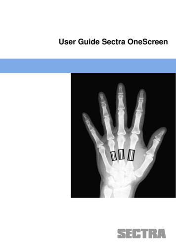
User Guide Sectra OneScreenAppendix B. Introduction to DXR-BMDB.1 Fundamentals of DXR-BMDThe Sectra OneScreen for Osteoporosis service estimates distal forearm BMD by Digital X-rayRadiogrammetry (DXR), which involves a radiogrammetric and textural analysis of the three middlemetacarpal bones1.To locate the bones in the radiograph, the DXR analysis engine utilizes a model-based algorithm thatrecognizes the diaphysis of the three middle metacarpals in the hand. After each diaphysis has beenidentified, regions of interest (ROI) for the three metacarpals are placed automatically with very highreproducibility (Figure 2).Figure 2: Outline of the three involved regions of interest.The heights of the ROIs are fixed to 2.0 cm, 1.8 cm, and 1.6 cm for the 2nd, 3rd, and 4th metacarpals,respectively.Within each ROI, the endosteal (inner) and periosteal (outer) edges are automatically found, therebysegmenting the bone into two cortical regions and one endosteal region. Along a profile across the bone,the two endosteal edge points are associated with the points of maximal intensity as illustrated in Figure3. The periosteal points are associated with the edge of the bone.1The Sectra OneScreen for Osteoporosis service is based on Sectra Osteoporosis Package . Sectra Osteoporosis Package applies the same image analysis algorithms for BMD estimation as was previously used by the Pronosco X-posure System V2.Copyright 2016, Sectra AB11
User Guide Sectra OneScreenRrMax. intensityLTWFigure 3: Outline of principles for determination of the basic radiogrammetrical quantities.The average cortical thickness (Ti) and bone width (Wi) are determined for each metacarpal i,accumulating over 90-200 measurements per centimeter bone for resolutions between 230 dpi and 508dpi. This e.g. implies that a total of 300-800 measurements contribute to the average cortical thickness ofa single bone.B.1.1 BMDA bone volume per projected area (VPA, cm) is computed for each metacarpal assuming a cylindricallyshaped bone:VPAi * Ti * (1- Ti / Wi)The total VPA for the metacarpals is defined as a weighted average:VPAmc (VPA2 VPA3 0.5 VPA4) / 2.5The fourth metacarpal bone is given a lower weight due to a lower precision and an inferior clinicalimportance. The final DXR-BMD is then calculated as:DXR-BMD c *VPAmc * (1-P)The scaling constant c is determined so that DXR-BMD on the average is equal to that of the mid-distalforearm region of the Hologic QDR 2000 densitometer (Hologic, Waltham, MA, USA). The constantadapts VPA to both the volumetric mineral density of compact bone and the typical shape characteristicsof the involved bones. P is an estimated three-dimensional porosity, aimed to be the fraction of thecortical bone volume that is not occupied by bone ([4], [7]).Copyright 2016, Sectra AB12
User Guide Sectra OneScreenAppendix C.DatabasesC.1Sectra OneScreen ReferenceGeneration of a DatabaseA reference database contains hand measurements obtained from a population of “normal” women ormen of one ethnic origin. Data for the reference databases was collected in compliance with the revisedHelsinki Declaration [5] and ICH Guidelines for Good Clinical Practice [6].The definition of “normal” women in the normative reference database was based on the following set ofInclusion and Exclusion Criteria.C.2 Inclusion Criteria Women who are of the targeted ethnical origin. Women who are generally healthy. Women aged between 20 and 79 years. Women who understand and have signed the Ethics Committee approved informed consentform.C.3 Exclusion Criteria Women who are pregnant. Women whose complete medical history shows any indication of a disease that may impact bonetissue, e.g. hyper/hypothyroidism, hyper/hypo-parathyroidism, diabetes (IDDM) or rheumatoidarthritis. Women with verified bone diseases apart from osteoporosis, such as osteomalacia, Paget’s bonedisease, myeloma or some cancers. Women with prolonged hospitalization or other extended immobilization. Women with immobilization and/or limited use of one or both arms within 6 months prior to theinvestigation. Women who have ever taken bisphosphonates or fluorides. Women who have taken estrogens, calcitonin, raloxifene or tamoxifen within the two yearsimmediately prior to enrolment in the study. Earlier use of these products is allowed if thewomen previously took the drug for less than one year. Topical administered estrogens areallowed. Women who are in chronic treatment with steroids, phenytoin or heparin. Women who are in chronic treatment with thyroxin in unstable doses (stable defined as nochange in dosage within the last 6 months). Women with a history of bilateral wrist fracture.C.4 Generation of Reference Values for T-scoresThe T-score shows the number of Standard Deviations between the patient’s BMD and the average ofpre-menopausal women within the population (Young Adult Woman). The statistical calculation of the Tscore is:Copyright 2016, Sectra AB13
User Guide Sectra OneScreenT X obs YAWSDYAWwhere Xobs is the observed BMD measurement. The reference level, YAW, and standard deviation, SDYAW,applied in the T-score calculation are the mean and standard deviation of BMD values obtained in youngadult women (YAW) from 20-39 years of age.C.5 Generation of Reference Values for Z-scoresThe Z-score shows the number of Standard Deviations between the patient’s BMD and the mean of agerelated women. Statistically, the Z-score is calculated as:Z X obs AgeSD Agewhere Xobs is the observed BMD measurement. The age dependent reference level is Age. The standarddeviation, SDAge, applied in the Z-score calculation is based on advanced regression analysis. Thereference level, mAge, is described in a second or third order polynomial in age. The standard deviation,SDAge, is calculated as a function of the reference level. Thus, the standard deviation is not constant for allages.The T-score parameters, YAW and SDYAW, and the Z-score parameters, Age and SDAge, are tabulated forspecific reference databases.Copyright 2016, Sectra AB14
User Guide Sectra OneScreenLABELProductSectra Osteoporosis PackageVersion2.0.6.0ManufacturerSectra ABTeknikringen 20, SE-583 30 al/contact/support/Contact0434Copyright & Legal Statement Sectra AB, Sweden, 2016All rights are reserved. Reproduction or transmission in whole or part, in any form or by any means,electronic, mechanical or otherwise, is prohibited without written consent of the copyright owner.Copyrights and all other proprietary rights in any software and related documentation ("Software")made available to you rest exclusively with Sectra AB. No title or ownership in the Software isconferred to you. Use of the Software is subject to the end user license conditions as are available onrequest.To the maximum extent permitted by law, you shall not decompile and/or reverse engineer thesoftware or any part thereof.Regulatory Clearance StatementThe quality system of Sectra AB [Sectra] conforms to ISO 9001, ISO 13485 and ISO 27001. All Sectramedical devices have obtained regulatory clearance for those markets where Sectra sells and deploysits devices, e.g. EEA, USA, Canada, Australia. For further regulatory information please contact Sectra.Trademark & Patent DisclaimerSectra, Sectra Imtec and the Sectra logotype, are registered trademarks of Sectra AB. FIMAG andImage Exchange Portal are registered trademarks of Burnbank Systems Ltd in the UK. IDS7, SectraBizTrack, Sectra CloudFlex, Sectra DoseTrack, Sectra IEP, Sectra Image Central, Sectra Image Lab,Sectra LiteView, Sectra UniView, Sectra One Connect, Sectra Open Archive, Sectra UserInfluence,and WISE are trademarks of Sectra Imaging IT Solutions AB. Sectra Preop Online and SectraOneScreen are trademarks of Sectra AB.Windows is a registered trademark of Microsoft Corporation in the United States and othercountries. Mac, Safari, and iPad are registered trademarks of Apple Inc. in the United States andother countries.All other names/products by or are registered trademarks of the respective manufacturer.The intellectual property of Sectra includes the following patents: U.S. patents 6,005,917;6,226,393; 6,411,729; 6,763,257; 7,162,623; 7,532,214; 7,660,461; 7,689,539; 7,830,381;7,936,930; 7,940,270; 8,041,129; 8,131,033; 8,295,620; 8,693,752; 8,880,143; 9,265,472. Japanesepatent 3646122. European patents 0818971; 1046374; 2512341. Swedish patents 0203545;0300951.
User Guide Sectra OneScreenPatents Pending in the U.S. and other countries.Copyright 2016, Sectra AB16
an imaging unit follows the Sectra OneScreen image acquisition guidelines provided by Sectra, and also on the image acquisition equipment used. It has been estimated that preliminary reports will on average be withheld in around 3 percent of cases and that a delivered preliminary report will on average be