Transcription
Downloaded from http://cshperspectives.cshlp.org/ at NYU MED CTR LIBRARY on November 1, 2017 - Published by Cold SpringHarbor Laboratory PressMechanical Properties of the Cytoskeletonand CellsAdrian F. Pegoraro,1 Paul Janmey,2 and David A. Weitz11Department of Physics and School of Engineering and Applied Sciences, Harvard University, Cambridge,Massachusetts 021382Institute for Medicine and Engineering and Department of Physiology, Perelman School of Medicine, and Departmentof Physics and Astronomy, University of Pennsylvania, Philadelphia, Pennsylvania 19104Correspondence: weitz@seas.harvard.eduSUMMARYThe cytoskeleton is the major mechanical structure of the cell; it is a complex, dynamicbiopolymer network comprising microtubules, actin, and intermediate filaments. Both theindividual filaments and the entire network are not simple elastic solids but are instead highlynonlinear structures. Appreciating the mechanics of biopolymer networks is key to understanding the mechanics of cells. Here, we review the mechanical properties of cytoskeletalpolymers and discuss the implications for the behavior of cells.Outline1 Introduction5 Rheology of networks alone2 Mechanical properties of the three classesof polymers6 Measuring cell stiffness and cautionary notes3 Single-filament mechanics7 ConclusionReferences4 Network mechanicsEditors: Thomas D. Pollard and Robert D. GoldmanAdditional Perspectives on The Cytoskeleton available at www.cshperspectives.orgCopyright # 2017 Cold Spring Harbor Laboratory Press; all rights reserved; doi: 10.1101/cshperspect.a022038Cite this article as Cold Spring Harb Perspect Biol 2017;9:a0220381
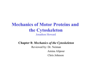
Downloaded from http://cshperspectives.cshlp.org/ at NYU MED CTR LIBRARY on November 1, 2017 - Published by Cold SpringHarbor Laboratory PressA.F. Pegoraro et al.1 INTRODUCTIONEukaryotic cells, especially those in multicellular organisms, are subjected to a variety of mechanical forces arisingfrom effects such as gravity, fluid flow, and active contraction generated by neighboring cells or resisted by theextracellular matrix. These forces are large enough to significantly deform a viscous fluid surrounded by a lipidmembrane such as a vesicle and, so, if the cell is to retainits original shape after the deforming force is released, or ifit is to be able to maintain its shape in response to such aforce, it must be at some level elastic. There are many molecular and structural mechanisms by which to make a cellsized object elastic or viscoelastic (e.g., placing a finitelystretchable membrane around a purely viscous fluid), butmost cell types are viscoelastic throughout their entire volume because they are filled with the three-dimensionalnetwork of protein filaments comprising the cytoskeleton(Schliwa 1986; Alberts 2015; Hardin et al. 2015). The filaments of the cytoskeleton are not simply mechanical supports but also the tracks along which motor proteins move,enzymes and substrates are localized, the lipid bilayers ofthe membrane are attached to the rest of the cell, and theassembly of signaling complexes is spatially organized(Wickstead and Gull 2011). Therefore, it is not always possible to isolate purely mechanical effects of cytoskeletalpolymers from their biochemical activities, but, withoutthese filaments, the eukaryotic cell would be too soft andfluid to maintain its shape and exert its function.The protein filaments comprising the cytoskeleton havesome similarities to the linear polymers within syntheticelastic materials such as the polyacrylamide in a gel or thepolyethylene in a plastic bag, but two structural features thatset them apart from synthetic polymers are a relatively largediameter and a much greater stiffness (MacKintosh andSchmidt 2010; Broedersz and MacKintosh 2014). Boththe intracellular and extracellular spaces of most organismscontain highly flexible polymers such as glycosaminoglycans and other polysaccharides. The cell interior alsocontains other long flexible objects such as nucleic acidpolymers and the membrane tubes of the endoplasmic reticulum, but the cytoskeleton is different in that its constituent filaments are orders of magnitude stiffer. The threemajor types of cytoskeletal polymer in most cells are microtubules (MTs), actin filaments (F-actin), and intermediatefilaments (IFs); they each differ strongly in the magnitude oftheir stiffness, but they are all sufficiently rigid that they canbe visualized as single extended filaments rather than tangled coils on the length scale accessible by light or electronmicroscopy, especially when they are arranged in filamentbundles. In many cases, these filaments are long and straightenough to span nearly the entire dimension of the cell.2The length and stiffness of cytoskeletal polymers generates several features of the networks they form that appearadvantageous from a biological perspective. For example,their elongated structure means that they can be assembledinto three-dimensional networks at much lower volumefractions than are required to form gels from more flexiblepolymers (Mofrad 2009; Kollmannsberger and Fabry 2011;Broedersz and MacKintosh 2014). Consequently, a network with the same rigidity or elastic modulus (i.e., itsresistance to deformation under applied force) as a 5%polyacrylamide gel can be made by cross-linked actin filaments with a concentration nearly two orders of magnitudelower. A second consequence of the length and rigidity ofcytoskeletal polymers is that the viscoelasticity of the networks they make is different from those of gels formed byflexible polymers, especially when the networks are deformed to relatively large strains. One such difference isseen in the strain stiffening of cytoskeletal networks, inwhich the stiffness of these networks increases the morethey are deformed—a feature that is lacking in flexiblepolymer gels such as polyacrylamide or gelatin and mighthelp cells limit their deformation when they are subjectedto abnormally large stresses. A further feature of cytoskeletal polymers is that they are all highly charged anionicpolyelectrolytes (Janmey et al. 2014). The large surfacecharge density of actin filaments, MTs, and IFs does notby itself have a major effect on their rigidity, but it doesinfluence the geometry of the networks that these polymersmake and how they interact with filament binding proteins.In addition to the creation of an elastic environmentwithin the cell interior, the cytoskeleton also plays important mechanical roles in linking the plasma membrane ofthe cell, as well as interior membranes such as the endoplasmic reticulum, to the rest of the cell, in limiting thediffusive motions of intracellular polymers that are largerthan the network mesh size, and possibly in controlling thepermeation of water and small solutes through the cytoskeleton. A first approximation to modeling the mechanicalproperties of the cytoskeleton and cell mechanics in generalis to consider the elements of the cytoskeleton as polymers,using experimental methods and theoretical models developed for traditional polymers but modified for the muchlarger, stiffer, and fragile biopolymers comprising the cytoskeleton. Within this framework, we use terms, concepts,and measurement techniques normally applied to inertmaterial to describe the mechanical properties of these networks (Fig. 1). An important difference between the cytoskeleton and simple polymer networks is the presence ofmotor proteins that move along actin filaments and MTs tocreate active materials that are out of thermodynamic equilibrium (Mizuno et al. 2007; Guo et al. 2014). The degree towhich the mechanics of live cells can adequately be modCite this article as Cold Spring Harb Perspect Biol 2017;9:a022038
Downloaded from http://cshperspectives.cshlp.org/ at NYU MED CTR LIBRARY on November 1, 2017 - Published by Cold SpringHarbor Laboratory PressMechanical Properties of the Cytoskeleton and CellsQuantities involved in mechanics measurementsChange in length, ΔLafter time, ΔτStress: σ Force, FStrain: γ .Strain: γ rateRelaxed length, L 0Elasticresponseσ G′γFAreaΔLL0ΔγΔτAreaViscousresponse.σ [G′′(Δτ) Δτ] γFigure 1. The terms applied to and the quantities measured in thestudy of rheology of biopolymer networks. G′ , elastic response; G′′ ,the viscous response. Biopolymers are viscoelastic and both G′ andG′′ are significant. (Adapted from Kasza et al. 2007, with permissionfrom Elsevier.)eled as polymer networks is by no means certain, but thisanalogy has potential to relate structural features of thecytoskeleton visualized by light, electron, and atomic forcemicroscopy to the mechanical properties of the cell. Thisreview will explore the mechanical properties of the cytoskeleton and cells from the perspective of polymer physicsand soft materials, summarize the structural features of thethree different cytoskeletal polymer types, and discuss howthey can be assembled into passive and active networks thatmediate cell mechanics.2 MECHANICAL PROPERTIES OF THE THREECLASSES OF POLYMERSLike any system of polymers, the mechanical properties ofcytoskeletal networks depend on the physical properties ofthe individual polymer strands, the structure and mechanical properties of the linkages between filaments, and thethree-dimensional geometry of the filament arrangement(Fabry et al. 2001; Chen et al. 2010). In most cell types, thecytoskeleton is formed by a three-dimensional compositenetwork of actin filaments (F-actin), MTs, and IFs, togetherwith the host of proteins that bind to the sides or ends ofthese linear polymers. Binding proteins for actin, IFs, andMTs regulate filament length, cross-link filaments to eachother, and apply forces to the filaments. The complex systems of cytoskeletal regulatory proteins have been extensively reviewed elsewhere.The three types of cytoskeletal filaments differ fromeach other, both chemically and physically, in ways thatpermit a wide range of material properties in the networksthey form. Among the most significant differences is thestiffness of each polymer type. The bending stiffness ofCite this article as Cold Spring Harb Perspect Biol 2017;9:a022038cytoskeletal filaments, usually quantified by its persistencelength, ℓp, ranges from a few hundred nanometers for IFs,to 10 mm for F-actin, to millimeters for MTs. Filamentstiffness, filament length, and the geometry of cross-linkingtogether determine the mechanical properties of cytoskeletal networks, and the unusually high stiffness of thesepolymers has motivated much theoretical and experimental work to relate the microscopic properties of the polymers to the macroscopic properties of the networks theyform. The presence of molecular motors that can generatemotions much larger than those produced by thermal energy and that can exert tension within networks creates newmaterial properties that are only beginning to be experimentally measured and theoretically understood (Mizunoet al. 2007).3 SINGLE-FILAMENT MECHANICSThe stiffness of polymer filaments can be quantified bytheir ℓp, which is defined as the length scale for the decayof the tangent–tangent correlation along the filament andis proportional to the stiffness of the polymer. This fundamental length has a simple physical interpretation: It is thetypical range over which the orientation of an isolatedpolymer remains correlated along its length. It can alsobe thought of as the maximum length over which the polymer will appear to be straight in the presence of the constant Brownian forces it experiences because of thermalenergy (kT) in a medium at finite temperature. If a polymercan be modeled as a uniform cylinder, ℓp kB/kT, in whichkB is the bending modulus of the filament. Usually ℓp iseasier to measure than kB, and the force needed to deform afilament is inferred from ℓp, but recent studies of the coupling between bending or stretching and twisting of F-actinsuggest that simultaneous application of torque, tension,or bending, as some actin-binding proteins appear to do,can have more profound effects on F-actin than suggestedby ℓp alone (De La Cruz et al. 2010; Yamaoka and Adachi2010). For complex filaments such as MTs, in which slidingbetween protofibrils can occur during MT bending, therelationship between ℓp and kB is not a simple proportionality, and some experiments and models predict a contourlength-dependent persistence length (Pampaloni et al.2006; Heussinger et al. 2010).The ratio between persistence length and the contourlength (ℓc) differentiates the polymer filaments into threeregimes. When ℓc . ℓp, the filaments are flexible (i.e., thermal energy can cause large transverse fluctuations), a filament then approximates a Gaussian polymer chain with aKuhn length—the length over which the polymer is fixedand beyond which it is free to rotate—of 2ℓp. In the limit ofℓp . ℓc, filaments are stiff and do not show transverse3
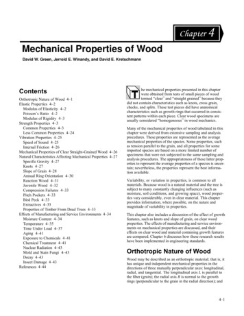
Downloaded from http://cshperspectives.cshlp.org/ at NYU MED CTR LIBRARY on November 1, 2017 - Published by Cold SpringHarbor Laboratory PressA.F. Pegoraro et al.thermal undulations but move in solution as rigid rodsdriven by thermal energy. Semiflexible filaments lie between these two limits, and ℓc and ℓp are on the same orderof magnitude.For filaments with a contour length of 10 mm, the threetypes of cytoskeleton filaments fall in these three stiffnessregimes, respectively. Intermediate filaments are the softestamong the three major types of cytoskeleton filaments.Their ℓp is reported to range from 200 nm to slightlymore than 1 mm and is IF-type-dependent (Schopfereret al. 2009; Beck et al. 2010). The small persistence lengthof the 10-nm diameter IFs containing many coiled-coildimer subunits per cross section is surprising as the persistence length of a single coiled-coil dimer such as tropomyosin is itself 100 nm (Sousa et al. 2010). The thinner,semiflexible F-actin is an order of magnitude stiffer thanIFs and has a persistence length ranging from 3 mm to17 mm (Gittes et al. 1993), which is comparable to thetypical filament length. MTs are considered to be stiff polymers, with ℓp .1 mm (Gittes et al. 1993). The characteristic ℓp and diameter of the different types of cytoskeletalfilaments are summarized in Fig. 2.Compared with IFs, MTs and F-actin are relatively brittle filaments and rupture under elongational strains lessthan 10% of their resting length. IFs, in contrast, canbear much larger extensional strain than MTs and F-actin.Several types of IFs have been stretched to .200% of theirresting length without breaking (Qin et al. 2009; Qin et al.2010). This large extensibility is thought to be attributableto the hierarchical structure of IFs that allows partial unfolding of subunits without breakage of the filament.The mechanical and rheological properties of networks(i.e., filaments that are linked together with different typesof cross-links) depend both on the response of individualpolymers to forces at the molecular level and on the way inwhich these single polymers are connected or otherwiseinteract with each other. The uniquely extended form ofcytoskeletal polymers results in a relationship betweenforce and extension that is different from that of purelyrigid rods or highly flexible coils. A single filament canrespond to both transverse and longitudinal forces by eitherbending or stretching/compressing. On length scalesshorter than the persistence length, bending can be described in mechanical (enthalpic) terms, as for elasticrods. In contrast, especially when the contour length isgreater than the persistence length, stretching and compression can involve both a purely elastic mechanical response analogous to that of macroscopic elastic rods, andan entropic response related to that of purely flexible coils.This entropic elastic response comes from the thermal fluctuations of the filament. Perhaps surprisingly, the longitudinal response can be dominated by entropy, even on length4Microtubulep 1 mm25 nmF-actinp 10 μm8 nmIFsp 1 μm10 nmFigure 2. Stiffness and structure of cytoskeleton filaments. Microtu-bules (MTs), F-actin filaments, and intermediate filaments (IFs) ofdiameter 10 mm are drawn on the same scale. Stiffer filaments withlarger persistence length (ℓ p) appear to be straighter. (Reprintedfrom Wen and Janmey 2013, with permission from Elsevier.)scales that are small compared with the persistence length.Thus, it can be misleading to model a filament as truly rodlike, even on short length scales compared with ℓp.The longitudinal single-filament response is often described in terms of a so-called force–extension relationshipin which the force required to extend a filament is measuredor calculated in terms of the degree of extension along aline. For filaments with the physical properties of cytoskeletal polymers, for which ℓp is on the order of ℓc, this relationship is not simple, especially at the large deformationsthat the cytoskeleton undergoes in vivo, and the full nonlinear force–extension curve has been calculated numerically (Storm et al. 2005). In the linear regime of relativelysmall extension, the force–extension relationships dependon whether the filament contour length is larger than thepersistence length. For ℓ ℓp, the force –extension relationship is approximated well by kT dℓ1ℓ2 t 1for ℓ ℓp ,ℓp ℓ4 ℓ dℓ 2(1)Cite this article as Cold Spring Harb Perspect Biol 2017;9:a022038
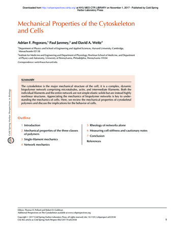
Downloaded from http://cshperspectives.cshlp.org/ at NYU MED CTR LIBRARY on November 1, 2017 - Published by Cold SpringHarbor Laboratory PressMechanical Properties of the Cytoskeleton and Cells4 NETWORK MECHANICSin which t is the tensile force needed to extend the restingend-to-end distance of the filament by a length dℓ.In contrast, for ℓ , ℓp, kTℓpdℓkDℓl20t 2 15 9 1 kDℓl0 dℓ 2kDℓl0ℓ4.1 Strain Stiffening of Cytoskeletal PolymerNetworksThe nonlinear force–extension relationship of semiflexiblepolymers, in which the force to extend the end-to-enddistance of the filament increases the more the filament isstretched, suggests that networks made from such filamentswill also become stiffer as they are deformed to largerstrains.Networks formed by cross-linked actin or IFs showstrain-dependent stiffening that is consistent with the nonlinear force–extension relationship of the individual polymers that make up the struts between nodes of suchnetworks. To measure these mechanical properties, a network is formed between two plates in a shear rheometer; byrotating one while the other is kept fixed, a shear strain isapplied to the network and the resultant restoring force canbe measured. With the strain, plate area, and restoring forceknown, it is possible to measure the shear modulus of thematerial between the plates. When using biopolymer networks, they increase their shear moduli as the shear strainincreases to relatively modest strains of a few percent foractin cross-linked by filamin A and 10%–20% for thesofter vimentin IFs, as shown in Figure 4 (Storm et al.2005; Wen and Janmey 2013). The magnitude of the shearmodulus of a vimentin network increases by more than anorder of magnitude between 1% and 80% strain (Lin et al.2010b). The strain stiffening of these networks is reversible,with the modulus returning to low levels when the shearstrain is reduced, unless the networks are strained to magnitudes large enough to break cross-links between fila-(2)for ℓ ℓp .A somewhat more accurate approximate form for the latterhas also been derived (Palmer and Boyce 2008). A moretractable approximate analytic expression derived from thistheory and applied to actin filaments concludes that theforce –extension relationship can be expressed as F kT1ℓp 4(1 r/ℓc )2 ℓc 6(1 r/ℓc ) ℓp , ℓc 2(1 r/ℓc )ℓp(3)in which F is the force and r is the end-to-end distance ofthe filament. This expression agrees well with the morecomplete expression for extensions within the range 1–0.3 ℓc/ℓp , r/ℓc , 1. The success of these different models is evident in being able to capture single-filament responses, as shown in Figure 3. From the single-filamentresponse, we can begin to understand the network response, in which geometry and filament connections playan important role in understanding how mechanics is determined for in vivo systems.1Force (pN)0.750.50.25BExact - ℓp 3 μmExact - ℓp 5 μmExact - ℓp 7.5 μm0.75Exact - ℓp 10 μmApproximation - ℓp 3 μmApproximation - ℓp 5 μmApproximation - ℓp 7.5 μmApproximation - ℓp 10 μmUndeformednetworkfilament(r rF 0)0.95r0 976 nmr0 rF 0 962 nm0.50.25c 00.941ExactApproximationForce (pN)A0.960.970.981.02 μm1.02 μm0.99r/ cc 1.0001.001.011.021.031.04λc r /r0Figure 3. (A) The effect of persistence length (ℓ p) on filament force–extension behavior, as computed usingnumerical solutions of the model developed by MacKintosh and colleagues (Storm et al. 2005) (symbols) comparedwith an analytic model approximation (lines) (Palmer and Boyce 2008) fixing contour length (ℓc) to ℓc 1.02 mm.(B) The effect of pretension on filament force–stretch behavior as computed by both methods. The end-to-enddistance of a filament at a given condition is given by r. (Reprinted from Palmer and Boyce 2008, with permissionfrom Elsevier.)Cite this article as Cold Spring Harb Perspect Biol 2017;9:a0220385
Downloaded from http://cshperspectives.cshlp.org/ at NYU MED CTR LIBRARY on November 1, 2017 - Published by Cold SpringHarbor Laboratory PressA.F. Pegoraro et al.300100250Vimentin80F-actin filamin AG′ (Pa)G′ 0.6Strain0.81Figure 4. Strain stiffening of cytoskeletal networks in vitro. Shear storage modulus measurements of F-actin cross-linked by filamin A, and vimentin, measured at frequencies near 1 Hz and a range of strain magnitudes. Vimentinnetworks can be strained much more than actin networks and also strain-stiffen to a much larger degree. (Adaptedfrom Storm et al. 2005; Wen and Janmey 2013, with permission from Elsevier.)ments or break the filaments themselves. In many settings,the dependence of shear modulus on increased stress isperhaps more biologically relevant than its dependenceon strain, and the dependence of shear modulus on imposed shear stress shows a characteristic power-law dependence that depends on the molecular mechanisms that leadto this nonlinear response (Fabry et al. 2001; Kollmannsberger and Fabry 2011).4.2 Mechanisms of Strain StiffeningThe molecular mechanism for strain stiffening in biopolymer gels (i.e., networks formed of cytoskeletal filaments) has been the subject of extensive theoretical work.Depending on the filament stiffness, network deformationcan be accommodated by bending, stretching, and reorientation of individual filaments (Vahabi et al. 2016; van Oosten et al. 2016).The strain stiffening of semiflexible polymer networkscan, within a limited range of strain, be accounted for by anentropic model that assumes that network deformation isuniform throughout the sample owing to affine deformation of each semiflexible polymer; affine stretching assumesthat the local strain at each point of the network matchesthe macroscopic strain of the network. The elasticity comesfrom the resistance of each thermally writhing polymeragainst the stretching, which effectively reduces the amplitude of thermal fluctuation and thus the thermal entropy ofthe filament (MacKintosh et al. 1995).An alternative explanation for strain stiffening based onfilament bending and enthalpic stretching has also beenproposed for stiff filament networks (Onck et al. 2005;van Dillen et al. 2008; Zagar et al. 2011) and can arise innetworks formed by filaments that themselves are linearly6elastic. In this model, because it is easier to bend stiff filaments than to stretch them, the filaments initially bend atsmall network deformations. With increasing strain, thebent filaments reorganize so that more and more filamentsalign along the direction of shear. The network deformation then comes mainly from the enthalpic stretching of thealigned stiff filaments. This transition from bending tostretching gives rise to strain stiffening. The bending, buckling, and reorganization of filaments cause nonaffine deformations in the network (Onck et al. 2005).These two strain-stiffening models predict differentnetwork deformations: The entropic model predicts uniform affine deformation, and the enthalpic model predictsheterogeneous nonaffine deformation in which individualfilament deformation does not match the average macroscopic behavior. Which theory works depends on the network structure and filament stiffness. It has been shownthat, as cross-linker concentration decreases, the deformation of an F-actin network transits from affine to nonaffine(Gardel et al. 2004). This change indicates that dense isotropic networks follow predictions of the entropic model,and the sparse networks follow predictions of the enthalpicmodel.For entropic networks, the effective contour length ofeach filament is determined by the distance between crosslinks; as the effective contour length decreases, networkmechanics are increasingly dominated by the mechanicalproperties of the individual filaments. The contour lengthis determined by the concentration of polymer cA and thedensity of cross-links, typically reported as the ratio R ofcross-links to monomers. The shear modulus at low strainis indeed found to scale with polymer concentration andcross-link density in the case of permanent cross-linksmade using, for example, the tetrafunctional biotin-bindCite this article as Cold Spring Harb Perspect Biol 2017;9:a022038
Downloaded from http://cshperspectives.cshlp.org/ at NYU MED CTR LIBRARY on November 1, 2017 - Published by Cold SpringHarbor Laboratory PressMechanical Properties of the Cytoskeleton and Cellsing protein avidin as the cross-linker of filaments labeledwith biotin, or an actin cross-linking protein such as scruin:G′ cA11/2 R(6x 15y)/5 ,(4)in which x characterizes how the cross-linker bundles thefibers, and y characterizes cross-linker efficiency. For theseentropic networks, the differential shear modulus K′ increases dramatically beyond a critical stress and is expectedto scale asymptotically as s3/2. This scaling derives fromcalculating the tension required to extend a single, thermally fluctuating filament—that is, the tension requiredto overcome the entropic motion of the fiber. Interestingly, the behavior of these entropic networks is self-similar;if the differential shear modulus is scaled by the zerostrain shear modulus and the applied stress is scaled bythe critical stress, all networks collapse onto a single universal stress-stiffening response curve. This universal curveis characteristic of affinely deforming networks of semiflexible polymers.4.3 Effect of Cross-LinksThe universal response curve and the predicted stress-stiffening behavior of entropic semiflexible polymers assumesthat the system has rigid, permanent cross-links; however,many physiological cross-links are dynamic, forming onlytransient bonds and thus can change the behavior of biopolymer networks. For example, the binding affinity of aactinin for F-actin is temperature-dependent; if the temperature of a cross-linked gel is increased from 8 C to 25 C,the network softens and becomes substantially more viscous as the effective number of cross-links is reduced owingto decreased binding (Tempel et al. 1996; Xu et al. 1998).The higher temperature leads to an increase in the rate of aactinin unbinding from the F-actin network and allowscross-links to rearrange over time, enabling the networkto remodel over long times when held at elevated temperatures (Sato et al. 1987). These dynamic cross-links thatallow remodeling are crucial for the normal physiologicalfunction of the cell and enable the cell to reorganize itscytoskeleton. Indeed, changes in binding affinity, andthus the temporal dynamics of biopolymer cross-linkers,has been directly tied to disease—a mutation of a-actininthat causes an increase in binding affinity for actin has beenfound to be responsible for some types of kidney disease(Yao et al. 2004; Weins et al. 2005).Although temperature is unlikely to be a large factor indetermining cross-link behavior in vivo, cells are constantlyexposed to varying mechanical forces in their environment.The mechanical loading of cytoskeletal networks can alsoCite this article as Cold Spring Harb Perspect Biol 2017;9:a022038change the binding dynamics of cross-links and thus affectcytoskeletal remodeling in response to force, as well as affecting force transmission through the cell. For example, inactin networks cross-linked with fascin, the forced unbinding of cross-links can occur at different rates of appliedstress and therefore lead to mechanically induced softeningof the system (Lieleg and Bausch 2007). Surprisingly, mechanical stress can also have the opposite affect—appliedstress can increase the binding time of cross-links and thusdelay network relaxation (Lieleg et al. 2009; Norstrom andGardel 2011; Yao et al. 2013). This “catch-bond” behaviordelays the onset of the gel-to-fluid transition and might beimportant for cells to resist mechanical deformation atphysiological timescales.Beyond changing the temporal dynamics of the system,cross-links themselves can also act like added mechanicalelements and thus dramatically change the rheologicalproperties of the network. For example, in the case of Factin cross-linked with filamin A (FLNa), the cross-linksthemselves act as flexible, extensible elements in the network; essentially the network comprises two differentsemiflexible polymers. This has several consequences fornetwork mechanics. The stiffness of the network is muchmore weakly dependent on cross-link concentration whencompared with networks with fixed cross-links; indeed, atlow stress, the elastic modulus of the network is only slightly above that of an uncross-linked solution of polymer(Gardel et al. 2006). Although an FLNa–F-actin networ
polymers and the membrane tubes of the endoplasmic re- . plasmic reticulum, to the rest of the cell, in limiting the diffusive motions of intracellular polymers that are larger thanthenetworkmeshsize,andpossiblyincontrollingthe . analogy has potential to relate structural features of the