Transcription
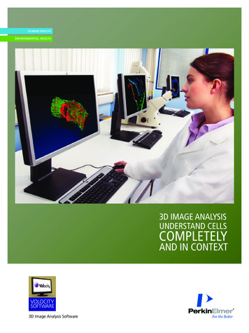
3D IMAGE ANALYSISUNDERSTAND CELLSCOMPLETELYAND IN CONTEXTVOLOCITYSOFTWARE3D Image Analysis Software
EXPLORE ANDVALIDATE YOURBIOLOGICALFINDINGS2
ADHESIONINVASIONMORPHOGENESISINVASIONGENE EXPRESSIONMOTILITYPROTEIN TRANSPORTLINEAGERAPID MEASUREMENT QUALITYEXPLOREVASCULAR GROWTHEMBRYOGENESISPRECISE RESULTSCELL CYCLE PROLIFERATIONMEASUREMITOSISINFECTIONPRECISION LINEAGE PROTEIN TRANSPORTGASTRULATION RECEPTOR ACTIVATIONQUALITYEXPLORECELL SIGNALING SYNAPTIC VESICLESMEASUREFISHAPOPTOSISSIGNAL Y REGULATIONEMBRYOGENESISRESLICE DATAGENE FERATIONINTERACTIVE RENDERINGRECEPTOR ACTIVATIONCELL SIGNALINGINFECTIONDIFFERENTIATIONVolocity SoftwareA UNIVERSALSOLUTION FOR3D IMAGEANALYSISTo get to that next important insight, you need to go where yourbiology leads you. The challenge is that biology lives in threedimensions. How can you know the shape of a structure unless youview it from every direction? How can you know its size unless you measure the volume? Howcan you tell whether two structures are close together or intertwined unless you can see bothcompletely and from every angle? Volocity 3D Image Analysis Software enables you to makediscoveries and answer complex questions that are beyond the limitations of 2D.Turn observations into understandingMoving seamlessly among restoration, visualization and quantitation, Volocity software isdesigned for true 3D analysis of fluorescence images. View your cells from every angle. Measureshapes, volumes and distances. Relate cellular structure to function with exceptional precisionand speed. Compare samples and identify trends. Produce publication-ready tables and charts.Take your image analysis to a new dimension with true 3D image analysis.A powerful solution for every labDesigned for ease of use and exceptional performance, Volocity software is compatible withmost confocal microscopy, widefield and high content screening systems – one 3D analysissolution and one learning curve for all your fluorescence imaging systems. With flexible licensingoptions, it can serve individual researchers as well as departments and entire organizations.Because it is also supported by world-class technical support, training and expertise, you cancount on Volocity software to accelerate your research through your entire 3D imaging process.Flexibility, ease of use and powerful performance: That’s why there are thousands of usersaround the globe and thousands of peer-reviewed papers referencing Volocity software – theuniversal solution for 3D image analysis.www.perkinelmer.com/volocity1
REVEAL MORE.RELATE MORE.REALIZE MORE.Whether you are studying cells, tissues or organisms, Volocitysoftware lets you see relationships you haven’t seen before andthen validate your observations. Because seeing is not enoughanymore, you have to prove it with quantitative results.Explore, interact and publish in 3DAcquire 3D image stacks from theinstrument of your choosing U ltraVIEW VoX – 3D Live Cell Imagingsystem powered by Volocity for seamlessacquisition to analysis O peretta – High Content Imaging system O ther fluorescence microscopesand imaging systems – Volocitysoftware supports files from mostconfocal, widefield and high contentimaging systemsVOLOCITY VISUALIZATIONGet a full picture of biological processes and uncover cellular relationships you can’t seewith 2D analysis, with rapid, interactive, high-resolution volume rendering of time-resolved,multichannel 3D data sets. I mport a wide range of file formats Drag and drop your image files into the software to interactively explore in 3D R otate, zoom and fly through rendered objects in real time C hoose from rendering options including solid surfaces, varying opacity and shadows S ee how the three dimensions intersect at any point E xplore structures and processes from any angle in fixed and dynamic experiments P repare stunning images for publication P roduce compelling movies for presentationTracking overlaid with 3D image datausing Volocity Quantitation.Cardiac tube of a Drosophila larva rendered usingVolocity Visualization.Volocity Visualization used toclearly show kinetochores as solidspheres within the nucleus.Show dynamic events in 3D over time andcreate publication-quality movies.2
Measure, analyze and relate your discoveriesVOLOCITY QUANTITATIONValidate and confirm your observations of cellular structure withaccurate 3D measurement and analysis. C ompare and relate cellular organelles within and between samplesfor a better understanding of intracellular and intercellular relationships P erform morphological analysis M easure fluorescence localization and colocalization M easure distances within and between organelles A utomatically track large numbers of cells R efine your experiment quickly and easily with immediate feedback O verlay measured objects on image data A utomatically measure or analyze multiple samples with batch processing U tilize specific analysis tools for quantifying colocalization, ratioedimages, FRAP and FRET C reate charts and graphs to identify trends, and export for publicationor further analysisSee your cells with stunning clarityVolocity Restoration – Quickly and easilyimprove the quality and resolution of yourwidefield and confocal images to gain greaterinsight. Reveal more detail for visualizationand achieve more accurate quantitation withdeconvolved data. C reate calculated PSFs or use a measuredPSF from your microscope B atch process multiple data sets F urther improve the quality of imagedata acquired on the UltraVIEW VoXand Operetta systemsMeasure multiple populations of intracellularand intercellular structures and show relationshipsbetween measured objects.Gain greater insight into your biologicalquestions – and share it soonerwww.perkinelmer.com/volocity3
A SOLUTION THAT’SFLEXIBLETO MEET YOUR LAB’S NEEDSVolocity software’s flexibility is unrivaled, allowing you to tailor your3D image analysis to the specific needs of your lab and your research.It has a range of powerful tools to choose from, flexible licensing tosuit every size of lab from individual researchers to entire organizations,and compatibility with a wide range of imaging instruments.Volocity Software – A comprehensive solution with benefits throughout your workflowSupports a wide range of file formats Save time with a single learning curve by using one solution for all your lab’s confocal and widefieldfluorescence microscopes and high content screening systemsBatch processing Increase productivity by avoiding repetitive actions and treating multiple data sets in the same way;compare measurements from multiple experiments to validate your resultsParallel processingA 64-bit Windows solutionSuitable for Mac OS X, 64-bit and 32-bit WindowsFlexible licensing optionsDistributed computing (optional)FRAP and Ratio plug-ins (optional) Utilize multiple processors for faster resultsSupport very large data setsChoose your preferred operating systemCombine and share software licenses across networks with the flexible, cross-platform Imaging License Serverto provide a cost-effective solution for individual labs and large institutesAccelerate processing-intensive tasks with the Imaging Computer Server and get your results fasterExtend the functionality of Volocity software for online FRAP and ratio acquisition and analysisQUANTITATIONPopulation-based and relative measurements Automatic tracking of large numbers of objects intime-resolved 2D and 3D Detection and measurement of objects in 2D and 3DChart and graph capabilitiesOverlaying of objects and tracks with 3D image data See results immediately so you can refine your protocol quickly and easilyWork in biological units and generate measurements your way – by cell, tissue or whatever you chooseImprove precision to understand samples in greater detailDisplay data and identify trends and patterns for a greater understanding soonerValidate and confirm results by gaining a better understanding of how they were derivedVISUALIZATIONRapid, interactive, high-resolution rendering ofmultichannel 3D volumesChoice of rendering optionsImages and movies for publication Rotate, zoom and fly through samples to explore your data sets in new waysChoose how to display your data for better understanding or to prepare for publicationShare and publish results as images, QuickTime , AVI and WMV movies, or QuickTime Virtual Reality moviesRESTORATION4Choice of restoration algorithms Create the highest-quality images with ultrafast improvement of resolution in X and Yor iterative restoration in X, Y and ZPSF flexibility across imaging technology Calculate PSFs for confocal, spinning disk, two-photon and widefield imaging modalities,or use a measured PSF
EXPERIENCE A WORLDOF EXPERTISE,SUPPORT ANDEXCELLENCEBacked by a global leader, Volocity software extends beyond advancedsoftware to give you a comprehensive solution – one that includes worldclass technical support, training and expertise.Critical knowledge and services to accelerateyour research O ur technical support desk provides customerswith an efficient, fast response to queries andproblems O ur expert imaging team understands yourscience and provides specialist applicationssupport and training to get you to where youwant to be A range of online resources, including technicalnotes, product manuals, tutorials, publicationnotes, citations library and case studies, areavailable on our website to support you A 12-month software maintenance agreement(SMA) ensures that you have access to the latestversions of Volocity software T he ability to purchase additional SMAs after theinitial 12-month period saves on upgradesA complete collection of cellular imagingsolutions to choose from C olumbus Image Data Management andAnalysis System – A universal solution for datamanagement and analysis that allows you toquickly and safely store and share images acrossa network. Drag and drop images from Volocitysoftware into the Columbus system for storageand access to 2D analysis, so you can measurechanges in cell properties. Or drag and dropimages from the Columbus system into Volocitysoftware for 3D visualization and analysis.www.perkinelmer.com/volocity O peretta High Content Imaging System – Anintuitive system designed for biologists who arelooking for the verification that high contentanalysis affords. It’s the efficient, effective wayto analyze and quantify your samples, and moveyour research forward. O pera High Content Screening System – Theultimate in high throughput, speed and resolution,the Opera system is ideal for flexible, scalableassay development and robust screening. U ltraVIEW VoX 3D Live Cell Imaging System– The only 3D spinning disk solution that takesyou from acquisition to analysis for multipleapplications. Designed for high speed, maximumsample protection and unprecedented 3D results,the system is also highly configurable to yourresearch needs. And with high-performanceVolocity software at its core, you get a fullyintegrated software solution to turn imagesinto results.Try the Volocity software for free. Simplyvisit www.perkinelmer.com/volocitydemo.Download the Volocity demo software and seefor yourself how easy it can be to interact withand analyze your images in 3D.5
Results. Smarter. Faster.With a growing emphasis on translational insight, it is moreimportant than ever to be able to examine the molecularmechanisms of disease and translate your in vitro modelsinto in vivo results. PerkinElmer offers leading solutions andrenowned expertise in assays, imaging and informatics thatwill help you bring it all together. Whether working in a well,cells or small animals, now you can focus on your science, gaininsight sooner and succeed tific referencesTo view all publications that reference the use of Volocitysoftware, please visit the PerkinElmer Life SciencesCitations Library, which currently contains over 4,500publications referencing the use of PerkinElmer productsat tionACompleteSolutionAcknowledgments:Alzheimer’s disease plaque surrounded by microglial cells rendered using Volocity software.Data set courtesy of Professor Elizabeth Head, Sanders-Brown Center on Aging, Lexington.Sample showing dividing human cancer cells before (upper half) and after (lower half) deconvolutionusing Volocity software. Image courtesy of Dr. Mark Petronczki, Cancer Research UK.Quantitative measurements of a polarized mammary epithelial acinus using Volocity software.Data set courtesy of Professor Charles Streuli and Dr. Nasreen Akhtar, University of Manchester.Cardiac tube of a Drosophila larva rendered using Volocity software. Images courtesy of Dr. NathalieLalevée and Dr. Sébastien Sénatore, Institut de Biologie du Développement de Marseille-Luminy.Live cell imaging of Drosophila S2 cells in anaphase. Image courtesy of Dr. E. Karsenti,EMBL, Heidelberg.Chicken embryonic fibroblasts before and after deconvolution using Volocity software.Image courtesy of Professor Philip Santangelo, Georgia Institute of Technology andEmory University, Atlanta.HeLa cell in telophase visualized using Volocity software. Image courtesy of Dr. Muriel Erent, McAinshLab, Centre for Mechanochemical Cell Biology, Warwick Medical School, The University of Warwick.PerkinElmer, Inc.940 Winter StreetWaltham, MA 02451 USAP: (800) 762-4000 or( 1) 203-925-4602www.perkinelmer.comFor a complete listing of our global offices, visit www.perkinelmer.com/ContactUsCopyright 2011, PerkinElmer, Inc. All rights reserved. PerkinElmer is a registered trademark of PerkinElmer, Inc. All other trademarks are the property of their respective owners.Printed in USASep. port DeskLearn more at www.perkinelmer.com/volocity009711 01ApplicationFocus
software lets you see relationships you haven’t seen before and then validate your observations. Because seeing is not enough anymore, you have to prove it with quantitative results. Explore, interact and publish in 3D VOLOCITY VISUALIZATION Get a full picture of biological processes and uncover cellular relationships you can’t see with 2D analysis, with rapid, interactive, high-resolution .