Transcription
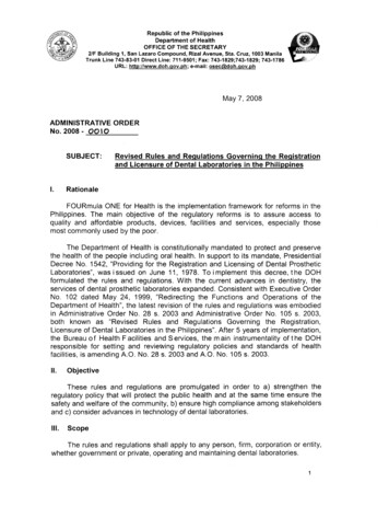
Neurosurg Focus 27 (1):E5, 2009Human cortical prostheses: lost in translation?Stephen I. Ryu, M.D.,1 and Krishna V. Shenoy, Ph.D. 2Department of Neurosurgery, Palo Alto Medical Foundation, Palo Alto; and 2Departments of ElectricalEngineering and Bioengineering, Neurosciences Program, Stanford University, Stanford, California1Direct brain control of a prosthetic system is the subject of much popular and scientific news. Neural technology and science have advanced to the point that proof-of-concept systems exist for cortically-controlled prosthesesin rats, monkeys, and even humans. However, realizing the dream of making such technology available to everyoneis still far off. Fortunately today there is great public and scientific interest in making this happen, but it will onlyoccur when the functional benefits of such systems outweigh the risks. In this article, the authors briefly summarizethe state of the art and then highlight many issues that will directly limit clinical translation, including system durability, system performance, and patient risk. Despite the challenges, scientists and clinicians are in the desirableposition of having both public and fiscal support to begin addressing these issues directly. The ultimate challengenow is to determine definitively whether these prosthetic systems will become clinical reality or forever unrealized.(DOI: 10.3171/2009.4.FOCUS0987)Key Wordsmotor brain-machine interface communication prosthesesRecently there have been several reports of humansdirectly interfacing with computers and controlling a cursor or performing other simple prostheticcontrol tasks. This has led to public interest in the prospect of fulfilling the predictions of a future where directman and machine communication will be commonplace.Although we have made tremendous advancements inthe development of electronic systems known as BMIs orBCIs, we find ourselves at an important threshold of determining whether their clinical potential will be realizedor not. With the public eye on this field, and significantfiscal support and encouragement, the field is now in aunique position to answer this question. In short, translation of human cortical prostheses will only result whentheir benefits clearly outweigh the risks. Here we discusssome of the obstacles that currently concern human cortical prostheses, which if unaddressed, may doom theirrealization.Motor and Communication ProsthesesExisting BMIs strive to restore normal function inAbbreviations used in this paper: BCI brain-computer interface;BMI brain-machine interface; bps bits per second; DBS deepbrain stimulating; EEG electroencephalography; M1 primarymotor cortex; NIH National Institutes of Health.Neurosurg Focus / Volume 27 / July 2009brain-computer interface performance riskpatients suffering from neurological impairments. Twosuccessful examples include cochlear prostheses, whichprovide surrogate electrical signals to the nerves processing auditory stimuli, and DBS devices that alter activity in the motor circuits to disrupt movement gone awry.Emerging BMIs aim to help paralyzed patients, such asthose with spinal cord injuries, who generally have intactcortical motor signals that are unable to arrive at end effectors such as the arm. Much research has been dedicated to studying the neural activity from various brainregions involved in movement, with the result that desiredmovements can now be estimated from cortical activity.Two types of prostheses, motor and communication, havetaken root (Fig. 1). Both types are similar, but differ intheir details. A motor prostheses taps into brain signals tocontinuously guide a paralyzed or prosthetic arm throughspace. This could be used to reach for objects or to painta picture where the movement path and speed are important. A communication prosthesis again uses brainsignals to guide a prosthesis, but in this case emphasizesjust the desired endpoint without concern for the movement path or speed. Selecting keys on a keyboard or dialing a phone are examples of where how you move is notas important as what you accomplish. Both functions areimportant for paralyzed patients, but most authors havefocused on one or the other type of prosthesis.1
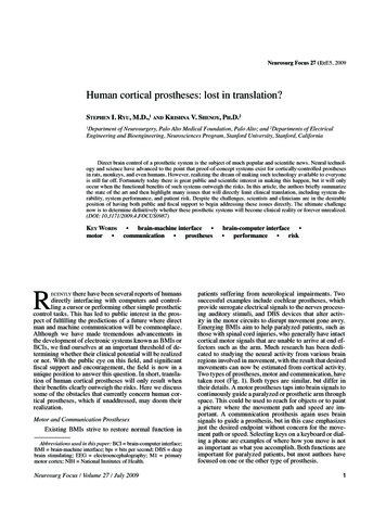
S. I. Ryu and K. V. ShenoyFig. 1. Concept sketch of cortical motor and communication prostheses. Neural signals are obtained from arrays of electrodesimplanted in various possible cortical areas (PMd, M1, PRR, MIP). These are then processed and interpreted to generate controlsignals. These can be used to reconstruct an arm trajectory (motor prosthesis) or to select a target from a menu (communicationprostheses). MIP medial intraparietal area; PMd dorsal premotor cortex; PMv ventral premotor cortex;s PRR parietalreach region; SMA supplementary motor area. Figure reproduced from Shenoy et al., Increasing the performance of corticallycontrolled prostheses, in Proceedings of the 28th IEEE EMBS Annual International Conference. New York City, USA, Aug30-Sept 3, 2006. IEEE, 2006, pp 6652–6655. Reproduced with permission, from IEEE, 2006. Copyright 2006, IEEE.A Brief History of Cortical ProsthesesIn the late 1960s, Fetz and collegues12–14 discoveredthat nonhuman primates could learn to regulate the firingrate of individual cortical neurons, which suggested theiruse to control a prosthesis.26 By the late 1990s, technological advances and a considerably better understandingof how cortical neurons contribute to limb movement15,49sparked renewed interest in developing clinically viablesystems. Isaacs and colleagues27 demonstrated that 2Dand 3D hand location could be reconstructed from theactivity of M1 neurons in rhesus monkeys, and others reported similar reconstructions from parietal cortex, dorsal premotor cortex, and M1.20,35,65In 1999 Chapin and colleagues7 reported a study in2trained rats that could move a lever to receive a rewardthrough brain activity alone. In 2002, Serruya and colleagues51 demonstrated 2D cursor control by rhesus monkeys using M1 neurons recorded from an implanted electrode array. Targets on a computer screen could be hitwithin 1–2 seconds using brain control only. That sameyear, Schwartz’s group60 demonstrated 3D cursor control by rhesus monkeys using M1 neurons. Targets couldbe hit on 70–80% of trials within 1.5–2.0 seconds. Thisgroup has since gone on to demonstrate that nonhumanprimates can use these 3D control signals to feed themselves with a robotic arm and also open and close a handlike gripper.57,63 Carmena and colleagues6 demonstrated2D cursor control and hand grasping force control withrhesus monkeys. These are examples of reconstructingNeurosurg Focus / Volume 27 / July 2009
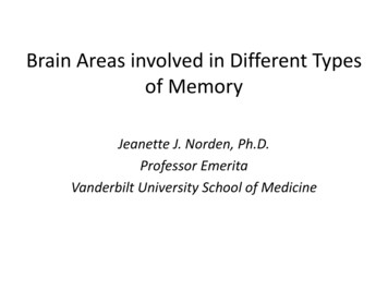
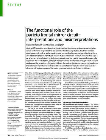
Toward human cortical prosthesesFig. 2. Photograph of the Utah electrode array (Blackrock Microsystems, Inc.), showing a silicon micromachined electrode array (arrowhead) with 96 needle-like electrodes. This is connected via a wirebundle to a connector port (arrow) that must be anchored to the subject.This port is then used to externally connect to each electrode. The thinsilver wires are for grounding (reference) purposes. Reprinted by permission from from Macmillan Publishers Ltd: Nature,23 2006.arm trajectory as appropriate for motor prostheses.Other groups have reasoned that if the desired targetlocation can be estimated directly from neural plan activity, the cursor can be positioned immediately on the desired key (in communication prostheses). Recent reportssuggest that there may be considerable performance benefit afforded by looking at the desired target and not theintermediate trajectory.20,38,46,53The Human Cortical Prostheses ExperienceIn the late 1990s, Kennedy and colleagues30,31 demonstrated a communication prosthesi that used one ortwo neurons from the motor cortex of locked-in humanpatients with amyotrophic lateral sclerosis to move a cursor across a virtual keyboard to type out messages. Thesepatients had glass cone electrodes implanted in the cortexthat had been treated with patient-derived neurotrophicfactors to encourage assimilation. Patients were able toslide cursors in 1D and 2D to type messages.There are also several implementations of commu-nication prostheses that enable target/menu selection bymodulating EEG waves.5,22,36,50,66 These techniques areattractive for their low risk. Electrocorticography recording from grids placed during epilepsy surgery have beenused as a potential source of signals for BCIs.34,48 Anothergroup described recording from a 32-wire microwire array in patients undergoing subthalamic nucleus stimulation. Ensembles from the subthalamic and thalamic motor nuclei could be used to control grip strength.41The first implanted cortical electrode array humanprosthesis was reported by Kim and colleagues23,32 who,in collaboration with Cyberkinetics NeurotechnologyInc., demonstrated 2D cursor control using M1 recordings. Performance was similar to their previous resultsusing the same electrode array technology in nonhumanprimates.51 This work was additionally encouraging inthat it demonstrated for the first time that studies in normal (uninjured) nonhuman primates could transfer to human patients with injuries.Lost in Translation?Taken together, these investigations provide compelling proof-of-concept demonstrations arguing that cortically controlled motor and communication prostheses arepossible. Although none of these devices have demonstrated a level of performance likely to be considered adequate for widespread use in humans, it was high enough tomotivate next-generation experiments and technologicaldesigns. Prostheses will be clinically viable when the anticipated quality of life improvement outweighs the potential risks. Only when prosthetic performance substantiallysurpasses what is possible with other techniques, and surgical risk is sufficiently mitigated, will invasive electrodebased cortical prostheses find more widespread use.We believe that there are 3 general areas that mustbe addressed immediately: system durability, system performance, and patient risks. Otherwise, these will likelybecome serious obstacles to clinical translation, potentially serious enough to halt the progress of human cortical prostheses.System DurabilityA prosthetic system must be durable to be clinicallyviable. Durability implies that it can be used in everydayFig. 3. Diagram showing neural recordings obtained from an unrestrained nonhuman primate over a 48-hour period afterimplantation of a chronic electrode array. The picture illustrates a box of recording electronics that was mounted on the subject’shead. Spike waveforms from these recordings show that waveforms changed from day to day which could not be explained byfluctuations in the signal path, indicating that the neurons seen were different. Figure reproduced from Shenoy et al., Increasingthe performance of cortically-controlled prostheses, in Proceedings of the 28th IEEE EMBS Annual International Conference. New York City, USA, Aug 30-Sept 3, 2006. IEEE, 2006, pp 6652–6655. Copyright 2006, IEEE.Neurosurg Focus / Volume 27 / July 20093
S. I. Ryu and K. V. ShenoyTABLE 1: Comparison of roughly normalized performance ofsystems performing target selection tasks*PerformerBpshuman typist (typical)10human typist (professional)25 EEG/ECoG prostheses0.5–1.3electrode-based prostheses (early)1.0–1.5electrode-based prostheses (fastest reported)6.5electrode-based prostheses (current theoretical)10WordsPerMinute2560 1.2–3.12.5–3.81625* As mentioned in the text, the determination of these numbers is byno means definitive because there are no standards; however, it is useful to look at these numbers to gauge desired performance. ECoG electrocorticography.life and not just within the confines of a controlled laboratory environment. To do this, the prosthetic system must:1) have a neural interface that will work consistently for aslong as possible; 2) be robust enough to be able to adapt tochanges in the population of neurons it is recording; and3) physically tolerate any real-life conditions to which itmay be subjected.Neural Interface IssuesThe brain interface is the point of contact betweenengineered and biological worlds. Electrode arrays areactively pursued as brain interfaces for the high qualityelectrical signals they provide, but they must be surgicallyimplanted into the brain tissue. These fine needle-like arrays of electrodes can be fashioned from individual wiresor on a micromachined substrate such as silicon (see the“Utah” array, Fig. 2). Many different electrodes have beensuccessfully used for cortical recording; however, mostof these were used for short spans of time, only severalmonths of data collection. As more long-term implants arebeing used, we are beginning to understand the lifespanof the current technologies.Some authors have reported obtaining good qualityrecordings from a few months to over 1 year from siliconelectrode arrays in nonhuman primates.59 In our experience with more than 15 Utah electrode array implants innonhuman primate frontal cortex, we achieved up to 3years (with 1 outlier array), but much more typically only6–12 months before signal quality on most electrodesdiminished substantially. Over 2 years of recording wasreported in 1 human trial participant with the same arraytechnology.24 The cause of this limited lifespan is not wellunderstood. Nonhuman primate pathology studies haveshown reactivity and gliosis around implanted electrodesranging from mild to concerning.16,42 We have noted fibroblastic tissue development encapsulating the explantedarray in some animals. Comparison of implantation techniques, including a pneumatic array inserter (commonlyused with Utah electrode arrays) and manual insertion(commonly used with microwire and Michigan arrays),has received some attention. In one study, mechanicallyinserted microwire arrays in rat auditory cortex func4tioned for 6 weeks while no manually inserted arraysdid.43 To address these possible biological and mechanical issues limiting electrode lifetime, there is considerabledevelopment aimed at producing advanced arrays withnovel materials, biocompatible coatings, and optimizedgeometries.33,52 Without more durable interfaces, corticalprosthetic systems will be severely impaired.System RobustnessEven with initially high-quality electrode interfaces,we can probably safely assume that the number of electrodes with excellent signal quality will degrade overtime. Prosthetic systems are being designed to counteror otherwise contend with this signal loss, else systemperformance will also degrade. Another issue regardsdynamic physical changes at the neural interface itself.The arrays are typically rigid structures that are grosslyfixed in the brain. However, because the brain deforms,there is micromotion between the electrode tip and braintissue.45 This gives rise to variations in the recordingsthat may be the result of tissue volume changes or abruptshifts associated with tissue acceleration/deceleration(such as a violent sneeze). We are currently characterizing these changes in recordings obtained 24 hours aday, 7 days a week from implanted arrays in nonhumanprimates.8,9,45 This will provide the data needed to designadaptive systems capable of compensating for these effects. (Fig. 3)Physical DurabilityMuch of the research done to date is with very delicate equipment inside a laboratory. A real prosthetic system must be physically durable. Most neurosurgeons haveseen an implant migrate to an inprobable location and hadto deal with traumatized implants. Clinical trials createdNeuroPort (Blackrock Microsystems, Inc. ), a reasonablydurable system, for human use, but it proved too bulkyand unwieldy for practical use. Other current systems arelarge devices that must be miniaturized for any practical use, but this brings up new considerations that havenot been addressed. Drawing from other examples, DBSdevices and cardiac pacers rely on electronics and powersupplies remote to the electrode, necessitating hardy butnevertheless accident-prone wires as well as a need forroutine replacement. Permanent linkages for power supplies for cochlear and phrenic nerve implants can be difficult to maintain. Programmable shunt valves are placedin strategic locations to allow for interrogation and adjustment. Finally, the unexpected should be expected inpatients who inadvertently fall, trip, and otherwise pushthe limits of manufacturing tolerances.System PerformanceThe performance of prostheses is a hot topic currently because it is difficult to quantify. Performance ismeasured by how well the motor prosthesis replicates amovement, while performance is measured in communication prostheses by how well information can be transmitted; there is no standard performance metric, however.Neurosurg Focus / Volume 27 / July 2009
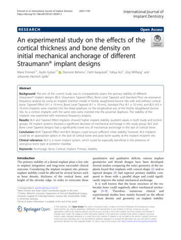
Toward human cortical prosthesesFig. 4. Diagram showing an overview of our nonhuman primate high-performance communication prosthesis experiment.The real-time prosthetic cursor placement task begins by fixating and touching central targets, followed by the appearance of aperipheral target to which the subject plans (but does not execute) a reach. A period of neural data following this target onset (Tgton) is set aside (Tskip). A period of neural data (Tint) is then analyzed to estimate the desired target; (P(m) refers to the target-mwith the highest probability; here, m 2), which could have appeared in one of 8 locations in this task, and after a brief computational decode and display rendering “overhead” period (Tdec rend), the predicted target is encircled. Figure reproduced fromShenoy et al., Increasing the performance of cortically-controlled prostheses, in Proceedings of the 28th IEEE EMBS AnnualInternational Conference. New York City, USA, Aug 30-Sept 3, 2006. IEEE, 2006, pp 6652–6655. Copyright 2006, IEEE.Motor prosthetics can be quantified by how closely theyreplicate true movements. In addition to the path taken,they must be quantified in terms of how quickly theymove. Communication prosthetics could be quantified bya commonly used information transfer metric known asbps. It also turns out that goal-oriented motor prosthetics(those which are essentially selecting targets) can also beroughly quantified with bps. Most current reported motor prosthetics operate roughly in the 1.0–1.5 bps range(Table 1). Quantification is vital because current noninvasive EEG-based BCIs operate in a similar range, but thesesystems incur little clinical risk. Though it is generallyagreed that the invasive electrodes provide better neuralsignals and from a smaller group of neurons, it is not settled that such signals are superior. Improved performancemust be objectively demonstrated.Here we describe some recent research in pushingthe performance limits of current prostheses. In addition,we discuss interesting optimizations that have developedfrom our research. Finally, we explore autonomous system design that may allow for “natural” operation of aprosthetic system.Neurosurg Focus / Volume 27 / July 2009Maximizing PerformanceWe have devoted much of our recent research effortto understanding the fundamental performance limitsof electrode-based neural prostheses and to establishing a principled design methodology to develop prostheses that can operate at these limits.46 We conducted aseries of experiments and computational simulations toinvestigate how quickly and accurately a communicationprosthesis could be driven by cortical activity. We usedsignals from the dorsal premotor cortex (PMd), as thosesignals can be correlated with the endpoint of a reach orselection. The subject was trained to plan to reach fortargets on the screen, which we could accurately andquickly determine based on neural activity. (Fig. 4) Weoptimized the algorithm and ultimately realized 6.5 bpsperformance – this is 4 times faster than previous prosthetic systems. This is equivalent to typing at a rate of 16 words per minute. This level of performance is veryencouraging as it helps supports both continuing prosthetic research and the potential of invasive electrodetechnology. We believe that even better performance can5
S. I. Ryu and K. V. ShenoyFig. 6. Illustration of brain states involved in autonomous reaching.We spend most of our time in the idle, or baseline state (gray). Fromhere, we switch to a different state where we plan a movement (blue).The movement can then be executed which is another brain state (red).Once completed we can then plan another move (blue arrow) or returnto the idling state (long gray arrow). One can also start planning andthen abort the move as indicated by the gray short return arrow. Automating a prosthetic involves determining the unique neural characteristics of these states to determine when these transitions occur. Our earlycomputational results suggest that this can be done. See the studies byAchtman et al., 2007, and Kemere et al., 2008.Fig. 5. Sixteen target optimal target placement (OTP) example. Redsquares are uniformly spaced targets around a circle, as an experimenter might use without OTP methods. Blue circles show an OTP solution. Figure from Cunningham et al., 200810 used with permission.be achieved, based on recent biological and algorithmicinsights.Neural OptimizationsIn the above study, we noted that our performancefell short by about 1.2 bps of our theoretically predictedperformance. We discovered that this was due in part toresponse changes of the individual neurons. Firing ratesbecame more or less intense depending on when in a sequence of trials the responses were measured. In otherwords, if this were a system used with human patients,depending on where in a word the subject was while typing a given letter, a neuron could respond slightly differently. We referred to this as neural response nonstationarity as opposed to having the same response for anyspecific condition. The underlying cause of this responsemodulation is not entirely clear, but presumably it is duein part to pushing the speed of planning in our prosthesesexperiments to some limit (that is, planning 3–4 reachesper second is perhaps close to the neurobiologically dictated limits). However, we can work around this by usingmathematical techniques to infer these effects from themeasured data, and then mitigate them. Our group introduced the use of factor analysis–based algorithms andwas able to decrease errors by up to 75% when applied todata from our previous experiment.47Optimal Target PlacementIn the 1870s, Christopher Sholes developed theQWERTY keyboard to optimize for minimal mechanicaldysfunction and maximum typing efficiency. Most keyboards or targets used in BCI research experiments havebeen created arbitrarily by the scientist—these layoutstended to be a circle of targets. However, both of these6layouts are independent of the response properties of theparticular neurons under observation and therefore couldlimit performance. Not surprisingly, any randomly selected population of neurons would have areas and directionsto which they would respond better or worse. By adjusting the location of the targets to optimize for these spatialconsiderations, could we improve performance? We developed a method to optimally place targets to maximizethe accuracy of the prosthesis. (Fig. 5) By just alteringthe location of the targets, we could increase performanceaccuracy by several percent.10 This has further utility astarget layouts can be modified over the life of the array toadjust for neural population drift.Autonomous System DesignAn often overlooked performance issue has to dowith system autonomy. We naturally spend time thinking of moving and not moving at all. An ideal prostheticshould be able to autonomously determine when an active prosthetic signal is present, or not, achieve “natural”function as opposed to having to turn the prostheses onand off. This can be done by determining the state of thebrain—“idling,” “thinking of moving/planning to move,”and “executing a movement”—and activating the prostheses based on this information. (Fig. 6) Such a systemwould have to deal with plans that are not executed andlong stretches of inactivity. We have developed hiddenMarkov model and maximum likelihood decoders to dojust this. 1,29 We have been able to determine the state ofthe brain accurately by observing the neural signals invarious states. In this way, we created a mechanism bywhich to switch a system on and off automatically.Given these recent advances, we believe it possibleto demonstrate, in future cortical prosthetic experiments,10-bps performance ( 25 words per minute). Performance quantification is a very important factor in assessing the value of the device, but is not the only criticalfactor to take into account. Other factors such as autonomous function must be considered. One additional exciting frontier is the ability of the patient to improve systemNeurosurg Focus / Volume 27 / July 2009
Toward human cortical prosthesesFig. 7. A working fully implantable miniaturized implant that integrates an electrode array with amplification and telemetriccircuits; shown in profile (a) and after being encased in polymer (b). Such a small implant would be necessary for long-termchronic recordings as well as to contribute to overall durability and feasibility of the system. Figure reproduced from Harrison etal., A wireless neural interface for chronic recording, in Biomedical Circuits and Systems Conference, 2008. IEEE, 2008, pp125–128. Copyright 2008, IEEE.performance—something that has been untestable untilrecently.Patient RisksA person’s well-being should never take second priority to the pursuit of interesting science. With increasedelectrode lifespan and durable devices, and as performance approaches acceptable functional restoration, wemust seriously address patient risks. This discussion ofrisk is not about another factor in making BCI a reality,so much as a frank discussion regarding whether invasiveBCIs are even worth pursuing as a practical solution.It is difficult to quantify the risk of these proposedprocedures. However, we can explore the potential issuesfor consideration. The risks involved with invasive BCIsfall into 2 categories, direct and indirect. The direct risksare related to the nature of the implant and any associatedprocedures. The indirect risks are related to the additionalpatient risk due to the lack of durability and performanceof the system.Surgical RiskSurgeons have implanted foreign objects into various parts of the body with varying success for manydecades. Neurosurgeons are familiar with the implantation of shunt tubing, vascular clips, pedicle screws, andneurostimulators. We can best estimate implanted cortical prosthetic systems risks based on our experience withsubdural grid placement for epilepsy and DBS for movement disorders. Complications from subdural recordinggrids for subacute recording (up to 21 days) have beendescribed. These include CSF leaks (in up to 31% of patients), hemorrhagic complications (in up to 14%), cerebral edema (in up to 14%), transient neurological deficits,and symptomatic pneumocephalus. 2,4,28,39,56 A review of179 DBS electrode implantations in 109 patients noted16 serious adverse events related to surgery in 14 patients (12.8%). There were 2 perioperative deaths (1.8%).There were also pulmonary embolisms, subcortical hemorrhages, chronic subdural hematomas, venous infarction, seizure, infections, CSF leaks, and skin erosion.The incidence of permanent injury was 4.6%.11,62 Im-Neurosurg Focus / Volume 27 / July 2009planted cortical prosthetics will probably incur a similarmorbidity profile which further emphasizes the need for aclear clinical benefit as is the case in DBS for movementdisorders.Infectious ComplicationsInfection is the greatest long-term concern for hardware implantation, and can range in severity from simpleskin infections to deep infections of the bone and brain.Although treatable, these infections may require prolongedantibiotic therapy and system explantation. Another concern is the ability of infection to track down hardware,resulting in brain infections which could directly damagethe viable cortex. Infection risk can never be completelyeliminated. Fortunately, we can learn how to mitigatesuch risks from the research in similar systems.Subdural electrode grids are short-term implants tunneled out of the scalp. These have been associated withan infection incidence of up to 8.7%, including wound infections (2.5%), meningitis (2.5%), osteomyelitis (0.8%),epdidural abscess, and empyema.28 Deep brain stimulators, in contrast, are completely internal systems that areimplanted for the long term. In published reports of hardware-related infections, the incidence ranges from 4.5 to6.1%.18,55 Although these infections are treatable, many ofthe infected patients required complete explantation ofthe system, resulting in loss of benefit.Until a completely implantable device is created,prosthetic systems may rely on a skull-mounted interface port (such as the NeuroPort); this skin interface is ofconcern for infection. There is another device called thebone-anchored hearing aid which requires attachment ofa metal post to the skull behind the ear, and is similarto such a port. The authors of a long-term review of 165patients with these implants noted a 21% incidence of local skin reactions, 18% loosening of the hardware, 8% severe reactions requiring treatment, and 1% skin necrosis.Overall 34% of the patients required revision surgery.3Implant-Related RisksImplanted hardware must be durable, and there arepractical considerations regarding wires and power asmentioned above. Clearly a small, fully implantable, self7
S. I. Ryu and K. V. Shenoypowered device is ideal to mitigate the risk of infection(Fig. 7). However, direct hardware complications havebeen reported for DBS devices, despite the small electrode and pacer package. A review of the literat
2. Photograph of the Utah electrode array (Blackrock Micro-systems, Inc.), showing a silicon micromachined electrode array (ar-rowhead) with 96 needle-like electrodes. This is connected via a wire bundle to a connector port (arrow) that must be anchored to the subject. This port is then used to externally connect to each electrode. The thin