Transcription
REVIEWSThe functional role of theparieto-frontal mirror circuit:interpretations and misinterpretationsGiacomo Rizzolatti*and Corrado Sinigaglia‡Abstract The parieto-frontal cortical circuit that is active during action observation is thecircuit with mirror properties that has been most extensively studied. Yet, there remainscontroversy on its role in social cognition and its contribution to understanding the actionsand intentions of other individuals. Recent studies in monkeys and humans have shed lighton what the parieto-frontal cortical circuit encodes and its possible functional relevance forcognition. We conclude that, although there are several mechanisms through which one canunderstand the behaviour of other individuals, the parieto-frontal mechanism is the only onethat allows an individual to understand the action of others ‘from the inside’ and gives theobserver a first-person grasp of the motor goals and intentions of other individuals.Mirror mechanismThe mechanism that unifiesperception and action,transforming sensoryrepresentations of thebehaviour of others intomotor representations ofthe same behaviour in theobserver’s brain.Motor actA movement with a specificmotor goal (for example,reaching, grasping andmanipulating). The successfulachievement of its goalrepresents the reinforcementof the motor act.*University of Parma,Department of Neuroscience,and the Italian Institute ofTechnology, Via Volturno 39,I‑43100 Parma, Italy.‡University of Milan,Department of Philosophy,Via Festa del Perdono 7,I‑20122 Milano, Italy.Correspondence to 8/nrn2805Published online10 March 2010One of the most intriguing and exciting developmentsin neuroscience in recent years has been the discovery ofa mechanism that unifies action perception and actionexecution1–3. The essence of this mechanism — calledthe mirror mechanism — is the following: each time anindividual observes another individual performingan action, a set of neurons that encode that action isactivated in the observer’s cortical motor system.The mirror mechanism is present in many corticalareas and brain centres of birds, monkeys and humans.The basic functions of these areas and centres vary considerably, from song production to the organization ofgoal-directed motor acts, to emotional processes. Thus,like other basic mechanisms (for example, excitatorypostsynaptic potentials), the functional role of the mirror mechanism depends on its anatomical location, withits function ranging from recognition of the song ofconspecifics in birds4,5 to empathy in humans6.The aim of this article is not to review the vastliterature on the mirror mechanism, but to focus on one specific circuit endowed with mirror properties: the parietofrontal action observation–action execution circuit. Thereason for this choice is twofold. First, the proposedinterpretation of the function of the parieto-frontalcircuit as a mechanism that enables individuals to understand the actions and intentions of others (mirror-basedaction understanding) represented a paradigm shift in theclassical view that these cognitive functions depend onhigher-level mental processes. Second, mostly as a reactionto this new perspective, there have been attempts tointerpret the functions of the action observation–actionexecution circuit in a way that minimizes or even denies itsrole in cognition. For these reasons, a review of the data onthe mirror mechanism in the action observation–actionexecution network seems timely and necessary.In this Review, we examine first what the parieto-frontalaction observation–action execution circuit encodes inmonkeys and humans and then discuss its possible functional relevance for cognition. After examining differentviews on these issues, we conclude that the parieto-frontal mechanism allows an individual to understand theactions of another individual ‘from the inside’ and givesthe observing individual a first-person grasp of the motorgoals and intentions of another individual.The parieto-frontal mirror networkThe monkey parieto-frontal network. The mirrormechanism was originally discovered in the ventralpremotor cortex of the macaque monkey (area F5)1–3.Single-neuron recordings showed that this area containsneurons — mirror neurons — that discharge both whena monkey executes a specific motor act and when itobserves another individual performing the same motoract. Mirror neurons do not fire in response to a simplepresentation of objects, including food. Most of them donot respond or respond only weakly to the observation ofthe experimenter performing a motor act (for example,grasping) without a target object 7.Area F5 has recently been divided into three sectors:F5c, F5p and F5a 8–9 (FIG. 1) . Mirror neurons were264 ApRIl 2010 VOluMe 11www.nature.com/reviews/neuro 2010 Macmillan Publishers Limited. All rights reserved
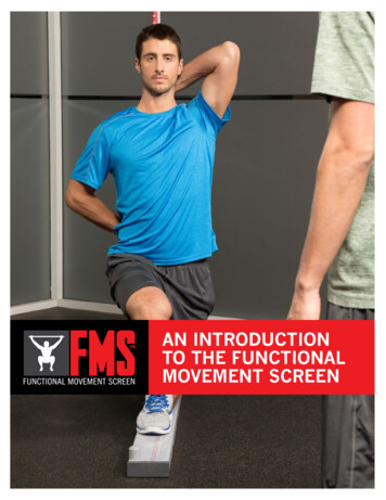
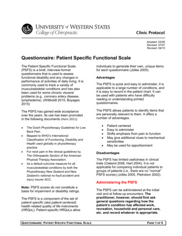
re 1 The parieto-frontal mirror network. Lateral view of the macaque brain.The coloured areas represent the areas of the s Neuroscienceneurons: the ventral premotor cortex (area F5), area PFG (located between parietalareas PF and PG) and the anterior intraparietal area (AIP). Note that the intraparietalsulcus (IPS) has been opened (light yellow) to show the areas inside. The parieto-frontalcircuit receives high-order visual information from areas located inside the superiortemporal sulcus (STS) and the inferior temporal lobe (IT). Neither of these temporalregions has motor properties. The parieto-frontal circuit is under control of the frontallobe (area F6 or pre-supplementary motor area and the ventral prefrontal cortex (VPF)).The inset provides an enlarged view of area F5, showing also its sectors (F5a and F5p)buried inside the arcuate sulcus. IAS, inferior limb of the arcuate sulcus; LIP, lateralintraparietal area; VIP, ventral intraparietal area.Mirror-based actionunderstandingThe comprehension of anobserved action based on theactivation of a motorprogramme in the observer’sbrain. The observed action isunderstood ‘from the inside’ asa motor possibility, rather than‘from the outside’ as a merevisual description.Superior temporal sulcus(STS). This sulcus separates thesuperior temporal gyrus fromthe middle temporal gyrus.Some of the areas in the STSencode biological motion.Although connected with theparietal areas of theparieto-frontal mirror network,STS areas cannot beconsidered mirror areasbecause of their lack of motorproperties.MovementA displacement of joints orbody parts without a specificgoal. It can be generatedspontaneously or producedartificially by electrical ormagnetic stimulation ofmotor areas.originally recorded in the cortical convexity that corresponds to F5c1–3. However, functional MRI (fMRI) datashowed that the other two areas also respond to observinga grasping action8.Mirror neurons are also present in the rostral partof the inferior parietal lobule (Ipl), particularly in areapFG10–12 and the anterior intraparietal area (AIp)9,13(FIG. 1) . Both these areas are heavily connected withF5: pFG mostly with F5c, and the AIp with F5a14. Botharea pFG and the AIp receive higher-order visual information from the cortex located inside the superior temporalsulcus (STS)13–14. STS areas, like mirror areas, encode biological motion, but they lack motor properties. They aretherefore not part of the mirror system in a strict sense.The AIp also receives connections from the middletemporal gyrus15. This input could provide the mirrorareas with information concerning object identity.Finally, area F5 is connected with area F6 — the presupplementary motor area (pre-SMA) — and with theprefrontal cortex (area 46)16. The prefrontal cortex is alsorichly connected with the AIp16. The frontal inputs control the selection of self-generated and stimulus-drivenactions according to the intentions of the agent17.It was recently shown that, in addition to areas pFG andAIp, two other areas of the parietal lobe contain mirrorneurons: the lateral intraparietal area and the ventralintraparietal area. The mirror properties of neurons inthese areas are not the focus of this Review but are brieflydiscussed in BOX 1.The human parieto-frontal network. There is convincing evidence that an action observation–action execution mirror circuit also exists in humans. This evidencecomes from brain imaging, transcranial magneticstimulation (TMS), electroencephalography (eeG) andmagnetoencephalography (MeG) studies.Brain imaging studies have shown that, as in the monkey, this action observation–action execution mirror circuit is formed by two main regions: the inferior section ofthe precentral gyrus plus the posterior part of the inferiorfrontal gyrus; and the inferior parietal lobule, including the cortex located inside the intraparietal sulcus18.Additional cortical areas (such as the dorsal premotor cortex and the superior parietal lobule) have also been occasionally found to be active during action observation andexecution19–21. Although it is possible that their activationis due to a mirror mechanism, it is equally possible that itreflects motor preparation. In support of this interpretation are single-neuron data from monkeys showing thatthese areas are involved in covert motor preparation22–23.As for the superior parietal lobule, although its activationis typically absent in studies in which the experimentersuse distal motor acts as visual stimuli, it is prominentwhen volunteers observe proximal arm movements thatare directed to a particular location in space24.Single-subject fMRI analyses have recently providedevidence that other cortical areas (for example, the primary and secondary somatosensory cortices and themiddle temporal cortex) also become active duringaction observation and action execution21. It has beensuggested21 that these activations outside of the ‘classical’ mirror areas are caused by additional mechanisms(for example, internal models) that are triggered by themirror mechanism. These activations would enrichthe information about the actions of other individualsthat the mirror mechanism provides.A tale of two populations. Some authors have recentlyargued that the activation of the same areas duringaction observation and action execution is not sufficient to prove the existence of the mirror mechanism inhumans25. Instead, they have suggested that, in humans,motor areas have distinct, segregated populations of visual and motor neurons, the visual neurons dischargingduring action observation and the motor neurons duringaction execution. They proposed to use the ‘repetition–suppression’ technique — that is, a technique based onthe progressive reduction of a physiological response torepeated stimuli to prove this point25. If mirror neuronsexist in humans, they should ‘adapt’ when the observation of a motor act is followed by the execution of thatmotor act, and vice versa.The ‘adaptation’ effects are, in general, difficult tointerpret 26. Adaptation occurs at the synaptic level andshould therefore be present only when informationrepeatedly reaches a neuron through the same or largelycommon pathways27. This input commonality is typicallyabsent when mirror neurons are activated during actionobservation and execution. During action observation,the input to the parieto-frontal circuit arrives from higherorder visual areas (for example, the STS)16 whereas, duringvoluntary movement, it mostly comes from the frontallobes17. The results of adaptation experiments thereforedepend on the design of the experimental paradigm andnATuRe ReVIewS NeuroscieNceVOluMe 11 ApRIl 2010 265 2010 Macmillan Publishers Limited. All rights reserved
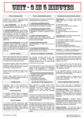
REVIEWSon the stimuli used. These considerations could explainwhy the results of repetition–suppression experimentshave been contradictory. Although some authors foundevidence of the mirror mechanism in the parietal28 or thefrontal nodes29, others obtained negative results30–31.Regardless of the empirical data that may help todefine some properties of the parieto-frontal mirrormechanism, the logic of the two-population story isflawed. Assuming that neurons in motor areas responding to action observation are merely visual neuronsimplies that motor areas contain a large number of ‘displaced’ visual neurons and that these neurons do notcommunicate with their ‘neighbour’ motor neurons.Both these assumptions are hard to reconcile with whatis known about the organization of the cerebral cortex.Most importantly, TMS studies have shown a clear congruence between the observed motor act and the activated motor representation32–36. Thus, if higher-ordersensory information describing a motor act reachesmotor neurons that encode that same motor act, thesemotor neurons are mirror neurons by definition.Humans do not differ from monkeys in this respect.What do parieto-frontal mirror neurons encode?Evidence for goal coding in monkeys. The crucialissue concerning the parieto-frontal mirror neuronsis their role in cognition. If this mirror mechanism isfundamental to understanding actions and intentions,the classical view — that the motor system has a roleonly in movement generation — has to be rejected andreplaced by the view that the motor system is also oneof the major players in cognitive functions. To addressthis fundamental issue, a preliminary problem mustfirst be solved: what do the parieto-frontal mirrorneurons encode when they discharge in response tothe observation of the actions of others?A way to solve this problem is to examine what mirror neurons encode when they discharge during motorbehaviour. what is recorded in single-neuron studiesduring both action execution and observation are actionpotentials — that is, neuronal output. Thus, having determined what neurons encode during the execution of anagent’s own motor act, one also knows what they encodewhen they are triggered by the agent’s observationof a motor behaviour of others.early experiments on area F5 found that most of themotor neurons in this area encode motor acts (that is,goal-related movements, such as grasping) rather thanmovements (that is, body-part displacements without aspecific goal, such as finger flexion)37–38. A recent studyprovided compelling evidence that this is the case39. Thisstudy describes single-neuron recordings from monkeysthat were trained to grasp objects using two types of pliers:normal pliers, which require typical grasping movementsof the hand, and ‘reverse’ pliers, which require hand movements executed in the reverse order (that is, first closingand then opening the fingers). The results showed thatF5 neurons discharged during the same phase of grasping in both conditions, regardless of whether this involvedopening or closing of the hand (FIG. 2).The functional properties of Ipl motor neurons aresimilar to those of F5 neurons: the goal of the executedmotor acts is the parameter that is encoded by Ipl neuronsthat fire during the execution of motor acts11,40–42.The mirror neurons in F5 and Ipl do not differ intheir motor properties from parieto-frontal motor neurons that do not have visual properties1–3. Thus, whenthey fire in response to motor act observation, they sendBox 1 Mirror neurons in other parietal areasThe cortical motor system in primates consists of a series of parieto-frontal circuits encoding different types of motorbehaviour, among which are hand grasping, mouth and head movements, arm reaching and various types of eyemovements. These circuits are activated by specific sets of sensory inputs that are conveyed to the parietal lobe fromsomatosensory, visual and auditory areas. In this Review, we focus on the mirror properties of the circuit formed in themonkey by parietal areas PFG and the anterior intraparietal area (AIP), and frontal area F5. Recently it was shown thattwo other areas of the parietal lobe also have mirror neurons: the lateral intraparietal area (LIP), which with the frontaleye field forms a circuit involved in the organization of eye movements; and the ventral intraparietal area (VIP), which isburied in the intraparietal sulcus and with frontal area F4 forms a circuit transforming somatosensory and visual stimulithat are presented around the monkey into head, mouth and arm movements (FIG. 1).The properties of mirror neurons located in area LIP have been described by Shepherd and colleagues97. They foundthat a set of LIP neurons fired both when the monkey looked in the neuron-preferred direction and when it saw anothermonkey looking in the same direction. Interestingly, another subset of LIP neurons that discharged when the recordedmonkey looked towards a certain direction was, by contrast, suppressed when the observed monkey looked in the samedirection. The authors suggested that LIP mirror neurons contribute to the sharing of attention between individuals.Mirror neurons in area VIP in the monkey have been described by Ishida and colleagues98. Previous studies showedthat VIP neurons encode tactile and visual stimuli delivered in the peripersonal space of the monkey99,100. Ishida andcolleagues showed that some of these neurons also respond to stimuli presented in the peripersonal space of anindividual located approximately 1 m from the monkey and facing it. Although motor responses were not studied bythis group, area VIP is connected with area F4, which represents peripersonal space and the neurons of which dischargeduring reaching movements. It is plausible, therefore, that neuronal responses that seem to be induced by visual stimuliactually represent potential motor acts directed towards specific body parts101–104. The study on VIP neurons is of greatinterest because it shows that the mirror mechanism of this area encodes body-directed rather than object-directedmotor acts, thus opening fascinating possibilities for individuals to encode the body of others.Thus, the function of mirror neurons is related to the motor properties of the areas in which they are located. This is alsotrue for adjacent areas located in the same cortical region.266 ApRIl 2010 VOluMe 11www.nature.com/reviews/neuro 2010 Macmillan Publishers Limited. All rights reserved
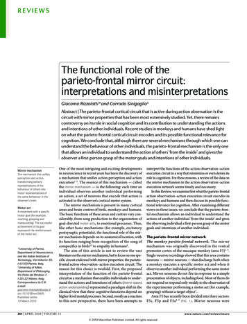
REVIEWSGoal and single-movement coding in humans. Inaccordance with early findings46–49, a series of new fMRIstudies provided strong evidence that the human parietofrontal mirror circuit encodes the goal of observed motoracts. Volunteers were instructed to observe video clips inwhich either a human or a robot arm grasped objects50.Despite differences in shape and kinematics betweenthe human and robot arms, the parieto-frontal mirrorcircuit was activated in both conditions. Another groupextended these results by investigating cortical activationin response to the observation of motor acts performedby a human hand, a robot hand or a tool51. Here, bilateral activation of a mirror network formed by intraparietal and ventral premotor cortex occured, regardlessof the effector. In addition, the observation of toolactions produced a specific activation of a rostral sectorof the left anterior supramarginal gyrus, suggestingthat this sector specifically evolved for tool use.Further evidence of goal encoding by the parietofrontal mirror circuit was obtained in an fMRI experiment in which two aplasic individuals, born withoutarms and hands, and control volunteers were asked towatch video clips showing hand actions52. All participants also performed actions with their feet, mouth and,in the case of controls, hands. The results showed thatthe parieto-frontal mirror circuit of aplasic individualsthat was active during movements of the feet and mouthwas also recruited by the observation of hand motor actsthat they have never executed but the motor goals ofwhich they could achieve using their feet or mouth.The issue of whether the human parieto-frontal mirror network encodes motor goals was also addressedby fMRI and TMS studies investigating the activationof motor areas in subjects listening to action-relatedaNormal pliersReverse pliersSpikefrequency (s–1)Reverse pliers1001.2 1 Sec1.10.90.8*Distance (cm)Spikefrequency (s–1)b Normal pliersDistance (cm)information about the goal of the observed motor acts.This information can be encoded with different degrees ofgenerality: some mirror neurons (strictly congruent mirror neurons) fire when the observed and executed motoracts are the same (for example, grasping with precisiongrip), whereas other mirror neurons (broadly congruentmirror neurons) fire when the observed motor act has thesame goal as the executed motor act (for example, grasping), but can be achieved in a different way (for example,with both precision and whole-hand grips)43–44.Recently, a single-neuron study investigated theeffect of the spatial relationships between an agent andan observer, comparing F5 mirror neuron responsesto motor acts performed near the monkey (in theperipersonal space) or outside its reach (in the extrapersonal space)45 (FIG. 3). The results showed that manyF5 mirror neurons were differentially modulated by thelocation of the observed motor act. Some neurons wereselective for actions executed in the monkey’s peripersonalspace, whereas others were selective for stimuli in theextrapersonal space. These findings indicate that mirrorneurons may encode the goal of the motor acts of anotherindividual in an observer-centred spatial framework,thus providing the observer with crucial information fororganizing their own future behaviour in cooperation orcompetition with the observed individuals.1001.21 Sec*1.11.05Figure 2 Goal coding in the monkey premotor cortex.a Schematic illustration of the experimental paradigm.Reviews NeuroscienceTo grasp the object with normalNaturepliers, themonkeyhasto close its hand (top) whereas, with the reverse pliers,the monkey has to open its hand (bottom). The arrowsindicate the direction of the motion of the tip of the pliers.b Activity of a neuron recorded from area F5. Rasters andhistograms illustrate the neuronal discharge during graspingwith normal pliers (left) and reverse pliers (right). Both rastersand histograms are aligned with the end of the tip closurephase (asterisks). The traces below each histogram indicatethe instantaneous hand position as recorded with apotentiometer and expressed as a function of the distancebetween the plier handles. Downwards traces indicate thatthe hand closes, whereas upwards traces indicate that thehand opens. Figure is reproduced, with permission, fromREF. 39 (2008) National Academy of Sciences.sounds. Hearing and categorizing animal vocalizationspreferentially activated the middle portion of the superior temporal gyri bilaterally (a region that is not relatedto motor act coding), whereas hearing and categorizing sounds of tools that were manipulated by handsactivated the parieto-frontal mirror circuit 53. Similarly,it was shown that listening to the sound of hand andmouth motor acts activated the parieto-frontal mirrornetwork54. This activation was somatotopically organized in the left premotor cortex and was congruent withthe motor somatotopy of hand and mouth actions.unlike in monkeys, the parieto-frontal mirror circuitof humans also becomes active during the observation ofindividual movements55–56. The initial evidence for thismechanism was based on TMS experiments which indicated that the observation of the movements of othersresults in an activation of the muscles involved in thenATuRe ReVIewS NeuroscieNceVOluMe 11 ApRIl 2010 267 2010 Macmillan Publishers Limited. All rights reserved
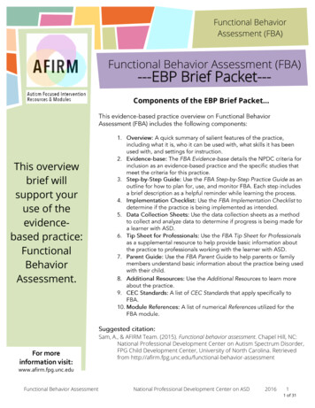
REVIEWSexecution of those movements32–36. Additional supportcomes from eeG and MeG studies showing that theobservation of movements without a goal desynchronizesthe rhythms recorded from motor areas57–64.Recently, it was shown that mirror coding mightdepend on the content of the observed behaviour. Motorevoked potentials (Meps) in response to TMS wererecorded from the right opponens pollicis (Op) musclein participants observing an experimenter either opening and closing normal and reverse pliers or using themto grasp objects65. The observation of tool movements(that is, opening and closing the pliers without graspinganything) activated a cortical representation of the handmovements involved in the observed motor behaviour.By contrast, the observation of the tool grasping actionactivated a cortical representation of the observed motorgoal, irrespective of the individual movements and theorder of movements required to achieve it. Together, thesefindings show that the human parieto-frontal mirrornetwork encodes both motor acts and movements.Understanding the actions of othersCognitive functions of the parieto-frontal network:evidence and criticisms. why should the motor system encode the goal of actions performed by others?From the discovery of mirror neurons, the interpretation of this finding was that they allow the observer tobSpike frequency (s–1)aunderstand directly the goal of the actions of others1–3:observing actions performed by another individual elicits a motor activation in the brain of the observer similarto that which occurs when the observer plans their ownactions, and the similarity between these two activationsallows the observer to understand the actions of otherswithout needing inferential processing 43–44.In support of this view, two studies showed that themeaning of the motor acts of other individuals couldbe understood in the absence of visual informationdescribing them. In one study, monkeys heard thesounds of a motor act (such as ripping a piece of paper)without seeing it 66; in the other study, the monkeysknew that behind a screen was an object and saw theexperimenter’s hand disappear behind the screen, butthey could not see any hand–object interaction67. Theresults showed that in both experiments F5 mirror neurons in the monkeys fired in the absence of visual information describing the motor act of the experimenter.The neuronal activation therefore underpinned thecomprehension of the goal of the motor act of the otherindividual, regardless of the sensory information thatdescribed that motor act.This interpretation of the function of the parieto-frontalmirror mechanism has been challenged with objectionsand alternative proposals68–71. A key criticism has beenadvanced by Csibra69. He argued that the interpretation ofNeuron 1100Neuron 2100100Spike frequency (s–1)1s1001s1s1001001sSpike frequency (s–1)Neuron 31001s1s1001sMirror neuron selective forthe extrapersonal space1001sMirror neuron selectivefor the peripersonal space1sClassical mirror neuronFigure 3 Mirror neuron responses in the monkey during observation of actions executed in the peripersonalNature Reviews Neuroscienceand extrapersonal space. a Schematic illustration of the experimental paradigm. The monkeyobservesanexperimenter grasping an object in its extrapersonal space (upper panel) and in its peripersonal space (middlepanel). The monkey grasps an object located in front of it (lower panel). b Responses of three neurons in the threeexperimental conditions. Neuron 1 responds selectively to the observation of action in the extrapersonal space,whereas neuron 2 responds to the observation of action in the peripersonal space. Neuron 3 does not exhibit spaceselectivity. All three neurons also discharge during action execution. Figure is reproduced, with permission, fromREF. 45 (2009) American Association for the Advancement of Science.268 ApRIl 2010 VOluMe 11www.nature.com/reviews/neuro 2010 Macmillan Publishers Limited. All rights reserved
REVIEWSTMS adaptation paradigmA paradigm by which specificneural populations withinthe stimulated corticalregion can be targeted.One population is ‘adapted’and will therefore be lessactive. Because transcranialmagnetic stimulation (TMS)targets less active neuralpopulations, the adaptedpopulation will be facilitatedmore strongly by TMSInferential reasoningThe capacity to attributeto an agent mental statesthat might account for theobserved motor action in termsof the reasons (for example,needs, desires and beliefs)underlying it.mirror neuron function in terms of action understandingcontains a “tension” between “the claim that the mirrormechanism reflects nothing else but faithful duplicationof the observed action” and “the claim that mirroring represents high-level interpretation of the observed action”.In other words, if mirror activity represents a copy of theobserved motor act, it is not sufficiently general to capturethe goal of that motor act; conversely, if it is sufficientlygeneral for goal understanding, it cannot be interpretedin terms of a direct matching mechanism between sensoryand motor representations (see also REFS 70,71).In the earlier studies on the mirror mechanism, itwas indeed not clearly specified that the parieto-frontalmirror mechanism in humans is involved in two kindsof sensory–motor transformation — one mapping theobserved movements onto the observer’s own motorrepresentation of those movements (movement mirroring), the other mapping the goal of the observed motoract onto the observer’s own motor representation of thatmotor act (goal mirroring), as described above. By matching individual movements, mirror processing provides arepresentation of body part movements that might servevarious functions (for example, imitation), but is devoidof any specific cognitive importance per se. By contrast,through matching the goal of the observed motor actwith a motor act that has the same goal, the observer isable to understand what the agent is doing. This is truenot only for the mirror neurons that are broadly congruent but also for those that are strictly congruent, becausethese neurons also do not encode the elementary aspectsof a movement (for example, its kinematics), but respondto the goal of the observed motor acts44,56.Typically, authors who play down or even deny theimportance of the motor system for cognitive
the mirror mechanism — is the following: each time an individual observes another individual performing an action, a set of neurons that encode that action is activated in the observer's cortical motor system. The mirror mechanism is present in many cortical areas and brain centres of birds, monkeys and humans.