Transcription
Transl. Stroke Res. (2016) 7:535–547DOI 10.1007/s12975-016-0496-0ORIGINAL ARTICLEDimethyl Fumarate and Monomethyl Fumarate PromotePost-Ischemic Recovery in MiceYang Yao 1 & Weimin Miao 2 & Zhijia Liu 1 & Wei Han 3 & Kaibin Shi 1 & Yi Shen 1 &Handong Li 1 & Qiang Liu 1,4 & Ying Fu 1 & DeRen Huang 5 & Fu-Dong Shi 1,4Received: 5 April 2016 / Revised: 16 August 2016 / Accepted: 18 August 2016 / Published online: 10 September 2016# The Author(s) 2016. This article is published with open access at Springerlink.comAbstract Oxidative stress plays an important role in cerebralischemia–reperfusion injury. Dimethyl fumarate (DMF) andits primary metabolite monomethyl fumarate (MMF) are antioxidant agents that can activate the nuclear factor erythroid2-related factor 2 (Nrf2)/heme oxygenase-1 (HO-1) pathwayand induce the expression of antioxidant proteins. Here, weevaluated the impact of DMF and MMF on ischemia-inducedbrain injury and whether the Nrf2 pathway mediates the effects provided by DMF and MMF in cerebral ischemia–reperfusion injury. Using a mouse model of transient focal brainischemia, we show that DMF and MMF significantly reduceneurological deficits, infarct volume, brain edema, and celldeath. Further, DMF and MMF suppress glial activation following brain ischemia. Importantly, the protection of DMFand MMF was mostly evident during the subacute stage andwas abolished in Nrf2 / mice, indicating that the Nrf2 pathway is required for the beneficial effects of DMF and MMF.Electronic supplementary material The online version of this article(doi:10.1007/s12975-016-0496-0) contains supplementary material,which is available to authorized users.* Fu-Dong Shifshi@tmu.edu.cn1Department of Neurology, Tianjin Neurological Institute, TianjinMedical University General Hospital, Tianjin 300052, China2The State Key Laboratory of Experimental Hematology, ChineseAcademy of Medical Sciences and Peking Union Medical College,288 Nanjing Road, Tianjin 300020, China3Department of Radiology, Tianjin Neurological Institute, TianjinMedical University General Hospital, Tianjin 300052, China4Department of Neurology, Barrow Neurological Institute, St.Joseph’s Hospital and Medical Center, Phoenix, AZ 85013, USA5Neurology and Neuroscience Associates, Unity Health Network,Akron, OH, USATogether, our data indicate that DMF and MMF have therapeutic potential in cerebral ischemia–reperfusion injury andtheir protective role is likely mediated by the Nrf2 pathway.Keywords Dimethyl fumarate . Monomethyl fumarate .Stroke . Nrf2 . HO-1AbbreviationsDMFDimethyl fumarateMMFMonomethyl fumarateNrf2Nuclear factor erythroid-2-related factor 2HO-1Heme oxygenase-1MCAO Middle cerebral artery occlusionDMSO Dimethyl sulfoxidemNSSModified neurological severity scoreTUNEL Terminal deoxynucleotidyl transferase biotindUTP nick end labelingTTC2,3,5-Tripenyltetrazolium chlorideSODSuperoxide dismutaseMDAMalondialdehydeGSHGlutathioneGFAPGlial fibrillary acidic proteinIba-1Ionized calcium-binding adapter moleculeIntroductionStroke is the second leading cause of death globally and composed of hemorrhagic and ischemic stroke, with the latterbeing more common and resulting from disruption of bloodsupply to the brain [1, 2]. Thrombolysis with tissue plasminogen activator (tPA) and mechanical thrombectomy [3, 4] iseffective at recanalization of an occluded artery, thus restoringcerebral circulation. However, reperfusion upon spontaneous
536or medically induced recanalization after cerebral ischemiacan cause tissue injury which in turn contributes to worseningneurological status as well as increased morbidity and mortality in patients with acute ischemic stroke [5]. Reperfusioninjury consists of a multi-step cascade with a wide range ofmechanisms, including disturbance of protein synthesis, oxidative stress, platelet activation, inflammatory immune responses, disruption of the blood–brain barrier (BBB), glialactivation, and neuronal apoptosis [6–8]. Some pharmacological agents have been studied to address ischemia–reperfusioninjury by blocking reactive oxygen species (ROS) and neuronal excitotoxicity [9, 10]. However, these agents have thus farfailed to demonstrate efficacy in clinical trials [11, 12]. Thecomplexity of the ischemia–reperfusion biological cascade,inadequate dosing of antioxidants, inappropriate targeting byantioxidants, and most importantly the narrow time window(from minutes to approximately 3 h) for targeting oxidativestress in clinical settings are among the possible reasons thathave led to the failure of these clinical trials [13–15]. It remains an urgent need to develop neuroprotective strategiestargeting multiple key steps in the biochemical cascade ofischemia–reperfusion injury.Dimethyl fumarate (DMF), derived from fumaric acid esters (FAE), represents a class of molecules exhibiting a multitude of biological effects including anti-oxidative stress andanti-apoptotic and immunomodulatory properties as well asproviding protection from microvascular dysfunction in a variety tissues [16]. DMF exerts immunomodulatory effects onT cell subsets, glial cells, via the reduction of proinflammatorycytokines such as IL-2, TNF-α, and ICAM in inflammatorycascades. DMF stabilizes and activates the transcription factornuclear factor (erythroid-derived 2)-like 2 (Nrf2), regulatingmany target genes such as HO-1, quinone oxidoreductase 1(NADPH), and nuclear factor kappa-light-chain-enhancer ofactivated B cells (NF-kB) [17, 18]. DMF is approved for thetreatment of relapsing multiple sclerosis in the US andEuropean countries.In a Parkinson’s disease model, DMF has been shown toameliorate dopaminergic neurotoxicity [19]. In intracerebralhemorrhage animal models, DMF induced Nrf2 target genes,reduced cerebral edema and inflammation, improved hematoma resolution, and enhanced neurological recovery [20, 21].Using a preconditioning, acute ischemic stroke model, Kunzeet al. demonstrated that prophylactic treatment with DMF didnot change the size of cerebral infarct, but was able to attenuate edema formation [22]. The unpredictable onset of strokeextremely limits the prophylactic use of DMF in clinical practice. The immediate impact of DMF on acute ischemic strokeand its post-conditioning role in treating acute ischemic strokehave not been assessed.In the present study, we examined a potential therapeuticrole for DMF in the acute and subacute stages following middle cerebral artery occlusion (MCAO) and reperfusion injury.Transl. Stroke Res. (2016) 7:535–547Given the significant biological difference between DMF andits major metabolite, monomethyl fumarate (MMF) [23, 24],comparisons between DMF and MMF were carried out. Ourdata demonstrated that DMF and MMF exert their protectiverole by reducing infarct size during the subacute stage ofstroke, but not immediately following MCAO. This protectionis likely due to increased Nrf2 activity.Materials and MethodsAnimalsNrf2 knockout mouse line was generously provided by Dr.Thomas W. Kensler of the University of Pittsburgh [25].Mice were backcrossed to the C57BL/6 background for morethan 10 generations. Heterozygous (Nrf2 / ) mice were usedto produce homozygous (Nrf2–/–) and wild-type (WT) littermates. Animals (20 to 25 g, 8 to 10 weeks old) had access tofood and water ad libitum and were housed under controlledconditions (23 2 C, 12-h light/dark periods). Adequate measures were taken to minimize the number of experiment animals used and to ensure minimal pain or discomfort in animals.All mice were randomly assigned to the different experimentalgroups. Animal exclusion criteria were as follows: mice diedwithin the observation period and subarachnoid or intracerebral hemorrhage macroscopically or by magnetic resonanceimaging were excluded. All animal experiments and procedures were approved by the Animal Experiments EthicalCommittee of Tianjin Medical University General Hospital.Middle Cerebral Artery Occlusion ModelFocal cerebral ischemia was modeled by occluding the leftmiddle cerebral artery (MCAO), based on the methods described by Longa et al. [26]. Briefly, the mice were anesthetized with chloral hydrate (30 mg/kg, intraperitoneal injection). A midline neck incision was then made to expose theleft common carotid artery, the external carotid artery, and theinternal carotid artery, which were all then isolated and ligated.A monofilament coated with silicone rubber (Xinong, 1418A,Beijing, China) was inserted into the internal carotid artery (9–10 mm) through the common carotid artery, to the beginningof the middle cerebral artery (MCA). A laser Doppler approach was used to monitor MCA occlusion and reperfusionas we previously described [27]. For this procedure, a smallincision was made in the skin overlying the temporalis muscleand a 0.7-mm flexible laser Doppler probe (model P10) waspositioned on the superior portion of the temporal bone (6 mmlateral and 2 mm posterior from the bregma). One hour afterthe induction of ischemia, the monofilament was removed torestore blood flow. Relative cerebral blood flow had to rise toat least 50 % of preischemic levels for the mice to be included
Transl. Stroke Res. (2016) 7:535–547in the study and subjected to further analyses. The body temperature of the mice was maintained at 37.0 0.5 C duringsurgery, and the mice were kept in a well-ventilated room at25 3 C in individual cages, with the provision of food andwater, until they regained full consciousness.DMF and MMF AdministrationDMF (Sigma-Aldrich, Steinheim, Germany) was dissolved in 10 % dimethyl sulfoxide (DMSO). Given theevidence of a dose-dependent effect in antioxidant strategy in general [13] and with DMF [28–30], we performeddose-finding experiments with oral gavage administrationof DMF in a mouse MCAO model (Supplemental Fig. 1).Subsequently, DMF was given at 30 or 45 mg/kg bodyweight, twice a day, for seven consecutive days with thefirst dose given 15 min before reperfusion (Fig. 1a). MMF(Sigma-Aldrich, Steinheim, Germany) was dissolved in0.01 M phosphate-buffered saline (PBS) and administeredintraperitoneally (i.p.) at a dosage of 30 or 45 mg/kg bodyweight, twice a day, for seven consecutive days with thefirst dose given 15 min before reperfusion (Fig. 1a).To discern a potential post-ischemic role for both DMF andMMF, WT mice were divided into five groups: vehicle (MCAO PBS, n 50), DMF 30 mg/kg (MCAO DMF 30 mg/kg,n 50), DMF 45 mg (MCAO DMF 45 mg/kg, n 50), MMF30 mg/kg (MCAO MMF 30 mg/kg, n 50), and MMF45 mg/kg (MCAO MMF 45 mg/kg, n 50). Assessmentsincluded neurobehavioral testing and infarct volume assessment using MRI in vivo and 2,3,5-tripenyltetrazolium chloride(TTC) staining in vitro at days 1, 3, and 7 post-ischemia as wellas brain edema and tissue pathology (Fig. 1a)To test whether the Nrf2/HO-1 pathway mediates the effects provided by DMF and MMF in cerebral ischemia reperfusion injury, Nrf2 / mice with MCAO were randomly divided into three groups: control (Nrf2 / PBS, n 18), DMF45 mg/kg (Nrf2 / DMF 45 mg/kg, n 18), and MMF45 mg/kg (Nrf2 / DMF 45 mg/kg, n 18). Assessmentsincluded neurobehavioral testing as well as brain edema andtissue pathology assessment (Fig. 1a).The arbitrary time points after MCAO in this study represent the following stages of stroke [27, 31]: 1 day, acutestage; 2–7 days, subacute stage.Neurobehavioral MonitoringA battery of neurobehavioral tests was performed beforeMCAO and on days 1, 3, and 7 after MCAO induction bytwo investigators who were blinded to the experimental groupassignment. These tests were summarized and expressed asthe modified neurological severity score (mNSS), a compositeof motor, sensory, reflex, and balance tests. Neurological function was graded on a scale of 0–18 as previously described537[32], with the higher score, the more severe impairment fromthe ischemia–reperfusion injury.2,3,5-Tripenyltetrazolium Chloride Assessment of InfarctSizeBrain tissues of mice were obtained at days 1, 3, and 7 postischemia and immediately sliced into coronal sections (2 mmthick) from the rostral to the caudal frontal tip using scalpels.The sections were stained with 1.5 % TTC (Sigma-Aldrich,USA), followed by immersion in normal saline at 37 C for20 min. Brain sections were then fixed in 4 % paraformaldehyde at 4 C overnight before being photographed. With thisstaining method, viable tissues stain deep red based on intactmitochondrial function, while infarcts remain white. The infarcted regions in each section were evaluated using ImagePro Plus v 4.0 image analysis software (Media Cybernetics,Washington, DC, USA). The total infarct volume was calculated as the sum of the infarct volume of each section. Theinfarct volume percentage was calculated as follows: ([total contralateral hemispheric volume] [total ipsilateral hemispheric stained volume])/(total contralateral hemisphericvolume) 100 % [33].Magnetic Resonance ImagingMagnetic resonance imaging (MRI) data were acquired usinga 3.0-Tesla clinical MR system (Discovery MR750, GeneralElectric, Milwaukee, WI, USA), using an eight-channelphased-array head coil. Coronal T2-weighted turbo spin echo(10 continuous slices, repetition time (TR) 2980 ms, echotime (TE) 78 ms, section thickness 1 mm, in-plane resolution 1 mm2) was used for lesion detection. The lesion borderson each slice were traced manually by using Image-Pro Plus(Media Cybernetics, Rockville, MD, USA) to measure thelesion area, then summed and multiplied by the slice thicknessto determine the lesion volume.Brain Edema MeasurementAt day 3 after MCAO, mice were sacrificed and brains wereharvested. Each brain was carefully divided into three parts asleft hemisphere, right hemisphere, and cerebellum. Tissueswere quickly weighed on an electronic analytical balance toobtain the wet weight. The brain tissues were then dried for72 h at 100 C to obtain the dry weight. Brain water contentcalculation was achieved using the following formula: (wetweight dry weight) / wet weight 100 %.ImmunostainingThe extent of cell death was assessed using a terminaldeoxynucleotidyl transferase biotin-dUTP nick end
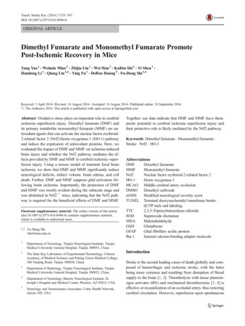
538Transl. Stroke Res. (2016) 7:535–547Fig. 1 DMF and MMF improve neurological deficits and reduce braininfarct volume and cerebral edema in mice with MCAO. a Schematicexperimental design of DMF and MMF treatment for acute ischemiccerebral stroke. b Administration of DMF or MMF reduces infarctvolume determined by 2,3,5-tripenyltetrazolium chloride (TTC)staining. Brain sections from a representative mouse are shown fromeach group (vehicle control, DMF 30, 45 mg and MMF 30, 45 mg).Each column shows four TTC-stained coronal brain slices arranged incranial to caudal order. c DMF and MMF treatments reduce the infarctvolume due to MCAO during the subacute phase. Mice in the DMF- andMMF-treated groups had smaller volume of infarct on days 3 and 7 postischemia–reperfusion injury. Data shown are mean SEM at day 3;*p 0.05, **p 0.01 as compared to the vehicle-treated group;##p 0.01, as compared between the DMF 45 mg group and DMF30 mg group, or MMF 45 mg and MMF 30 mg group, n 10 pergroup. d Neurobehavioral improvement in mice with MCAO uponDMF or MMF treatment is time- and dose-dependent. Significantimprovement in mNSS scores is evident in mice treated with 45 mg/kgDMF or MMF (p 0.01) as compared to mice in the vehicle controlgroup on day 3 and day 7, but not on day 1 post-ischemia–reperfusioninjury. A lesser degree of improvement is observed in mice treated withDMF or MMF at a dose of 30 mg/kg body weight. Mean SEM;**p 0.01, as compared to those in the vehicle control group. e DMFand MMF attenuate cerebral edema associated with transient MCAO ina dose-dependent manner. Data depicted here are mean SEM; *p 0.05and **p 0.01 as compared to the vehicle-treated group; ##p 0.01 ascompared between DMF 45 mg/kg and DMF 30 mg/kg group or MMF45 mg/kg and MMF 30 mg/kg group; n 10 per grouplabeling (TUNEL) kit (Roche, USA). Once stained, thespecimens were analyzed under a florescence microscope(Nikon C-HGFI, Japan). The total number of nuclei andTUNEL-positive cells were counted in four random fieldsof a 20 view of the edge of the infarct, and the ratio ofapoptotic cells to nuclei was calculated as apoptotic cellsin percent. Paraffin sections from mouse brains sacrificed24 h after MCAO, cut to a thickness of 5 μm, were firstdeparaffinized in xylene and subsequently rehydrated withvarious grades of ethanol. After rehydrating the sections,nonspecific binding was blocked by incubating the sections in 10 % bovine serum albumin for 20 min. Thesections were then incubated overnight at room temperature with anti-glial fibrillary acidic protein (GFAP) oranti-ionized calcium-binding adapter molecule (Iba-1) antibodies to identify astrocytes and microglia, respectively.Finally, mounting media containing DAPI was appliedand a coverslip was placed over the sections. The stained
Transl. Stroke Res. (2016) 7:535–547539sections were examined and analyzed with a fluorescencemicroscope (Olympus, Tokyo, Japan).expression levels of the messenger RNAs (mRNAs) were thenreported as fold changes vs control.Determination of Indicators of Oxidative StressStatistical AnalysisBrain tissues were collected at day 3 after MCAO. Superoxidedismutase (SOD) activity as well as the levels ofmalondialdehyde (MDA) and glutathione (GSH) were measuredas indicators of oxidative stress [34]. The brains were washed,weighed, and then homogenized in ice-cold saline (nine volumes) for 20 min to prepare a 10 % (w/v) homogenate. Thehomogenate was then centrifuged at 4000 rpm/min for 10 minat 4 C. SOD activity and levels of MDA and GSH were measured as described previously [34, 35]. Data were calculated inreference to the protein concentration in each sample.All data were analyzed by SPSS 18.0 software and expressedas mean SEM. Sample size per group was determined usinga priori sample size calculation (G*Power version 3.1). Toachieve α 0.05 at β 0.2 (power 80 %) with a mean 20 %standard deviation, results from sample size calculation showthat n 6–10 mice per group was appropriate. Statistical differences were measured by unpaired two-tailed Student t testfor comparison of two groups or ANOVA followed byBonferroni post hoc test for multiple group comparisons.Values of p 0.05 were considered significant.Western Blot AnalysisResultsOn day 3 post-MCAO, ipsilateral hemispheres were homogenized in RIPA lysis buffer (Sigma, USA) and 1 mmol/Lphenylmethanesulfonyl fluoride (PMSF) (Sigma, USA). Aftercentrifugation, the supernatants were collected as total proteins.Proteins were loaded and transferred to a PVDF membrane(Millipore, USA). After being blocked, membranes were incubated overnight at 4 C with anti-Nrf2 (1:1000, Millipore), antiHO-1 (1:1000, Millipore), or rabbit polyclonal antibodies.Membranes were then incubated for 1 h at room temperaturewith horseradish peroxidase (HRP)-labeled goat anti-rabbit secondary antibody (1:4000, Vector, Burlingame, USA). Themembranes were placed into a gel imaging system (Bio-Rad,ChemiDoc XRS, USA) and then exposed. The intensity ofblots was quantified using the Quantity One Analysis software(Bio-Rad, USA). β-Actin was used as an internal control.RNA Isolation and Real-Time PCRTotal RNA was extracted from the ischemic hemisphere usingTRIzol Reagent (Invitrogen, Carlsbad, CA, USA) accordingto the manufacturer’s instructions at day 3 after MCAO. Theconcentration of RNA was quantified by ultraviolet spectrophotometry at 260/280 nm. cDNA was transcribed usingTransScript First-Strand cDNA Synthesis SuperMix Kit(TransGen; catalog no. AT301) in accordance with the manufacturer’s instructions. PCR was performed on the Opticon 2Real-Time PCR Detection System (Bio-Rad) using the following primers: Nrf2 forward GGTTGCCCACATTCCCAAAC, Nrf2 reverse TCCTGCCAAACTTGCTCCAT;HO-1 forward CGACAGCATGTCCCAGGATT, HO-1 reverse CTGGGTTCTGCTTGTTTCGC; and β-actin forwardAAATCGTGCGTGACATCAAAGA, β-actin reverseGGCCATCTCCTGCTCGAA; SYBR Green PCR MasterMix (Roche) was also used. Samples were run in duplicateand normalized to β-actin using the 2–ΔΔCt method. TheImprovement in Neurological Deficits at Subacute Stagein MCAO Mice Treated with DMF or MMFOn day 1 post-MCAO ischemia–reperfusion injury, there wasno statistical difference in mNSS between groups of MCAOmice treated with vehicle control, DMF (30, 45 mg/kg), orMMF (30, 45 mg/kg) groups. Surprisingly, on day 3 postMCAO ischemia–reperfusion injury, mice in both of theDMF (30 and 45 mg/kg)-treated groups exhibited significantly reduced mNSS. A similar impact was evident in mice receiving MMF (30 and 45 mg/kg) treatment. The improvementof neurobehavioral function with DMF or MMF treatmentwas dose-dependent with a better outcome in mice treatedwith the relatively higher dose of DMF (45 mg/kg) or MMF(45 mg/kg) as compared with those treated with the lowerdose of DMF (30 mg/kg) or MMF (30 mg/kg) body weight,respectively (Fig. 1d). Further, the reduction of mNSS wassustained on day 7 post-MCAO ischemia–reperfusion injury.DMF and MMF Reduce Infarct VolumeThe infarct volume of brain tissue was measured on days 1, 3,and 7 post-MCAO ischemia–reperfusion injury with TTCstaining (Fig. 1b). On day 1 post-MCAO, there was no statistical difference in the volume of the infarct in mice treatedwith vehicle and DMF (30, 45 mg per kg body weight)(Supplemental Fig. 2). However, a significant reduction ofinfarct volume was observed in MCAO mice treated withDMF on day 3 post-MCAO. The reduction of infarct volumewith DMF treatment was sustained in mice on day 7 postMCAO (Fig. 1b, c). A dose effect was observed with thehigher dose of DMF at 45 mg/kg body weight having a greatereffect on the infarct volume reduction. To further confirm theprotective role of DMF, we examined its primary metabolite
540MMF. A similar impact on acute ischemic infarct volume wasobserved with MMF in a dose-dependent manner (Fig. 1b, c).Administration of DMF (30, 45 mg/kg) or MMF (30, 45 mg/kg) significantly decreased the percentage of the infarct volume from 49.36 5.33 % in the vehicle group to 39.69 6.65 % (p 0.05), 30.64 2.85 % (p 0.01), 38.05 3.60 %(p 0.01), or 28.21 2.58 % (p 0.01) on day 7 post-ischemia–reperfusion injury, respectively.Brain EdemaThe brain water content (BWC) in mice of the vehicle groupwas 19.6 2.5 %, while the DMF-treated (30, 45 mg/kg) andMMF-treated (30, 45 mg/kg) groups had decreased BWC of11.47 2.3 % (p 0.05, as compared with vehicle group), 8.1 1.9 % (p 0.01), 11.42 3.52 % (p 0.05), or 6.42 2.51 % (p 0.01), respectively (Fig. 1e).Longitudinal Examination of Infarct Volume with MRITo further monitor the evolution of ischemic brain lesions following MCAO ischemia–reperfusion injury, MRI wasemployed to image the lesions in vivo on days 1, 3, and 7post-MCAO ischemia–reperfusion injury. T2-weighted imageswere used to calculate the volume of ischemic lesions. On day 1post-MCAO ischemia–reperfusion injury, there was no difference in the volume of ischemic lesion in the vehicle-treatedcontrol group as compared to those in the DMF (30 mg or45 mg/kg)-treated groups. However, on day 3 post-ischemia,the volume of ischemic lesion was significantly reduced in micetreated with DMF at a dose of 30 or 45 mg per kg body weight.The DMF-induced ischemic volume reduction on T2-weightedimages was sustained on following MRI examinations at day 7post-MCAO ischemia–reperfusion injury (Fig. 2).DMF and MMF Inhibit Neural ApoptosisA TUNEL assay was used to assess neuronal apoptosis.TUNEL staining was performed in brain tissue sectionsobtained at day 3 after MCAO. In the sham group, nodiscernable neural apoptosis was seen. In the vehiclegroup, TUNEL-positive cells were densely distributed inthe ischemic cortex (53.4 2.61 %). The number of apoptotic cells was decreased in mice treated with DMF30 mg (44.1 2.64 %, p 0.05 as compared to that inthe vehicle-treated group), DMF 45 mg (29.2 1.3 %,p 0.01), MMF 30 mg (40.7 0.77 %, p 0.05), orMMF 45 mg/kg body weight (22.2 2.12 %, p 0.01).These results suggest a protective role of DMF or MMFagainst apoptosis of cells in the central nervous system(CNS) arising from ischemia–reperfusion injury (Fig. 3).Transl. Stroke Res. (2016) 7:535–547DMF and MMF Treatment Leads to Suppressed GlialActivationNeural cell death in cerebral ischemia–reperfusion injury isassociated with astrocytosis and microgliosis [36]. To test thehypothesis that cytoprotective properties of DMF and MMFmight diminish the glial activation elicited by transient ischemiaand reperfusion, immunohistochemistry studies were performed. As shown in Fig. 4, the numbers of GFAP-positiveand Iba-1-positive cells were decreased in the ischemic regionof the MCAO mice as compared to the sham group. Comparedwith the vehicle group (27.4 2.90 %), the number of GFAPpositive cells was decreased in the DMF 30 mg (20.3 2.47 %,p 0.05), DMF 45 mg (16 3.08 %, p 0.01), MMF 30 mg(20.5 2.23 %, p 0.05), and MMF 45 mg/kg body weight(17 2.71 %, p 0.01) treated groups. The number of Iba-1bearing cells was also decreased in the DMF 30 mg (27.6 2.11 %, p 0.05), DMF 45 mg (22.8 2.7 %, p 0.01), MMF30 mg (29.8 1.57 %, p 0.05), and MMF 45 mg/kg bodyweight (19.7 3.12 %, p 0.01) treated groups as comparedwith the vehicle control group (40.0 2.98 %) (Fig. 4).DMF and MMF Attenuate Ischemia–Reperfusion InjuryEvoked Intracellular Oxidative StressSOD is often regarded as the first line of defense against anupswing of ROS and is responsible for the conversion of superoxide to H2O2 in the cytoplasm and mitochondria [37].GSH plays a major role in the detoxification of peroxides[38]. Brain tissues with MCAO ischemia–reperfusion injuryhave decreased SOD activity and GSH levels. MDA, one ofthe products of membrane lipid peroxidation, reflects the degree of ROS-inflicted damage via membrane lipid peroxidation. Compared with the vehicle-treated group, there weresignificantly increased levels of SOD activity and GSH anddecreased levels of MDA in the groups treated with DMF andMMF (30 and 45 mg/kg) (Table 1). These data suggest thatDMF and MMF reduced MCAO-induced oxidative stress.DMF and MMF Promote Expression of Nrf2 and HO-1To identify whether Nrf2 signaling is involved in the neuroprotective effect of DMF and MMF in MCAO ischemia–reperfusion injury, we analyzed ischemic brain tissues by western blot and RT-PCR. Western blot analysis of cortical tissuesat day 3 post-ischemia reveals that DMF and MMF had nosignificant effects on Nrf2 levels in sham animals, but augmented the expression of Nrf2 and HO-1 protein upon DMFtreatment in mice with MCAO. The induction of Nrf2 andHO-1 expression by DMF was further confirmed by RTPCR analysis at the mRNA level. Moreover, increased levelsof Nrf2 and HO-1 were evident in mice with MCAO treatedwith MMF (30 or 45 mg/kg) (Fig. 5).
Transl. Stroke Res. (2016) 7:535–547541Fig. 2 Effects of DMF on infarct volume were sustained. Coronal MRIsections show T2 lesions in vivo on days 1, 3, and 7 after MCAO. Thevolume reduction in ischemic lesions with DMF was sustained. Datashown are mean SEM; *p 0.05, **p 0.01 as compared to thevehicle group; #p 0.05, ##p 0.01, as compared between DMF45 mg/kg and DMF 30 mg/kg group, n 10 per groupAbsence of DMF or MMF ProtectionAgainst Ischemia–Reperfusion Brain Injury in Nrf2Gene-Deficient Micethe vehicle and DMF 45 mg/kg-treated Nrf2 / groups. Thesefindings were further confirmed with MMF 45 mg/kg treatment in Nrf2 / mice with MCAO (Fig. 6a–d). Furthermore,neither levels of HO-1 mRNA nor its protein expression wasaltered upon DMF or MMF treatment in Nrf2–/– mice withMCAO ischemia–reperfusion injury (SupplementalFig. 3).These findings suggest that the Nrf2 pathway is essential for DMF and MMF to exert their protective role in brainischemia–reperfusion injury.To further determine the mechanism by which DMF andMMF exert neuroprotective effects, Nrf2 / mice were usedin the current study. Nrf2 / mice were treated with DMF(45 mg/kg) or MMF (45 mg/kg) after MCAO. There was nodifference in infarct volume, mNSS, or brain edema betweenFig. 3 DMF and MMF reducecell death caused by cerebralischemic–reperfusion injury.TUNEL staining shows a largenumber of apoptotic cells in brainsections at 72 h post-ischemia–reperfusion injury. a In thevehicle-treated group, numerousTUNEL-positive cells are presentin the ischemic cortex. b–e DMFor MMF treatment reduces thenumber of apoptotic cells. fQuantitative analysis of TUNELpositive cells shows DMF- andMMF-induced protection fromcell death and its dose-dependentpattern. Data are presented asmean SEM; *p 0.05,**p 0.01, compared to thevehicle group; ##p 0.01, ascompared between DMF 45 mgand DMF 30 mg group, orbetween MMF 45 mg and MMF30 mg group; n 8 per group
542Transl. Stroke Res. (2016) 7:535–547Fig. 4 DMF or MMF suppresses glial activation associated withtransient MCAO. a Brain slices from mice with MCAO stained withanti-glial fibrillary acidic protein (GFAP) antibody. b Brain slicesstained with anti-ionized calcium-binding adapter molecule (Iba-1)antibody. Brain tissue sections obtained from mice 72 h after transientMCAO show increased astrocytosis and microgliosis (vehicle control ina, b, respectively). Treatment with DMF 30, 45 mg/kg or MMF 30,45 mg/kg body weight reduces the expression of GFAP and Iba-1. cQuantitative analysis of GFAP-bearing cells and d that of Iba-1-bearingcells in cerebral tissues from mice with transient MCAO treated withDMF or MMF. The percentages of GFAP- and Iba-1-expressing cellsare decreased by DMF or MMF treatment in a dose-dependent manner.Data are presented as mean SEM; *p 0.05, **p 0.01 as compared tothe vehicle group; ##p 0.01 as compared between DMF 45 mg vs DMF30 mg or MMF 45 mg vs MMF 30 mg groups; n 8 per groupDiscussionThe beneficial impact of DMF treatme
Medical University General Hospital, Tianjin 300052, China 4 Department of Neurology, Barrow Neurological Institute, St. Joseph's Hospital and Medical Center, Phoenix, AZ 85013, USA 5 Neurology and Neuroscience Associates, Unity Health Network, Akron, OH, USA Transl. Stroke Res. (2016) 7:535-547 DOI 10.1007/s12975-016-0496-