Transcription
IVIGRC White PaperIntersociety Venous Insurance and Government RelationsCommittee (IVIGRC)Treatment of Superficial Treatment of Venous Disease of the Lower Leg and PelvisBackgroundIn the last 10 years the diagnosis and treatment of venous disease has advanced more thanin the last 100 years. Ultrasound, endovenous ablation devices, foam sclerotherapy andtumescent anesthesia have greatly improved patient care and have moved treatment fromthe operating room to the office or radiology suite. This has created challenges forinsurers. Medical necessity policy for the treatment of chronic venous disease (CVD) hasbecome fragmented and inconsistent across the U.S. and among private insurers, andMedicare. As with any medical specialty, those who are most committed to that specialtygenerally provide the best care. Commitment includes some form of training, a practicefocused in that area, and continuing education through attendance at meetings and otherCME. The American College of Phlebology (ACP) the American Venous Forum (AVF), theSociety of Interventional Radiology (SIR) and other organizations have been at theforefront of advancing education, research and appropriate treatment of venous disease.Dedicated venous physicians representing these organizations formed the IntersocietyVenous Insurance and Government Relations Committee (IVIGRC).In 2011, the Society for Vascular Surgery and the American Venous Forum1undertook a comprehensive summary of all the available venous research and graded it byrelevance and quality of data. Their goal was to analyze all the available evidence-basedmedicine and create rational guidelines for treatment of venous disease of the lower limbsand pelvis. This 70 page review by Gloviczki et al was well received by the medicalcommunity across specialties treating venous disease.The IVIGRC has accepted the challenge of taking this review and formatting it in adocument that would be more suitable to the insurance industry. It reflects the evidencebased recommendations in the Gloviczki paper and other studies. Recommendations arebased on a consensus of a panel of experts where the evidence based research is sparse. Itis focused on those interventions that have the most policy variation in the insuranceindustry or where policies substantially deviate from the evidence based researchavailable.We acknowledge that all carriers are free to determine coverage guidelines etc. based upontheir own independent review of the literature and resources like Cochrane and others.However, we suggest that evidence based medical necessity should not vary greatly basedon geography or insurer. We would like to introduce the concept of “medically significantvenous insufficiency “or “evidence-based medical significance“. This eliminates confusionaround terms like “cosmetic” or not medically necessary”. The medical evidence shoulddetermine the definition of medically significant venous insufficiency using a combinationof CEAP and VCSS (discussed below). We would propose that payers retain the evidencebased definition of medical significance, but choose at what level it becomes either a1
“covered benefit” or a “non-covered benefit”. Insurers could establish different benefitlevels for their various premium options. In this way the evidence-based medical criteriawould still be consistent across the industry.The IVIGRC understands the importance of delivering quality care in a cost effectivemanner and welcomes the opportunity to work with the insurance industry in whateverway possible to achieve those ends.In the following pages are our medical necessity guidelines in a summary format.The recommendations of the IVIGRC (and the Gloviczki paper) have been determined bythe method suggested by the Grading of Recommendations Assessment, Development, andEvaluation system (GRADE) working group. (www.gradeworkinggroup.org)For each guideline, the letter A, B, or C marks the quality of current evidence as high,medium or low quality. The grade of recommendation of a guideline can be strong (1) orweak (2), depending on the risk and burden of a particular diagnostic test or a therapeuticprocedure to the patient vs. the expected benefit. The words “we recommend” are used forGRADE 1—strong recommendations—if the benefits clearly outweigh risks and burdens,or vice versa; the words “we suggest” are used for GRADE 2—weak recommendations—when the benefits are closely balanced with risks and burdens. Where current evidence isweak or lacking, the degree of consensus of the committee reflects the grade with thequality of the recommendation adjusted accordingly.Following the summary are the common ICD9 and CPT codes for venous disease. This isfollowed by the accompanying appendix Benchmark Evidence Based Policy for Treatmentof Chronic Venous Disease and Varicose Veins which provides the detailed review andreferences for these recommendations.2
Summary of Guidelines for Treatment of Venous DiseaseIndications for TreatmentTreatment of asymptomatic varicose veins is not medically necessary. GRADE 1AIndications for treatment include pain (achiness, heaviness), edema, varix hemorrhage,recurrent superficial phlebitis, stasis dermatitis, or ulceration.Patients should be evaluated using the CEAP classification and the Venous Clinical SeverityScore (VCSS). We would define medically necessary as a CEAP classification of C2 or higherand a VCSS score of 5 or higher. GRADE 1AWe suggest the treatment of some CEAP C2 patients with isolated varices, by medicalcompression hose alone may an acceptable form of treatment. GRADE 2CWe recommend against compression therapy being considered the primary treatment ofsymptomatic varicose veins (class C2) in those patients who are candidates for saphenousvein ablation GRADE 1B.In AdditionAll patients being considered for treatment must have a duplex ultrasound. GreatSaphenous Vein (GSV), Small Saphenous Vein (SSV, Anterior Accessory of the GreatSaphenous Vein (AAGSV) and Posterior Accessory of the Great Saphenous Vein(PAGSV) ) incompetence must have a reflux time 500 msec. “Pathologic” perforatingveins includes those with outward flow of 500 ms, with a diameter of 3.5 mm, locatedbeneath a healed or open venous ulcer. GRADE 1BWe suggest all noninvasive vascular diagnostic studies be performed by a qualifiedphysician or by a qualified technologist under the general supervision of a qualifiedphysician. These individuals should have passed some form of credentialing examinationand hold a certificate from a nationally recognized credentialing organization such as theAmerican Registry of Diagnostic Medical Sonographers (ARDMS) or CardiovascularCredentialing International (CCI) GRADE 2CWe suggest a follow up ultrasound examination CPT code 93971 after endovenous thermalablation or ultrasound guided chemical ablation to confirm non compressibility andabsence of reflux in the treated area. GRADE 2C3
Treatment of Great or Small Saphenous VeinsWe recommend endovenous thermal ablation (laser and radiofrequency) is the preferredtreatment for saphenous and accessory saphenous (GSV, SSV, AAGSV, PAGSV) veinincompetence. GRADE 1BWe recommend open surgery is appropriate in veins not amenable to endovenousprocedures but otherwise is not recommended because of increased pain, convalescenttime, and morbidity. GRADE 1BWhen open surgery of the great saphenous vein is performed we suggest it should includehigh ligation and invagination stripping to the level of the knee. GRADE 2BWhen open surgery of the small saphenous vein is performed we recommend it includehigh ligation at the knee crease and selective invagination of the proximal portion. GRADE1BTreatment of Circumflex Veins and Other Non Truncal VeinsThe treatment of other non-truncal, tributary varicose vein reflux (circumflex veins(anterior and posterior thigh) and intersaphenous vein) is more complex. The medicalrecord should reflect that these veins are incompetent, and note their size, presence orabsence of tortuosity, and depth relationship to the skin, i.e. accessible or not accessible byphlebectomy.We recommend varicose (visible) tributary veins can be treated by stab phlebectomy,liquid sclerotherapy or foam chemical ablation. GRADE 1BWe suggest (non visible) tributary veins be treated by ultrasound guided liquidsclerotherapy or foam chemical ablation. GRADE 2BTreatment of Perforator VeinsWe recommend against selective treatment of incompetent perforating veins in patientswith simple varicose veins (CEAP class 2). GRADE 1BWe suggest treatment of “pathologic” perforating veins located beneath a healed or openvenous ulcer (CEAP class 5-6). GRADE 2BFor treatment of “pathologic" perforating veins we suggest the SEPS procedure,ultrasound-guided chemical ablation or thermal ablations. GRADE 2C4
Coding ReferenceCPT/HCPCS 377997589476942SELECTIVE CATHETER PLACEMENT, VENOUS SYSTEM, FIRST ORDER BRANCHSINGLE OR MULTIPLE INJECTIONS OF SCLEROSING SOLUTIONS, SPIDER VEINS(TELANGIECTASIA); LIMB OR TRUNKINJECTION OF SCLEROSING SOLUTION; SINGLE VEININJECTION OF SCLEROSING SOLUTION; MULTIPLE VEINS, SAME LEGENDOVENOUS ABLATION THERAPY OF INCOMPETENT VEIN, EXTREMITY,INCLUSIVE OF ALL IMAGING GUIDANCE AND MONITORING, PERCUTANEOUS,RADIOFREQUENCY; FIRST VEIN TREATEDENDOVENOUS ABLATION THERAPY OF INCOMPETENT VEIN, EXTREMITY,INCLUSIVE OF ALL IMAGING GUIDANCE AND MONITORING, PERCUTANEOUS,RADIOFREQUENCY; SECOND AND SUBSEQUENT VEINS TREATED IN A SINGLEEXTREMITY, EACH THROUGH SEPARATE ACCESS SITES (LIST SEPARATELY INADDITION TO CODE FOR PRIMARY PROCEDURE)ENDOVENOUS ABLATION THERAPY OF INCOMPETENT VEIN, EXTREMITY,INCLUSIVE OF ALL IMAGING GUIDANCE AND MONITORING, PERCUTANEOUS,LASER; FIRST VEIN TREATEDENDOVENOUS ABLATION THERAPY OF INCOMPETENT VEIN, EXTREMITY,INCLUSIVE OF ALL IMAGING GUIDANCE AND MONITORING, PERCUTANEOUS,LASER; SECOND AND SUBSEQUENT VEINS TREATED IN A SINGLE EXTREMITY,EACH THROUGH SEPARATE ACCESS SITES (LIST SEPARATELY IN ADDITION TOCODE FOR PRIMARY PROCEDURE)TRANSCATHETER OCCLUSION OR EMBOLIZATION, PERCUTANEOUS, ANYMETHOD, NON-CENTRAL NERVOUS SYSTEM, NON-HEAD OR NECKENDOSCOPY, SURGICAL, WITH LIGATION OF PERFORATOR VEINS, SUBFASCIAL(SEPS)LIGATION AND DIVISION OF LONG SAPHENOUS VEIN AT SAPHENOFEMORALJUNCTION, OR DISTAL INTERRUPTIONSLIGATION, DIVISION, AND STRIPPING, SHORT SAPHENOUS VEINLIGATION, DIVISION, AND STRIPPING, LONG (GREATER) SAPHENOUS VEINSFROM SAPHENOFEMORAL JUNCTION TO KNEE OR BELOWLIGATION AND DIVISION AND COMPLETE STRIPPING OF LONG OR SHORTSAPHENOUS VEINS WITH RADICAL EXCISION OF ULCER AND SKIN GRAFTAND/OR INTERRUPTION OF COMMUNICATING VEINS OF LOWER LEG, WITHEXCISION OF DEEP FASCIALIGATION OF PERFORATOR VEINS, SUBFASCIAL, RADICAL (LINTON TYPE), WITHOR WITHOUT SKIN GRAFT, OPENSTAB PHLEBECTOMY OF VARICOSE VEINS, ONE EXTREMITY; 10-20 STABINCISIONSSTAB PHLEBECTOMY OF VARICOSE VEINS, ONE EXTREMITY; MORE THAN 20INCISIONSLIGATION AND DIVISION OF SHORT SAPHENOUS VEIN AT SAPHENOPOPLITEALJUNCTION (SEPARATE PROCEDURE)LIGATION, DIVISION, AND/OR EXCISION OF VARICOSE VEIN CLUSTER(S), ONELEG. FOR BOTH LEGS, REPORT WITH A MODIFIER 50.UNLISTED PROCEDURE, VASCULAR SURGERYTRANSCATHETER THERAPY, EMBOLIZATION, ANY METHOD RADIOLOGICALSUPERVISION AND INTERPRETATIONULTRASONIC GUIDANCE FOR NEEDLE PLACEMENT (EG, BIOPSY, ASPIRATION,5
93770939659397093971INJECTION, LOCALIZATION DEVICE), IMAGING SUPERVISION ANDINTERPRETATIONDETERMINATION OF VENOUS PRESSURENONINVASIVE PHYSIOLOGIC STUDIES OF EXTREMITY VEINS, COMPLETEBILATERAL STUDY (EG, DOPPLER WAVEFORM ANALYSIS WITH RESPONSES TOCOMPRESSION AND OTHER MANEUVERS, PHLEBORHEOGRAPHY, IMPEDANCEPLETHYSMOGRAPHY)LOWER EXTREMITY VENOUS DUPLEX ULTRASOUND - BILATERALDUPLEX SCAN OF EXTREMITY VEINS INCLUDING RESPONSES TO COMPRESSIONAND OTHER MANEUVERS; UNILATERAL OR LIMITED STUDYICD-9 9.81459.89NEVUS, NON-NEOPLASTIC (SPIDER VEINS)TELANGIECTASIA, TELANGIECTASISPHLEBITIS AND THROMBOPHLEBITIS OF SUPERFICIAL VESSELS OF LOWEREXTREMITIESPHLEBITIS AND THROMBOPHLEBITIS OF LOWER EXTREMITIES UNSPECIFIEDVARICOSE VEINS OF LOWER EXTREMITIES WITH ULCERVARICOSE VEINS OF LOWER EXTREMITIES WITH INFLAMMATIONVARICOSE VEINS OF LOWER EXTREMITIES WITH ULCER AND INFLAMMATIONVARICOSE VEINS OF LOWER EXTREMITIES WITH OTHER COMPLICATIONSVULVAR VARICOSITIES OF PIRENIUM (SPECIFICALLY)POSTPHLEBETIC SYNDROME WITHOUT COMPLICATIONSPOSTPHLEBETIC SYNDROME WITH ULCERPOSTPHLEBETIC SYNDROME WITH INFLAMMATIONPOSTPHLEBETIC SYNDROME WITH ULCER AND INFLAMMATIONPOSTPHLEBETIC SYNDROME WITH OTHER COMPLICATIONCHRONIC VENOUS HYPERTENSION WITH ULCERCHRONIC VENOUS HYPERTENSION WITH INFLAMMATIONCHRONIC VENOUS HYPERTENSION WITH ULCER AND INFLAMMATIONVENOUS(PERIPHERAL) INSUFFICIENCY, NSPECIFIEDOTHER SPECIFIED DISORDERS OF CIRCULATORY SYSTEM (PHLEBOSCLEROSIS,VENOFIBROSIS, COLLATERAL CIRCULATION[VENOUS], ANY SITE)6
AppendixBenchmark Evidence Based Policy for Treatment ofChronic Venous Disease and Varicose VeinsArticle OutlineI.II.IntroductionMethodology of guidelinesIII.DefinitionsIV.The scope of the problemV.AnatomyVI.Diagnostic evaluationVII.Classification of CVDVIII.Outcome assessmentIX.TreatmentA. Compression treatmentB. Open venous surgeryC. Endovenous Thermal Ablations Saphenous VeinD. Liquid SclerotherapyE. Ultrasound Guided Chemical Ablation Saphenous Vein with FoamX.Perforating VeinsXI.ConclusionsXII.References7
I. Introduction:The purpose of this policy document is to update providers and third party payors with the mostcurrent evidence-based guidelines for care of chronic venous disease and varicose veins.Evidence-based medicine involves utilizing the best available scientific information to makedecisions about patient care.14 It has been used successfully in recent years to guide indicationsfor therapy, validate new techniques for efficacy and cost control, and to develop reliableoutcome assessment methods in many areas of clinical practice. Chronic venous disease andvaricose veins have seen recent advances in minimally invasive therapies and clinical researchthat are leading more patients to seek treatment. This in turn has led to more proceduresperformed by physicians from a variety of specialties, and advances in industry resulting in newtechnology. Academic interest in venous disease has focused on the analysis of data for validityand scientific interest, as well as analysis of treatment outcomes.In the US and Europe, varicose veins are found in more than 20% of the population, withapproximately 5% of patients exhibiting signs of chronic venous disease, including edema andskin changes. Around 1% of patients have active or healed venous ulcers.57 In the US alone,according to the San Diego epidemiologic study, in excess of 33 men and women between 40and 80 years old have varicose veins, with more than 2 million suffering from advanced chronicvenous disease with skin changes or ulcers.1 Each year in the US alone, more than 20,000patients are newly diagnosed with venous ulcers.3The Bonn Vein Study,59 a large European population based study, enrolled 3072 adults aged 18to 79. In this group, uncomplicated varicose veins were identified in 14.3%, with symptoms ofmore advanced chronic venous disease including edema or skin changes found in 49.1% of menand 62.1% of women.Many cases of varicose veins are due to primary venous disease, caused in some cases by anintrinsic vein wall abnormality, although the etiology can be multifactorial. Labropoulos54 wrotethat primary varicose veins can arise from local or multifocal weakness of the vein wall thatoccurs with or without saphenous valvular incompetence. Varicosities can result from secondarycauses, including deep vein thrombosis (DVT) or obstruction, superficial thrombophlebitis, orarteriovenous fistula. Varicose veins may also be congenital and manifest as a venousmalformation376. It has been shown that primary varicose veins can progress to chronic venousdisease with severe symptoms, including venous ulcers. In 1948 Bauer63 reported that 58% of hispatients with symptoms of severe CVD had primary venous disease without a history of deepvein thrombosis (DVT). The North American subfascial endoscopic perforator surgery (SEPS)registry includes more patients with advanced CVD resulting from primary venous disease thanpost-thrombotic syndrome (70% vs 30%).62Varicose veins and the complications of chronic venous disease are associated with a high directcost to the patient and society as a whole. Chronic pain, refractory swelling and the open sores of8
venous ulcers are associated with disability, loss of working days and lower quality of life(QOL), loss of working days. In the United States, the direct medical cost of CVD has beenestimated to be between 150 million and 1 billion annually.3, 4 In the United Kingdom, 2% ofthe annual national health care budget is spent on treating venous ulcers.1Varicose veins and chronic venous disease are prevalent in the adult population of the US.Advances in scientific technology have resulted in new minimally invasive endovascular surgicaltechniques, changing the way physicians care for patients with venous disease. Patientacceptance of office-based, outpatient procedures has been very strong, and clinical outcomesfrom these procedures are positive. More interventions for chronic venous disease are beingperformed every year, and interest in these procedures has grown among patients, physicians,device manufacturers, and third party payors.376II. Methodology of guidelinesGuidelines for the care of patients with varicose veins, as recommended here, are based onscientific evidence. The need for adopting evidence-based guidelines and reporting standards forvenous diseases has been recognized by leaders in the field for some time. 15,16,17, 18, 19, 20 Thecurrent guidelines have been formulated by a Venous Guideline Committee, who reviewed theliterature, including consensus documents and guidelines already in existence,21, 22, 23, 24, 25, 26, 27,28, 29, 30, 31as well as meta-analyses,6, 7, 8, 9, 10, 11, 12, 32, 33, 34, 35, 36, 37, 38, 39, 40, 41, 42 reports13, 43, 44, 45, 46and recommendations from the American Venous Forum.47The guidelines offered here are based on the Grading of Recommendations Assessment,Development, and Evaluation (GRADE) system, as it was described by Guyatt et al (Table I). 48For each guideline, the letter A, B, or C marks the level of current evidence. The grade ofrecommendation of a guideline can be strong (1) or weak (2), depending on the risk and burdenof a particular diagnostic test or a therapeutic procedure to the patient vs the expected benefit.The words “we recommend” are used for GRADE 1—strong recommendations—if the benefitsclearly outweigh risks and burdens, or vice versa; the words “we suggest” are used for GRADE2—weak recommendations—when the benefits are closely balanced with risks and burdens376.III. DefinitionsCurrently accepted terminology for the superficial, perforating, and deep veins of the leg andpelvis are used.49, 50 Definitions of varicose and spider veins as well as other manifestations ofCVD follow recommendations of the CEAP classification and the recent update on venousterminology of the International Committee of the AVF.51, 52Varicose veins of the lower limbs are dilated subcutaneous veins that are 3 mm in diametermeasured in the upright position.53 Synonyms include varix, varices, and varicosities. Varicositycan involve the main axial superficial veins—the great saphenous vein (GSV) or the smallsaphenous vein (SSV)—or any other superficial vein tributaries of the lower limbs.9
Varicosities are manifestations of chronic venous disease (CVD).51, 52 CVD includes medicalconditions of long duration, involving morphologic and functional abnormalities of the venoussystem manifested by symptoms and/or signs, indicating the need for investigation and care. Theterm chronic venous disorder is reserved for the full spectrum of venous abnormalities andincludes dilated intradermal veins and venules between 1 and 3 mm in diameter (spider veins,reticular veins, telangiectasia; CEAP class C1).Varicose veins can progress to a more advanced form of chronic venous dysfunction such aschronic venous insufficiency (CVI).55, 56 In CVI, increased ambulatory venous hypertensioninitiates a series of changes in the subcutaneous tissue and the skin: activation of the endothelialcells, extravasation of macromolecules and red blood cells, diapedesis of leukocytes, tissueedema, and chronic inflammatory changes most frequently noted at and above the ankles.41, 53Limb swelling, pigmentation, lipodermatosclerosis, eczema, or venous ulcerations can develop inthese patients.IV. The scope of the problemIn the US and Europe, varicose veins are found in more than 20% of the population, withapproximately 5% of patients exhibiting signs of chronic venous disease, including edema andskin changes. Around 1% of patients have active or healed venous ulcers.57 In the US alone,according to the San Diego epidemiologic study, in excess of 33 men and women between 40and 80 years old have varicose veins, with more than 2 million suffering from advanced chronicvenous disease with skin changes or ulcers.1 Each year in the US alone, more than 20,000patients are newly diagnosed with venous ulcers.3The Bonn Vein Study,59 a large European population based study, enrolled 3072 adults aged 18to 79. In this group, uncomplicated varicose veins were identified in 14.3%, with symptoms ofmore advanced chronic venous disease including edema or skin changes found in 49.1% of menand 62.1% of women.Varicose veins and the complications of chronic venous disease are associated with a high directcost to the patient and society as a whole. Chronic pain, refractory swelling and the open sores ofvenous ulcers are associated with disability, loss of working days and lower quality of life(QOL), loss of working days. In the United States, the direct medical cost of CVD has beenestimated to be between 150 million and 1 billion annually.3, 4 In the United Kingdom, 2% ofthe annual national health care budget is spent on treating venous ulcers.1V. AnatomyNew venous terminology has recently been developed and is in use by vascular societies aroundthe world.47, 49, 61 The success of assigning uniform names to common veins was accompanied bynew information on anatomy obtained with duplex ultrasonography, three-dimensional computedtomography (CT), and magnetic resonance (MR) imaging; all these resulted in betterunderstanding of the anatomy of veins and the pathology of CVD.33, 6210
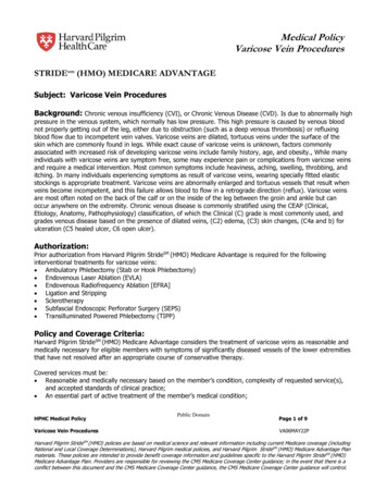
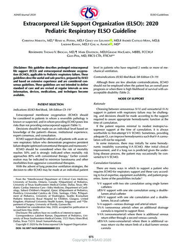
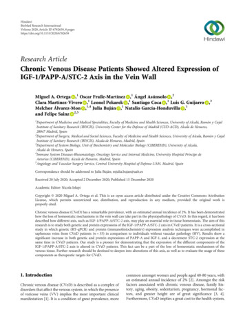
Superficial veinsSuperficial veins of the lower limbs are those located between the deep fascia, covering themuscles of the limb, and the skin. The main superficial veins are the great saphenous vein (GSV)and the small saphenous vein (SSV). The GSV originates from the medial superficial veins of thedorsum of the foot and ascends in front of the medial malleolus along the medial border of thetibia, next to the saphenous nerve (Fig 1). There are posterior and anterior accessory saphenousveins in the calf and the thigh. The saphenofemoral junction (SFJ) is the confluence ofsuperficial inguinal veins, comprising the GSV and the superficial circumflex iliac, superficialepigastric, and external pudendal veins. The GSV in the thigh lies in the saphenoussubcompartment of the superficial compartment, between the saphenous fascia and the deepfascia.(Used with permission of Mayo Foundation for Medical Education and Research.) Fig 1. Medial superficial and perforating veins of the lower limb.11
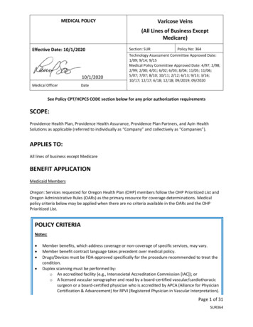
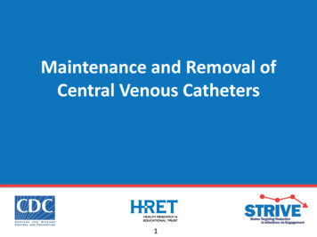
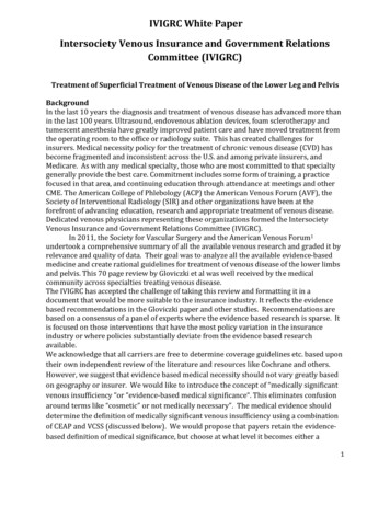
The SSV is the most important posterior superficial vein of the leg (Fig 2). It originates from thelateral side of the foot and drains blood into the popliteal vein, joining it usually just proximal tothe knee crease. The intersaphenous vein (vein of Giacomini), which runs in the posterior thigh,connects the SSV with the GSV.65(Used with permission of Mayo Foundation for Medical Education and Research.) Fig 2. Posterior superficial and perforating veins of the leg.Deep veinsDeep veins accompany the main arteries of the limb and pelvis. The deep veins of the calf(anterior, posterior tibial, and peroneal veins) are paired structures, and the popliteal and femoralveins may also be paired. The gastrocnemius and soleal veins are important deep tributaries. The12
old term superficial femoral vein has been replaced by the new term femoral vein.52 The femoralvein connects the popliteal to the common femoral vein.The pelvic veins include the external, internal, and common iliac veins, which drain into theinferior vena cava (IVC). Large gonadal veins drain into the IVC on the right and the left renalvein on the left.Perforating veinsPerforating veins connect the superficial to the deep venous system (Fig 1). They pass throughthe deep fascia that separates the superficial compartment from the deep. Communicating veinsconnect veins within the same system. The most important leg perforating veins are the medialcalf perforators.66 The posterior tibial perforating veins (formerly called Cockett perforators)connect the posterior accessory GSV of the calf (formerly called the posterior arch vein) with theposterior tibial veins and form the lower, middle, and upper groups. They are located just behindthe medial malleolus (lower), at 7 to 9 cm (middle) and at 10 to 12 cm (upper) from the loweredge of the malleolus. The distance between these perforators and the medial edge of the tibia is2 to 4 cm.66 (Fig 1). Paratibial perforators connect the main GSV trunk with the posterior tibialveins. In the distal thigh, perforators of the femoral canal usually connect directly the GSV to thefemoral vein.VI. Diagnostic EvaluationA. Clinical ExaminationPatients with varicose veins may present with no symptoms at all; the varices are then ofcosmetic concern only, with an underlying psychologic impact. Psychologic concerns related tothe cosmetic appearance of varicose veins will, however, reduce a patient's QOL in many cases.Symptoms related to varicose veins or more advanced CVD include tingling, aching, burning,pain, muscle cramps, swelling, sensations of throbbing or heaviness, itching skin, restless legs,leg tiredness, and fatigue.70 Although not pathognomonic, these symptoms suggest CVD,particularly if they are exacerbated by heat or dependency noted during the course of the day andrelieved by resting or elevating the legs or by wearing elastic stockings or bandages.51Pain during and after exercise that is relieved with rest and leg elevation (venous claudication)can also be caused by venous outflow obstruction caused by previous DVT or by narrowing orobstruction of the common iliac veins (May-Thurner syndrome).69, 70, 71 Diffuse pain is morefrequently associated with axial venous reflux, whereas poor venous circulation in bulgingvaricose veins usually causes local pain376.B. Duplex scanning13
Duplex Doppler scanning is recommended as the first diagnostic test for all patients withsuspected CVD.5,79 The test is safe, noninvasive, cost-effective, and reliable. It is excellent forthe evaluation of infrainguinal venous obstruction and valvular incompetence.81 It alsodifferentiates between acute venous thrombosis and chronic venous changes.82,83Technique of the examinationEvaluation of reflux in the deep and superficial veins with duplex scanning should be performedwith the patient upright, with the leg rotated outward, heel on the ground, and weight taken onthe opposite limb.5 The supine position gives both false-positive and false-negative results ofreflux.84The examination is started below the inguinal ligament, and the veins are examined in 3- to 5-cmintervals. For a complete examination, all deep veins of the leg are examined, including thecommon femoral, femoral, deep femoral, popliteal, peroneal, soleal, gastrocnemial, anterior, andposterior tibial veins. The superficial veins are then evaluated, including the GSV, the SSV, theaccessory saphenous veins, and the perforating veins.The four components that should be included in a complete duplex scanning examination forCVD are (1) visibility, (2) compressibility, (3) venous flow, including measurement of theduration of reflux, and (4) augmentation. Asymmetry in flow velocity, lack of respiratoryvariations in venous flow, and waveform patterns at rest and during flow augmentation in thecommon femoral veins indicate proximal obstruction. Reflux can be elicited in two ways:increased intra-abdominal pressure using a Valsalva maneuver for the common femoral vein orthe SFJ, or by manual compression and release of the limb distal to the point of examination. Thefirst is more appropriate for evaluation of reflux in the common femoral vein and at the SFJ,whereas compression and release is the preferred technique more distally on the limb.84The cutoff value for abnormally reversed venous flow (reflux) in the saphenous, tibial, and deepfemoral veins has been 500 ms.81 International consensus documents previously recommended0.5 seconds as a cutoff value for all veins to use for lower limb venous incompetence.5, 22, 86 Thisvalue is, however, longer, 1 second, for the femoral and popliteal veins. 81 The Committeerecommends 500 ms as the cutoff value for saphenous, tibial, deep femoral, and perforating veinincompetence, and 1 second for femoral and popliteal vein incompetence.Perforating veins should be evaluated in patients with advanced disease, usually in those withhealed or active venous ulcers (CEAP class C5-C6) or in those with recurrent varicose veinsafter previous interventions. The SVS/AVF Guideline Committee definition of clinically relevantperforating veins includes those with outward flow of 500 ms, with a diameter of 3.5 mm,located beneath a healed or open venous ulcer (CEAP class C5-C6).5, 81, 88, 89VII. Classification of CVD - Clinical CEAP14 p
We suggest a follow up ultrasound examination CPT code 93971 after endov enous thermal . 459.81 VENOUS(PERIPHERAL) INSUFFICIENCY, NSPECIFIED 459.89 OTHER SPECIFIED DISORDERS OF CIRCULATORY SYSTEM (PHLEBOSCLEROSIS, VENOFIBROSIS, COLLATERAL CIRCULATION[VENOUS], ANY SITE) 7