Transcription
HindawiBioMed Research InternationalVolume 2020, Article ID 6782659, 8 pageshttps://doi.org/10.1155/2020/6782659Research ArticleChronic Venous Disease Patients Showed Altered Expression ofIGF-1/PAPP-A/STC-2 Axis in the Vein WallMiguel A. Ortega ,1 Oscar Fraile-Martínez ,1 Ángel Asúnsolo ,2Clara Martínez-Vivero ,1 Leonel Pekarek ,1 Santiago Coca ,1 Luis G. Guijarro ,3Melchor Álvarez-Mon ,1,4 Julia Buján ,1 Natalio García-Honduvilla ,1and Felipe Sainz 2,51Department of Medicine and Medical Specialities, Faculty of Medicine and Health Sciences, University of Alcalá, Ramón y CajalInstitute of Sanitary Research (IRYCIS), University Center for the Defense of Madrid (CUD-ACD), Alcalá de Henares,28047 Madrid, Spain2Department of Surgery, Medical and Social Sciences, Faculty of Medicine and Health Sciences, University of Alcala, Ramón y CajalInstitute of Sanitary Research (IRYCIS), Alcala de Henares, Madrid, Spain3Department of System Biology, Unit of Biochemistry and Molecular Biology (CIBEREHD), University of Alcala,Alcalá de Henares, Spain4Immune System Diseases-Rheumatology, Oncology Service and Internal Medicine, University Hospital Príncipe deAsturias (CIBEREHD), Alcalá de Henares, Madrid, Spain5Angiology and Vascular Surgery Service, Central University Hospital of Defense-UAH, Madrid, SpainCorrespondence should be addressed to Julia Buján; mjulia.bujan@uah.esReceived 20 July 2020; Accepted 2 December 2020; Published 15 December 2020Academic Editor: Nicola IelapiCopyright 2020 Miguel A. Ortega et al. This is an open access article distributed under the Creative Commons AttributionLicense, which permits unrestricted use, distribution, and reproduction in any medium, provided the original work isproperly cited.Chronic venous disease (CVeD) has a remarkable prevalence, with an estimated annual incidence of 2%. It has been demonstratedhow the loss of homeostatic mechanisms in the vein wall can take part in the physiopathology of CVeD. In this regard, it has beendescribed how different axis, such as IGF-1/PAPP-A/STC-2 axis, may play an essential role in tissue homeostasis. The aim of thisresearch is to study both genetic and protein expressions of the IGF-1/PAPP-A/STC-2 axis in CVeD patients. It is a cross-sectionalstudy in which genetic (RT-qPCR) and protein (immunohistochemistry) expression analysis techniques were accomplished insaphenous veins from CVeD patients (n 35) in comparison to individuals without vascular pathology (HV). Results show asignificant increase in both genetic and protein expressions of PAPP-A and IGF-1, and a decrement STC-2 expression at thesame time in CVeD patients. Our study is a pioneer for demonstrating that the expression of the different components of theIGF-1/PAPP-A/STC-2 axis is altered in CVeD patients. This fact can be a part of the loss of homeostatic mechanisms of thevenous tissue. Further research should be destined to deepen into alterations of this axis, as well as to evaluate the usage of thesecomponents as therapeutic targets for CVeD.1. IntroductionChronic venous disease (CVeD) is described as a complex ofdisorders that affect the venous system, in which the presenceof varicose veins (VV) implies the most important clinicalmanifestation [1]. It is a condition of great prevalence, morecommon amongst women and people aged 40-80 years, withan estimated annual incidence of 2% [2]. Amongst the riskfactors associated with chronic venous disease, family history, aging, obesity, sedentarism, pregnancy, hormonal factors, and greater height are of great significance [3, 4].Furthermore, CVeD implies a great cost to the health system,
2especially in occidental societies [3]; consequently, it is necessary to keep expanding the knowledge of this disease towardsa more appropriate management.Usually, CVeD affects lower limbs, producing venoushypertension which the difficulties of the venous bloodreturn to the heart, having been described as the valvular failure as a key factor of this process [5, 6]. Several authors propose that vein wall alterations can precede those processes.The mechanisms, described in regard to varicose veins, areof great importance: remodelling of the extracellular matrix[7], cellular hypoxia mechanisms [8], and multiple hemodynamic changes which, amongst other effects, conduce to secondary inflammation and oxidative stress, globally affectingto the different layers of the vein wall [9–11]. Therefore, itis known that a change in the normal homeostasis of the tissue conforming the vein wall occurs. Hereof, it has beendescribed the potential determinant role of different axis suchas the IGF-1/PAPP-A/STC-2 axis in tissue homeostasis [12].Insulin-like growth factor-1 (IGF-1) is a growth factorimplied in a great variety of metabolic processes of proliferation, growth, and cellular differentiation. Its levels are directlycorrelated to growth hormone (GH) levels, accomplishing allthese effects through bounding to IGF type 1 receptor(IGF1R) [13]. Insulin and IGF-1 share a great structural similarity, even though they bound to different receptors; cellularsignalisation mediated by both of them is almost identical,highlighting their interaction with some factors like Akt orFoxO [14].The role of the pregnancy-associated plasma protein A(PAPP-A) has been described as a negative regulator of thelevels of IGFBPs, especially of IGFBP 2, 4, and 5. PAPP-Aindirectly controls the effects of IGF-1 from the sectioningof IGFBPs, potentiating their cellular effects by increasingtheir bioavailability [15]. The role of PAPP-A in differentvascular conditions, such as atherosclerosis, has been demonstrated, having been associated with an increase of inflammation and endothelial dysfunction along with proliferation andmigration of smooth muscular cells [16], processes that are aswell important for the pathogenesis of varicose veins. Inaddition, PAPP-A is a protein which can be determined inserum, where its usage as a biomarker for several vascularpathologies has been demonstrated [17–19]. On the otherhand, stanniocalcin 2 (STC-2) has been depicted as an inhibitor of the proteolytic activity of PAPP-A, having been demonstrated multiple alterations of the PAPP-A/Stc-2 axis in agreat variety of conditions, such as diabetes mellitus, cancer,and cardiovascular disorders [12].Due to all of these reasons, this study is aimed at analyzing the expression of the different components of this axis inthe vein wall from CVeD patients in comparison to the control patients without venous disease (HV), using immunohistochemistry techniques (IHC) and real-time PCR (RTqPCR).2. Patients and Methods2.1. Study Design. This is a cross-sectional study of 35patients clinically diagnosed with CVeD (47,000 [27,00068,000] years old) in comparison to 27 control patients with-BioMed Research Internationalout venous disease (HV) (45,000 [23,000-66,000] years old),p 0:198. In the CVeD group, we had 22 women and 13men, while in the HV group, we had 20 women and 7 men,p 0:349. We observed that in CVeD patients, there were22 patients with a family history of CVeD, while in HV, therewere 18 patients, p 0:021.The inclusion criteria were the following: women andmen diagnosed with CVeD, with a body mass index(BMI) less than or equal to 25; with and without venousreflux in the greater saphenous vein; signed informed consent; and commitment to undergo follow-up during thepre- and postoperative periods, as well as to provide tissuesamples. The exclusion criteria were patients with venousmalformations or arterial insufficiency, patients withoutaccess to their clinical history, patients with pathologiesthat could affect the cardiovascular system (infectious diseases, diabetes, hypertension, and dyslipidemia), patientswith toxic habits, and those uncertain of participating inthe monitoring. The clinical diagnosis of CVeD and theevaluation of venous reflux were based on a noninvasivecolor Doppler ultrasound (7.5 to 10 MHz) of the superficial and deep vein systems. The saphenous vein segmentsfor the HV group were verified during organ extractionfor bypass surgery.This study was conducted according to basic ethical principles (autonomy, harmless, benefit, and distributive justice);its development follows the standards of Good Clinical Practice and the principles enunciated in the last Declaration ofHelsinki (2013) and the Oviedo Convention (1997). The project was approved by the ethics committee of the Gómez-UllaMilitary Hospital (37/17).2.2. Sample Processing. The totality of the mayor saphenousvein was extracted via saphenectomy. These fragments wereintroduced in two sterile tubes, one containing a RNA latersolution (Ambion, Austin, TX, USA) and another containingMEM medium (Minimum Essential Medium) with 1% ofantibiotic/antimycotic (Thermo Fisher Scientific, Waltham,MA, USA). Samples were processed in a Telstar AV 30/70Müller class II 220 V 50 MHz laminar flow bell (Grupo Telstar SA, Terrassa, Spain) in a sterile environment. Samplescollected in the RNAlater solution were kept at -80 C untilbeen processed for the analysis of gene expression. Samplespreserved in MEM were used to carry out histological studies.Samples were washed and hydrated multiple times withMEM without antibiotic to remove the erythrocytes andisolate the venous tissue. Subsequently, the venous tissuewas sectioned in fragments and samples were preservedin F13 fixative (60% ethanol, 20% methanol, 7% polyethylene glycol, and 13% distilled water). After the time neededto fix it, samples were dehydrated in increasing concentrations of alcohol and were subjected to paraffin inclusionprocess. Once the tissue is impregnated, a paraffin blockis confectioned. From these blocks, fine five-micron sections were made in a HM 350 s rotation microtome(Thermo Fisher Scientific, Waltham, MA, USA) and collected in slides impregnated with a 10% polylysine solution in order to accomplish the adherence of the sectionsto the slides.
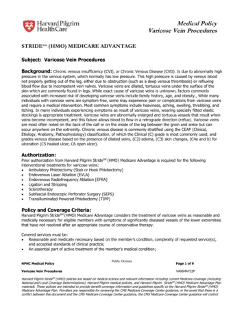
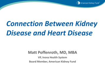
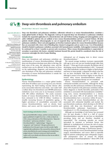
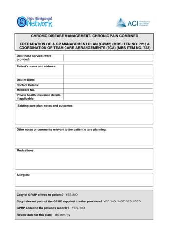
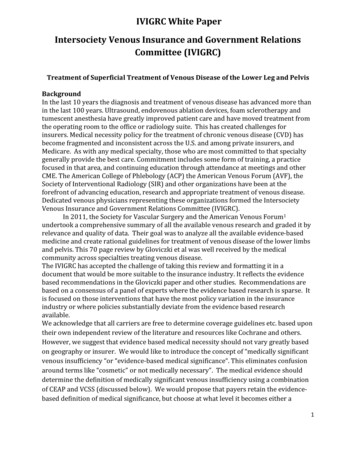
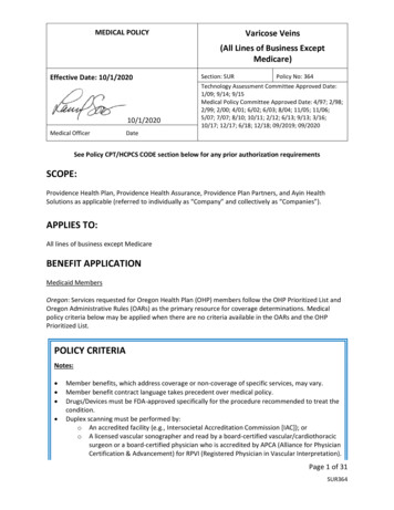
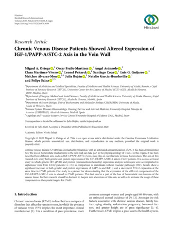
BioMed Research International2.3. Genetic Expression Study. The expression of the genes ofinterest was studied by real-time PCR method (RT-qPCR), inwhich the amount of cDNA was quantified in each of them.The RNA extraction was carried out by the guanidinephenol-chloroform isothiocyanate method, following theprocedure described by Ortega et al. [20]. Used primers weredesigned by the Primer-BLAST tool [21] and the AutoDimerapplication [22]. The StepOnePlus system was used withthe relative standard curve method to carry out the quantitative PCR (qPCR). 5 μL from each sample, previously dilutedin nucleases free water, was mixed with 10 μL of the intercalant agent IQ SYBR Green Supermix (Bio-Rad Laboratories), 1 μL of forward primer, 1 μL of reverse primer, and3 μL of free DNases and RNases water in a MicroAmp 96well plate (Applied Biosystems-Life Technologies), obtaininga final volume of 20 μL. Final results were normalized andcompared to the GAPDH constitutive gene expression(Table 1). Data obtained from each gene were interpolatedin a standard curve. In the 96-well plate, samples were triplicated and duplicated in a standard curve, and the tworemaining wells were filled with negative controls.2.4. Immunohistochemistry Studies. The antigen-antibodyreaction was detected by the ABC method (avidin-biotincomplex) with peroxidase or alkaline phosphatase as chromogen, in accordance with the protocol described byOrtega et al. [23]. The blocking of nonspecific boundingsites was carried out with 3% bovine serum albumin(BSA) and PBS overnight at 4 C (Table 2(a)). The incubation with the secondary antibody bounded to biotin wasdiluted in PBS for one and a half hour at room temperature(Table 2(b)).An incubation with the ExtrAvidin -Peroxidase avidinperoxidase conjugate (Sigma-Aldrich, St. Louis, MO, USA)was carried out for 60 minutes at room temperature (dilutedto 1 : 1200 on PBS). The incubation was revealed with thediaminobenzidine chromogenic substrate (DAB Kit, SK4100) (Vector Laboratories, Burlingame, CA, USA). Thechromogenic substrate was prepared immediately beforeexposition (5 mL of distilled water, 2 drops of tampon, 4drops of DAB, and 2 drops of hydrogen peroxide). This technique allows a brown colored stain. In order to obtain contrast with the nuclei stain, incubation in Carazzi’shaematoxylin was carried out for 5-15 minutes mount inaqueous medium with Plasdone. In every genetic study, sections from the same tissue were used as negative control, inwhich the incubation with the primary antibody wassubstituted with incubation with blocking solution.2.5. Statistical Analysis in Interpretation of Results. For thepurpose of the statistical analysis, the GraphPad Prism 5.1 statistical program was used, and the Mann–WhitneyU and χ2 Pearson’s test were applied. The data areexpressed as the median with interquartile range (IQR).Error bars on the figures are expressed with IQR. The significance values were set at p 0:05, p 0:005 , and p 0:001.The preparations were examined under a Zeiss Axiophotoptical microscope (Carl Zeiss, Oberkochen, Germany).3Given the important role of the proteins involved, the assessment of the histological results was performed by the intensity of expression for immunohistochemical staining with ascore of 1 to 3. Henceforth, histological samples frompatients were classified as negative (0) or positive (1-3) IRSscore method [24]. For each established group of subjects, 7randomly selected microscopy fields were examined in eachof the 5 sections. Subjects were classified as positive whenthe average proportion of the labelled sample was greater orequal to 5% of the total sample. This was done by calculatingthe total percentage of tissue marked in each microscopy fieldto obtain the value of mean for the study sample as describedby Cristóbal et al. [25]. The observation and quantification ofthe samples were independently performed by twoinvestigators.3. Results3.1. Increased Expression of IGF-1 in the Vein Wall of CVeDPatients. Genetic expression study of IGF-1 showed no significant differences between the study groups, although itdid show an upward trend in CVeD patients (CVeD 2:250½0:630 4:013 vs. HV 2:014 ½1:036 3:068 , p 0:5559)(Figure 1(a)).In contrast, a significant increase in the IGF-1 protein expression was observed in the vein wall from CVeD patients(CVeD 2:000 ½0:500 3:000 vs. HV 1:000 ½0:000 2:250 , p 0:0104). The microscopic analysis showed how theexpression of IGF-1 was located in the three tunics of the veinwall (Figure 1(b)). An increased intensity was observed in thesmooth muscular fibres and extracellular matrix of the veinwall from CVeD patients (Figure 1(c)).3.2. CVeD Patients Show a Significant Increase of PAPP-A.Genetic expression study of PAPP-A in the vein wall showeda significant increase in CVeD patients in comparison toHV (CVeD 10:230 ½7:846 11:726 vs:HV 9:089 ½7:324 10:013 , p 0:0029) (Figure 2(a)).Protein expression of PAPP-A was significantly increasedin the vein wall from CVeD patients (CVeD 1:500 ½0:250 3:000 vs:HV 0:500 ½0:000 1:500 , p 0:0002). PAPPA was located in the three tunics of the vein wall from CVeDpatients (Figure 2(b)). It is worth noting how the expressionof PAPP-A was intensely located in the smooth muscular fibresof the vein wall from these patients (Figure 2(c)).3.3. Significant Decrease in the Expression of STC-2 amongstCVeD Patients. A significant decrease in the genetic expressionof STC-2 was observed amongst CVeD patients in comparisonto HV by RT-qPCR (CVeD 7:190 ½3:296 11:324 vs:HV 10:845 ½9:079 13:389 , p 0:0001) (Figure 3(a)).Protein expression study performed by immunohistochemistry techniques showed in a similar fashion asignificant decrease (CVeD 0:250 ½0:000 1:000 vs:HV 1:000 ½0:000 2:500 , p 0:0056). The microscopic analysis showed SCT-2 was mainly located in the medium tunic ofthe vein wall from HV patients (Figure 3(c), asterisk).
4BioMed Research InternationalTable 1: Primer sequences used in RT-qPCR and temperature (Tm).GeneGAPDHIGF-1STC-2PAPP-ASec. Fwd (5 ′ 3 ′ )Sec. Rev (5 ′ 3 ′ CTGCCCCTCAGCTTGA60695152Table 2: Primary (A) and secondary (B) antibodies used in the immunohistochemical studies performed, showing the dilutions used and theprotocol rotocol specificationsIGF-1PAPP-ASTC-2RabbitMouseRabbit1 : 1001 : 5001 : 300Abcam (ab263903)Abcam (ab52030)Abcam (ab261915)10 mM sodium citrate, pH 6 before incubation with blocking solution0.1% Triton in PBS, 10 minutes before incubation with blocking ol specificationsIgG (mouse)IgG (rabbit)GoatMouse1 : 3001 : 1000Sigma (F2012/045k6072)Sigma (RG-96/B5283)——4. DiscussionCVeD causes a severe alteration in the homeostasis of thevein wall, unchaining the activation of both cellular and systemic responses [11]. It has been found that this process isassociated with different pathologic mechanisms which arekey in the development of the disease such as an increase inthe process of apoptosis and cellular death [26], deregulationin the synthesis of some cytokines [27], and even with somecell signaling pathways and changes in the extracellularmatrix [28].The impact that IGF-1 signaling can have on the agingprocess has long been known. Various animal models ranging from yeasts to human beings have demonstrated thatlower levels of IGF-1 are associated with an increased longevity and a deceleration of the aging process [29]. However, it isknown that the effects and levels of IGF-1 both at a physiologic level and a pathologic level depend on tissue, gender,and age of the subject [30], and it has also been demonstratedhow the alteration of its signaling is associated with theappearance of several aging-associated diseases [31].Previous studies have shown the existence of an increasein IGF-1 levels and its receptor, IGF-1R in varicose veins, incomparison to control veins, thus demarcating the importantrole it can play in the pathogenesis of the disease [32]. Moreover, the role of IGF-1 regarding the activation of thePI3K/Akt/mTOR pathway has been demonstrated and howthese components were jointly involved in aging process, aswell as its associated diseases [33]. Jia et al. [34] reportedhow IGF-1-mediated signaling promoted the phosphorylation of PI3K and Akt in smooth muscular cells of saphenousveins, promoting their proliferation in vitro. Along the samelines, our previous results showed that the PI3K/Akt/mTORpathway was altered in CVeD patients, showing a direct association between greater involvement of this pathway andboth premature and asynchronous aging of the venous tissue[28]. There are also more studies which show how IGF-1 promotes the activation of different signaling pathways, such asMAPK [35]. It has been demonstrated the existence of agreater activation of some components in this pathway, suchas ERK1/2 in CVeD patients, especially in those who presentvenous reflux [36]. Our results show how the increase of IGF1 could be associated with these statements, collaboratingwith the detriment suffered by venous tissue.Amongst the different regulatory mechanisms of IGF-1,pregnancy-associated plasma protein A (PAPP-A) is one ofthe most studied elements vitally important in the regulationof this factor [37]. Although PAPP-A was initially found andidentified in trophoblasts of the placenta from pregnantwomen, it has been demonstrated how it is also expressedin a vast range of cells and tissues, including fibroblasts andsmooth muscular cells, both of great relevance in the pathogenesis of the varicose vein [7, 38].PAPP-A is a protein which undergoes very strict regulation in the cell. On the one hand, it is known that itsexpression can be modulated by the synthesis of some proinflammatory cytokines, especially by IL-1β and TNF-α [37].The role of inflammation in CVeDs has been extensivelystudied. It is known that the inflammatory process accompanies the development of the diseases as a consequence ofthe tissue damage associated with the situation of venoushypertension and valvular incompetence, observing anincrease in leukocyte infiltration, as well as in the synthesisof proinflammatory cytokines [39]. It has been demonstratedthat in atheromatous coronary arteries, this macrophagemediated proinflammatory environment induces the
BioMed Research International5(b)CVeD(d)I(a)Relative quantity mRNA of e 1: (a) Expression of mRNA for IGF-1 in CVeD (chronic venous disease) and HV (healthy controls) in arbitrary units. (b–d) Imagesshowing immunostaining for IGF-1 in CVD patients in the three venous wall robes (asterisk) and CV. I: intimate robe; M: middle robe; A:adventitious robe.(b)CVeDHVI(a)Relative quantity mRNA of A320Figure 2: (a) Expression of mRNA for PAPP-A in CVeD (chronic venous disease) and HV (healthy controls) in arbitrary units. (b–d) Imagesshowing immunostaining for PAPP-A in CVD patients in the three tunics of the venous wall (asterisk) and CV. I: intimate robe; M: middlerobe; A: adventitious robe. p 0:005.expression and secretion of PAPP-A in both endothelial andsmooth muscular cells [40]. In addition, as previously mentioned, PAPP-A increases the bioavailability of IGF-1 andthe signaling mediated by its receptor in an autocrine andparacrine manner in the tissue [41], having been describedthe role of IGF-1 in the stimulation of the inflammatoryresponse and the synthesis of cytokines [42].Our results suggest that in the vein wall from CVeDpatients, the inflammation associated with this conditioncould be acting similarly by increasing the expression ofPAPP-A, in a positive feedback process. This fact is involvedin promoting other main pathogenic processes, such as endothelial dysfunction [43].On the other hand, there are different mechanisms whichnegatively regulate PAPP-A levels in the cell, those that arecarried out by a series of proteins depicted as stanniocalcins1 and 2 [44]. Interestingly, it is known that Stc-2, contrarilyto Stc-1, covalently bounds to PAPP-A, performing bothessential functions in the regulation of calcium levels and cellular response to different conditions as oxidative stress orendoplasmic reticulum stress, hence being fundamental incellular homeostasis [45, 46]. We previously demonstratedthe existence of an increase in oxidative stress damagemarkers and in the lipidic peroxidation process in CVeDpatients [11]. Our results show how decreasing Stc-2 levelscould support this fact, in addition to implying the loss of
BioMed Research InternationalRelative quantity mRNA of I⁎MA320(c)Figure 3: (a) Expression of mRNA for STC-2 in CVeD (chronic venous disease) and HV (healthy controls) in arbitrary units. (b, c) Imagesshowing the immunostaining for STC-2 in CVD patients in the three tunics of the venous wall (asterisk) and CV. I: intimate robe; M: middlerobe; A: adventitious robe. p 0:001.an inhibition mechanism of the proteolytic activity of PAPPA and, therefore, of the signaling regulation mediated byIGF-1 in the venous tissue that appears in CVeD patients.The analysis of the serum levels of these components andtheir possible use as biomarkers is also currently in progress.It is important to note that circulating PAPP-A can be foundin 2 different forms: a first form associated with pregnancy,covalently bound to the proteolytically inactive eosinophilmajor basic protein (proMPB), and a second form producedby vascular cells that are not covalently bound to proMBPand that do exhibit proteolytic activity, which this distinctionis being important for its measurement and interpretationunder different conditions [47]. The presence of Stc-2 inserum has also been identified, as well as IGFBP-4, which isregulated by PAPP-A [48]. Although PAPP-A also modulates other IGFBPs, such as IGFBP 2 and 5, IGFBP-4 exclusively appears to be regulated by the proteolytic activityof PAPP-A. Taking into account all of these reasons, theStc-2/PAPP-A/IGF-1 axis is necessary to know all thephysiopathologic mechanisms involved in vascular diseases[49–54].5. ConclusionsOur study is pioneering in showing how CVeD patientshave a significant increase of PAPP-A expression in theirvein wall. These markers may be quite relevant as CVeDmarkers. Further studies should be oriented to the measurement and assessment of different components of thisaxis which may serve as serum biomarkers in venous disease patients. However, our study is the first to highlightthe presence and importance of IGF-1/PAPP-A/STC-2markers in CVeD.
BioMed Research InternationalData AvailabilityThe data used to support the findings of the present study areavailable from the corresponding author upon request.Conflicts of InterestThe authors declare that they have no conflicts of interest.Authors’ ContributionsMiguel A Ortega and Oscar Fraile-Martínez contributedequally in this work. Julia Buján, Natalio García-Honduvilla,and Felipe Sainz shared senior authorship in this work.AcknowledgmentsThis work was supported by grants from the National Institute of Health Carlos III (FIS-PI18/00912 and B2017/BMD3804 MITIC-CM).References[1] R. T. Eberhardt and J. D. Raffetto, “Chronic venous insufficiency,” Circulation, vol. 130, no. 4, pp. 333–346, 2014.[2] E. Rabe, G. Berboth, and F. Pannier, “Epidemiology of chronicvenous diseases,” Wiener Medizinische Wochenschrift,vol. 166, no. 9-10, pp. 260–263, 2016.[3] M. McArdle and E. A. Hernandez-Vila, “Management ofchronic venous disease,” Texas Heart Institute Journal,vol. 44, no. 5, pp. 347–349, 2017.[4] M. A. Ortega, M. A. Saez, O. Fraile-Martínez et al., “Increasedangiogenesis and lymphangiogenesis in the placental villi ofwomen with chronic venous disease during pregnancy,” International Journal of Molecular Sciences, vol. 21, no. 7, p. 2487,2020.[5] J. V. White and C. Ryjewski, “Chronic venous insufficiency,”Perspectives in Vascular Surgery and Endovascular Therapy,vol. 17, no. 4, pp. 319–327, 2005.[6] Y. J. Youn and J. Lee, “Chronic venous insufficiency and varicose veins of the lower extremities,” The Korean Journal ofInternal Medicine, vol. 34, no. 2, pp. 269–283, 2019.[7] Y. Chen, W. Peng, J. D. Raffetto, and R. A. Khalil, “Matrixmetalloproteinases in remodeling of lower extremity veinsand chronic venous disease,” Progress in Molecular Biologyand Translational Science, vol. 147, pp. 267–299, 2017.[8] M. A. Ortega, B. Romero, Á. Asúnsolo et al., “Behavior ofsmooth muscle cells under hypoxic conditions: possible implications on the varicose vein endothelium,” BioMed ResearchInternational, vol. 2018, Article ID 7156150, 9 pages, 2018.[9] J. D. Raffetto and F. Mannello, “Pathophysiology of chronicvenous disease,” Int Angiol, vol. 33, no. 3, pp. 212–221, 2014.[10] D. Saribal, E. M. Kanber, F. S. Hocaoglu-Emre, and M. C.Akyolcu, “Effects of the oxidative stress and genetic changesin varicose vein patients,” Phlebology, vol. 34, no. 6, pp. 406–413, 2019.[11] M. A. Ortega, B. Romero, Á. Asúnsolo et al., “Patients withincompetent valves in chronic venous insufficiency showincreased systematic lipid peroxidation and cellular oxidativestress markers,” Oxidative Medicine and Cellular Longevity,vol. 2019, Article ID 5164576, 9 pages, 2019.7[12] R. Hjortebjerg, “IGFBP-4 and PAPP-A in normal physiologyand disease,” Growth Hormone & IGF Research, vol. 41,pp. 7–22, 2018.[13] Martinelli CE Jr, R. J. Custódio, and M. H. Aguiar-Oliveira,“Physiology of the GH-IGF axis,” Arquivos Brasileiros deEndocrinologia e Metabologia, vol. 52, no. 5, pp. 717–725,2008.[14] T. Sasako and K. Ueki, “Insulin/IGF-1 signaling and aging,”Nihon Rinsho, vol. 74, no. 9, pp. 1435–1440, 2016.[15] P. Monget, “La PAPP-A et le systeme des IGFs,” Annales d'endocrinologie, vol. 77, no. 2, pp. 90–96, 2016.[16] X. H. Yu, L. H. He, J. H. Gao, D. W. Zhang, and X. L. Zheng,“Pregnancy-associated plasma protein-A in atherosclerosis:molecular marker, mechanistic insight, and therapeutic target,” Atherosclerosis, vol. 278, pp. 250–258, 2018.[17] M. Lodh, B. Goswami, A. Parida, S. Patra, and A. Saxena,“Assessment of serum leptin, pregnancy-associated plasmaprotein A and CRP levels as indicators of plaque vulnerabilityin patients with acute coronary syndrome,” CardiovascularJournal of Africa, vol. 23, no. 6, pp. 330–335, 2012.[18] H. Duan, G. Zhao, B. Xu, S. Hu, and J. Li, “Maternal serumPLGF, PAPPA, β-hCG and AFP levels in early second trimester as predictors of preeclampsia,” Clinical Lababoratory,vol. 63, no. 05, pp. 921–925, 2017.[19] H. Gutiérrez-Leonard, E. Martínez-Lara, A. E. Fierro-Macías,V. M. Mena-Burciaga, and M. D. Ronquillo-Sánchez, “Pregnancy-associated plasma protein-A (PAPP-A) as a possiblebiomarker in patients with coronary artery disease,” Ir J MedSci, vol. 186, no. 3, pp. 597–605, 2017.[20] M. A. Ortega, B. Romero, Á. Asúnsolo et al., “Pregnancy-associated venous insufficiency course with placental and systemicoxidative stress,” Journal of Cellular and Molecular Medicine,vol. 24, no. 7, pp. 4157–4170, 2020.[21] M. A. Ortega, Á. Asúnsolo, M. J. Álvarez-Rocha et al.,“Remodelling of collagen fibres in the placentas of women withvenous insufficiency during pregnancy,” Histol Histopathol,vol. 33, no. 6, pp. 567–576, 2018.[22] P. M. Vallone and J. M. Butler, “AutoDimer: a screening toolfor primer-dimer and hairpin structures,” BioTechniques,vol. 37, no. 2, pp. 226–231, 2004.[23] M. A. Ortega, M. Á. Saez, Á. Asúnsolo et al., “Upregulation ofVEGF and PEDF in placentas of women with lower extremityvenous insufficiency during pregnancy and its implication invillous calcification,” BioMed Research International,vol. 2019, Article ID 5320902, 8 pages, 2019.[24] P. Sanmartín-Salinas, M. V. Toledo-Lobo, F. Noguerales-Fraguas, M. E. Fernández-Contreras, and L. G. Guijarro, “Overexpression of insulin receptor substrate
Research Article Chronic Venous Disease Patients Showed Altered Expression of IGF-1/PAPP-A/STC-2 Axis in the Vein Wall Miguel A. Ortega ,1 Oscar Fraile-Martínez ,1 Ángel Asúnsolo ,2 Clara Martínez-Vivero ,1 Leonel Pekarek ,1 Santiago Coca ,1 Luis G. Guijarro ,3 Melchor Álvarez-Mon ,1,4 Julia Buján ,1 Natalio García-Honduvilla ,1 and Felipe Sainz 2,5 1Department of Medicine and Medical .