Transcription
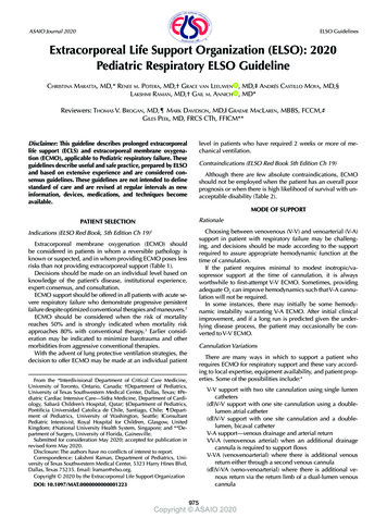
ASAIO Journal 2020 ELSO GuidelinesExtracorporeal Life Support Organization (ELSO): 2020Pediatric Respiratory ELSO GuidelineCHRISTINA MARATTA, MD,* RENEE M. POTERA, MD,† GRACE VAN LEEUWEN , MD,‡ ANDRÉS CASTILLO MOYA, MD,§LAKSHMI RAMAN, MD,† GAIL M. ANNICH , MD*Reviewers: THOMAS V. BROGAN, MD,¶ MARK DAVIDSON, MD,‖ GRAEME MACLAREN, MBBS, FCCM,#GILES PEEK, MD, FRCS CTh, FFICM**level in patients who have required 2 weeks or more of mechanical ventilation.Disclaimer: This guideline describes prolonged extracorporeallife support (ECLS) and extracorporeal membrane oxygenation (ECMO), applicable to Pediatric respiratory failure. Theseguidelines describe useful and safe practice, prepared by ELSOand based on extensive experience and are considered consensus guidelines. These guidelines are not intended to definestandard of care and are revised at regular intervals as newinformation, devices, medications, and techniques becomeavailable.Contraindications (ELSO Red Book 5th Edition Ch 19)Although there are few absolute contraindications, ECMOshould not be employed when the patient has an overall poorprognosis or when there is high likelihood of survival with unacceptable disability (Table 2).MODE OF SUPPORTRationalePATIENT SELECTIONChoosing between venovenous (V-V) and venoarterial (V-A)support in patient with respiratory failure may be challenging, and decisions should be made according to the supportrequired to assure appropriate hemodynamic function at thetime of cannulation.If the patient requires minimal to modest inotropic/vasopressor support at the time of cannulation, it is alwaysworthwhile to first-attempt V-V ECMO. Sometimes, providingadequate O2 can improve hemodynamics such that V-A cannulation will not be required.In some instances, there may initially be some hemodynamic instability warranting V-A ECMO. After initial clinicalimprovement, and if a long run is predicted given the underlying disease process, the patient may occasionally be converted to V-V ECMO.Indications (ELSO Red Book, 5th Edition Ch 19)1Extracorporeal membrane oxygenation (ECMO) shouldbe considered in patients in whom a reversible pathology isknown or suspected, and in whom providing ECMO poses lessrisks than not providing extracorporeal support (Table 1).Decisions should be made on an individual level based onknowledge of the patient’s disease, institutional experience,expert consensus, and consultation.ECMO support should be offered in all patients with acute severe respiratory failure who demonstrate progressive persistentfailure despite optimized conventional therapies and maneuvers.2ECMO should be considered when the risk of mortalityreaches 50% and is strongly indicated when mortality riskapproaches 80% with conventional therapy.3 Earlier consideration may be indicated to minimize barotrauma and othermorbidities from aggressive conventional therapies.With the advent of lung protective ventilation strategies, thedecision to offer ECMO may be made at an individual patientCannulation VariationsThere are many ways in which to support a patient whorequires ECMO for respiratory support and these vary according to local expertise, equipment availability, and patient properties. Some of the possibilities include:4From the *Interdivisional Department of Critical Care Medicine,University of Toronto, Ontario, Canada; †Department of Pediatrics,University of Texas Southwestern Medical Center, Dallas, Texas; ‡Pediatric Cardiac Intensive Care—Sidra Medicine, Department of Cardiology, Sabará Children’s Hospital, Qatar; §Department of Pediatrics,Pontificia Universidad Catolica de Chile, Santiago, Chile; ¶Department of Pediatrics, University of Washington, Seattle; ‖ConsultantPediatric Intensivist, Royal Hospital for Children, Glasgow, UnitedKingdom; #National University Health System, Singapore; and **Department of Surgery, University of Florida, Gainesville.Submitted for consideration May 2020; accepted for publication inrevised form May 2020.Disclosure: The authors have no conflicts of interest to report.Correspondence: Lakshmi Raman, Department of Pediatrics, University of Texas Southwestern Medical Center, 5323 Harry Hines Blvd,Dallas, Texas 75235. Email: lraman@elso.org.Copyright 2020 by the Extracorporeal Life Support OrganizationV-V support with two site cannulation using single lumencatheters(dl)V-V support with one site cannulation using a doublelumen atrial catheter(dl)V-V support with one site cannulation and a doublelumen, bicaval catheterV-A support—venous drainage and arterial returnVV-A (venovenous arterial) when an additional drainagecannula is required to support flowsV-VA (venovenoarterial) where there is additional venousreturn either through a second venous cannula(dl)V-VA (veno-venoarterial) where there is additional venous return via the return limb of a dual-lumen venouscannulaDOI: 10.1097/MAT.0000000000001223975Copyright ASAIO 2020
9762020 PEDIATRIC RESPIRATORY ELSO GUIDELINEIn ECMO centers that cannot access double-lumen cannulae, single-cannula two-vessel (femoral drainage and internaljugular return) V-V ECMO is appropriate. In small children,this may be more difficult as small femoral vessels may limitcannula size that prove inadequate to provide full ECMO support. On occasion, drainage from the jugular vein and returnthrough the smaller femoral vein may provide better support.V-V support may be challenging in small children because ofbody habitus variability resulting in challenges with bicaval catheter placement as one size does not fit all. Additionally, availability of dual lumen catheters is not universal to each ECMOcenter. As such, in patients where the femoral vessels cannothouse cannulae large enough to fully support complete respiratory nor cardiorespiratory failure, V-A cannulation through theneck may be the most appropriate option. However, many centers have reported success in supporting these children with (dl)V-V and two cannulae ECMO techniques, therefore, the typeand site of cannulation is a decision made on a case-by-casebasis by the ECMO team and those performing the cannulation.In older pediatric patients who require hemodynamic support, optimal ECMO support can be accomplished by the (dl)V-VA method: i.e., via the femoral artery plus a dual lumenvenous cannula in the internal jugular, or two-vessel venouscannulation of the internal jugular and femoral vein (V-VA support). This is usually considered in cases of severe refractorysepsis where upon return of myocardial function, the arterialcannula can be removed leaving the patient on V-V ECMO forthe rest of the run. In V-VA support, as in femoral V-A ECMO, it is essential toensure adequate oxygen delivery to the brain and heart.This should be undertaken by monitoring pulse oximetryplaced on the right hand (or ear), if possible, near infrared spectroscopy (NIRS) monitor on the forehead, and viaarterial blood gas analysis from a line in the right radialartery. The amount of flow from the femoral arterial cannula retrograde up the aorta in a VV-A approach will depend onnative cardiac function. Upon the return of native cardiacfunction, competition will begin to be observed and thepatient will be desaturated above the abdomen (Harlequin syndrome). To minimize differential cyanosis, theapproach can be to change the proximal venous drain,which is in the jugular vein to an arterial return cannulamaking it V-VA. The additional return cannula in the superior vena cava (SVC) will ensure that oxygenated bloodperfuses the upper body, notably the cerebral and coronary circulation. Differential cyanosis should be monitored for, with a right radial arterial line and NIRS whenpossible.Cannulation (ELSO Red Book 5th Edition Ch 20)Cannulation may be achieved by direct cut down, percutaneous cannulation or by Semi-Seldinger technique. A bolus ofheparin (25–100 U/kg) should be given before cannula placement, even if the patient is coagulopathic or bleeding.(dl)V-V access may be achieved with a double lumen catheter placed into the right atrium via the right internal jugularvein. Cannulation of the left internal jugular vein should onlybe undertaken with surgical expertise, as it carries a risk of SVCperforation.The tip of a dual-lumen catheter will vary depending onmanufacturer specifics.Fluoroscopy or echocardiography are highly recommendedwhen placing a bicaval dual-lumen cannula in children. This is toconfirm placement of the wire within the inferior vena cava (IVC)and to ensure that it hasn’t migrated into the hepatic vein, as itfrequently does in young patients. When placing a bicaval duallumen catheter, a modified cut down approach might be used asit allows for placement of the needle into the internal jugular veinand to facilitate the placement of the guide wire into the IVC.There are many considerations and challenges with selecting the appropriate mode, cannula size, and cannulationsite in pediatrics. Some additional considerations should begiven to the addition of a cephalad venous cannula for venousdrainage from the head, the addition of lower limb perfusioncannula for adequate distal perfusion if femoral arterial cannulation is used, as well as the potential for reconstruction of theneck vessels after cannulation (Table 3).Recirculation can be limited by using the femoral site fordrainage and the right internal jugular vein site for reinfusion.Circuit and Cannulae (see Separate Guideline)When cannulating a pediatric patient for ECMO, the teamshould attempt to place the largest venous drainage and largestreturn cannula possible to be able to achieve adequate maximum flows.Whenever possible, the team should choose cannulae thatwill provide complete respiratory support and meet metabolicdemands, as monitored by SvO2 and lactate.MANAGEMENT DURING ECMOCircuit Management (see Separate Guideline)Patient managementA. Blood flow (ELSO Red Book 5th Edition Ch 4): DuringV-V ECMO, oxygenated blood from the pump is returned tothe body where it is mixed with deoxygenated blood from systemic venous return. This results in a systemic arterial saturation of 75%–85%. As native lung function improves, the systemic arterial saturation increases.During V-A ECMO, the proportion of infused oxygenatedblood to native aortic blood is significantly higher, leading to anormal systemic arterial saturation, but the oxygenated bloodmay not be evenly distributed especially as native cardiacoutput returns leading to differential hypoxia (Table 4).B. Gas exchange (ELSO Red Book 5th Edition Ch 4): Atthe initiation of ECMO, whether it be V-V or V-A, hypercarbiashould be corrected slowly to avoid rapid changes in cerebralperfusion (e.g., over 4–8 hours).Carbon dioxide clearance is dependent on sweep gas flow.Oxygenation is primarily determined by circuit blood flowrate, total cardiac output, and hemoglobin levels (Table 5).C. Hemodynamics (ELSO Red Book 5th Edition Chapters 4,13, and 22):a. Venovenous ECMO V-V ECMO does not provide the patient with direct hemodynamic support, it will however provide improved conditions for ventricular function by providingCopyright ASAIO 2020
2020 PEDIATRIC RESPIRATORY ELSO GUIDELINEwell-oxygenated coronary artery blood flow. Therefore, vasoactive medications and infusions may be required to optimize cardiac output, blood pressure, and systemic vascularresistance.SvO2 may not be a reliable indicator of oxygen delivery dueto recirculation in V-V ECMO.Recirculation occurs when oxygenated blood returns directly to the ECMO circuit and not to the patient. This results inproportionally less oxygenated blood circulating to the patient.It is increased by higher circuit flow and lower cardiac output.Type and placement of cannulae affect recirculation as well.Recirculation may be higher in atrial double lumen or two-sitesingle cannulation techniques.Hypertension occurs frequently with V-V ECMO and maybe seen due to the diversion of venous blood into the ECMOcircuit resulting in low cardiac filling pressures.In the event of a cardiac arrest during a V-V ECMO run,compressions should be initiated when possible, as per Pediatric Advanced Life Support (PALS) algorithms. Vasoactive support should be initiated early. The V-V circuit should continuerunning during the resuscitation to support gas exchange inlungs that will most likely not be adequately supported by conventional mechanical ventilation or bagging. Cardiopulmonary resuscitation, although necessary, puts the patient at riskfor cannula displacement, vessel rupture, and air entrainmentinto the circuit. The priorities during a cardiac arrest while onV-V ECMO are: (1) appropriate resuscitation of the patient and(2) initiation of V-A ECMO support as soon as possible.b. Venoarterial ECMO V-A ECMO provides hemodynamicsupport, and the amount of support depends on flow providedby the size of the cannulae native cardiac output and systemicvascular resistance.Although the pulse pressure and mean arterial pressure maybe lower than usual, systemic perfusion is usually adequate.Inotropes and vasopressors should be weaned in V-A ECMO ifpossible as the perfusion improves with ECMO support.End-organ oxygen delivery is best assessed by mixed venous blood saturation, lactate, urine output, NIRS, and peripheral perfusion in V-A ECMO. Increasing pump flows may helpachieve better perfusion and oxygen delivery in V-A ECMO,however, this may be at the expense of increased hemolysis.If systemic oxygen delivery is inadequate, the pump flowshould be increased based on the size of the cannula until perfusion is adequate. Extra blood or crystalloid to optimize cardiac preload and/or oxygen carrying capacity may be requiredto achieve end organ oxygen delivery.In V-A ECMO, the loss of pulsatile flow may incur an increase in renin and angiotensin. Hypertension may also reflectfluid overload, pain, or agitation. Systemic vasodilators, diuretics, or sedation may all be used to treat hypertension depending on the etiology.D. Ventilator management (ELSO Red Book 5th EditionChapters 21 and 22): Regardless whether V-V or V-A ECMOis employed for primary respiratory failure, the goal should belung rest via reduction of barotrauma/volutrauma and minimization of oxygen toxicity.Typical rest settings should include low-normal peak inspiratory pressure ( 25) and fraction of inspired oxygen 50%.Positive end-expiratory pressure should be between 5 and15 cm H2O and titrated to lung recruitment and distention of977large airways to promote secretion clearance, whereas the patient remains unable to self-recruit. Low or no respiratory ratesmay be needed.The use of lung recruitment maneuvers is controversial.5Spontaneous breathing should be allowed, as this enhancesrecovery and lung rehabilitation.5 However, strong spontaneous breathing can produce high transpulmonary pressuresand result in patient self-inflicted lung injury (P-SILI).6 Adjusting sweep as flow to improve ventilation may help.Sweep gas should be adjusted to maintain the blood PaCO2at 40–45 mm Hg, or permissively higher in patients withchronic lung disease.7If a pneumothorax develops, the ventilator settings can befurther reduced at the risk of incurring atelectasis. Consideration should be given to manage the patient with CPAP only orno positive pressure. Due to the risk of bleeding, the decisionto place a chest tube for a pneumothorax should be consideredcarefully by senior clinicians, with hemodynamic compromisebeing the only absolute indication for placement.iNO may be considered in patients with severe right heartfailure and pulmonary hypertension, to promote forward flowand ventilation/perfusion matching, although it is not typicallyneeded during V-V ECMO runs. Positive end-expiratory pressure on ventilator rest settings should be optimized to minimize right heart strain and prostaglandins may be consideredto maintain ductal patency in neonates.Extubation while on ECMO is an option to consider as itallows for decreased sedation, improved neurologic assessments, and increased rehabilitation. However, this must becarefully considered in young children in whom it may bedifficult to adequately clear secretions and in whom wakefulness must be weighed against the risk of cannula kinking anddislodgement.E. Fluid management (ELSO Red Book 5th Edition Ch 22):The ultimate goal of management is to target dry weight.Once hemodynamic stability is achieved, diuretics may beinstituted, with the addition of hemofiltration if needed. If thepatient has significant acute kidney injury, continuous hemofiltration may be added to the extracorporeal circuit to maintainfluid and electrolyte balance.Early renal support with hemofiltration may aid in providingoptimal nutritional and metabolic support, whilst ensuringfluid overload is avoided or reversed.F. Sedation (ELSO Red Book 5th Edition Ch 22): The patientshould be adequately sedated during cannulation and for thefirst 12 to 24 hours to avoid air embolism, minimize the metabolic rate, ensure cannulation sites are secure, and to reducerisk of cannula site bleeding.It is rarely necessary to paralyze the patient except duringcannula placement, or to achieve adequate venous drainagewhile other measures are enacted to enhance this.Because sedation during V-V ECMO is often needed to tolerate endotracheal intubation, conversion to tracheostomymay be considered to minimize sedation and immobilizationespecially if a prolonged ECMO run is anticipated.Sedation should be titrated to the patient’s level of anxiety,discomfort and eventually, it should be titrated such that thepatient is able to perform self-recruitment and interact synchronously with the ventilator.Copyright ASAIO 2020
9782020 PEDIATRIC RESPIRATORY ELSO GUIDELINETable 1. Common Conditions Requiring ECMO (ELSO RedBook, 5th Edition, Chapters 18 and 19)Conditions for Respiratory ECLS: Acute respiratory distress syndrome Viral or bacterial pneumonia Aspiration pneumonia Status asthmaticus Mediastinal masses Pulmonary hemorrhage Severe air leak Bridge to transplant Perioperative support to airway surgery Temporary lung nonfunction (e.g., extensive bronchoalveolar lavage)ECLS, extracorporeal life support; ECMO, extracorporeal membrane oxygenation.Sedation and analgesia should be held long enough to safelyperform a neurologic exam daily, if possible.Minimizing sedation will increase mobilization, promotespontaneous ventilation for pulmonary recruitment and lungrecovery, and decrease withdrawal.8,9G. Nutrition (ELSO Red Book 5th Edition Ch 22): Enteralfeeding should be promoted to reduce the need for total parenteral nutrition.Daily caloric goals should approximate those for critical illness.Even if the gut is nonfunctional, the team should promotebowel health with an effective laxative regimen. A significantcomplication that is often the harbinger of impending clinicaldecline is gastrointestinal bleeding and feeding intolerance.H. Infection (ELSO Red Book 5th Edition Ch 22): Routinecircuit cultures and prophylactic antibiotics are not indicated,beyond treating an underlying infection.The patient will be unable to mount a fever during ECMOdue to circuit temperature control, therefore a high index ofsuspicion must be maintained.I. Neurology (ELSO Red Book 5th Edition Chapters 14and 22 and Neuromonitoring Guidelines): Routine headTable 2. Relative Contraindications, Conditions With PoorPrognosis (ELSO Red Book, 5th Edition, Chapter 19)Conditions rendering patient unlikely to benefit from ECLS: Large intracranial bleed with mass effect or need forneurosurgical intervention Hypoxic cardiac arrest without adequate CPR Irreversible underlying cardiac or lung pathology (and not atransplant candidate) Pulmonary hypertension and chronic lung disease Chronic multiorgan dysfunction Incurable malignancy Allogenic bone marrow recipients with pulmonary infiltratesConditions with worse prognosis in respiratory ECLS: Hepatic or renal failure Pertussis infection in infants Fungal pneumonia ImmunodeficiencyRelative contraindications: Vessel anomalies or having previously been clipped or ligatedfor prior ECMO Localized site infectionCPR, cardiopulmonary resuscitation; ECLS, extracorporeal lifesupport; ECMO, extracorporeal membrane oxygenation.Table 3. Cannulation ComplicationsCannulation complications: Cannula malposition Bleeding Vessel damage Adjacent organ injury Air embolism Right atrial injury Pericardial effusionTable 4. Blood Flow RateOxygen DeliveryNeonatesChildren6 ml/kg/min4-5 ml/kg/minECMO Blood Flow Rate100–150 ml/kg/min80–120 ml/kg/minECMO, extracorporeal membrane oxygenation.ultrasounds should be performed in neonates at ECMO initiation, and then daily or second-daily thereafter.Daily neurologic assessments should be performed inpatients while on ECMO. If there are any concerns for bleeding or infarct, computed tomography is indicated.NIRS and EEG may also be used to monitor neurologic function, particularly in sedated patients.J. Anticoagulation (ELSO Red Book 5th Edition Ch 7 andAnticoagulation Guidelines): Most children require a generalalgorithm with a customized approach for titration of heparinor direct thrombin inhibitors. Anticoagulation management,especially in neonates, can be challenging due to developmental differences in hemostasis, low pump flows, and hightransfusion requirements.WEANING OFF ECMOWeaning V-V ECMO (ELSO Red Book 5th Edition Ch 24)Improving compliance and radiographic appearance of thelungs are indications of recovery. Additionally, improvement inoxygenation and ventilation may be observed without changesto the circuit or ventilator.When management is carried out using lowest blood flowto provide adequate support and low ventilator settings,weaning will occur naturally as patient improves. A trial offmay be indicated sooner, in circumstances such as uncontrolled bleeding.Although weaning, it is imperative not to oversedate the patient because of dyspnea. Providers must distinguish dyspneaand tachypnea without distress in an unbothered patientversus dyspnea with distress. The removal or depression ofrespiratory drive may hinder the ability to wean off ECMOTable 5. Ventilation and OxygenationFactors that DetermineVentilationSweep gas flow rateMembrane propertiesand surface areaInlet PCO2Copyright ASAIO 2020Factors that Determine OxygenationMembrane integrity (presence of clots)Blood flow rateMembrane properties and surface areaO2 concentration
9792020 PEDIATRIC RESPIRATORY ELSO GUIDELINEsupport. The provider may have to tolerate higher than normalrange CO2 given residual chronic lung disease while weaning,and the sweep gas may have to be adjusted to allow for higherCO2 before the wean. During decannulation, some patientsmay not tolerate muscle paralysis as it may cause inadvertenthypercarbia and failure to wean off ECMO.Trial off (ELSO Red Book 5th Edition Chapters 16 and 34)When trialing off V-V support, adjust the ventilator to acceptable settings to test gas exchange independent of ECMOsupport. Maintain blood flow and anticoagulation, stop thesweep gas, and cap off the oxygenator.Patient arterial blood gases, oxygen saturation, end-tidalCO2, and clinical exam should be assessed for tolerance.If lung function is adequate at safe ventilator settings for anhour or more, the patient may be ready for decannulation.The patient may remain on the circuit without sweep gasor oxygen for up to 24–48 hours to ensure that the wean istolerated in patients whose ability to maintain native gas exchange is questioned. It should be noted that this practiceshould be limited to few patients with very questionable trials off ECMO.If there is no uncertainty about the need for further ECLS, itis best to remove the cannulae after the trial off has successfully finished.Stopping ECMO for Nonrecovery, Nonbridging toTransplant (ELSO Red Book 5th Edition Ch 70)ECMO should be discontinued when there is no hope forhealthy survival (severe brain damage, no lung recovery, andno hope of organ transplant), as decided by the multidisciplinary team caring for the patient.This possibility of stopping should be explained to the familywhen consent for ECMO is obtained. A reasonable deadlinefor organ recovery or replacement should be set early in thecourse.For lung failure, patients may be supported for a prolongedperiod (days to months) awaiting recovery. The managementof these patients often requires consultation with other ECMOcenters that have had similar experiences.Follow up (ELSO Red Book 5th Edition Chapters19 and 22 and Neuromonitoring guidelines)All ECMO survivors should be followed up regularly. Followup with appropriate specialists is indicated for survivors whodemonstrate chronic lung disease or significant kidney injury.Neurodevelopmental abnormalities have been described inup to 50% of ECMO survivors.Patients should have a thorough neurologic evaluation.Survivors require a follow-up plan that consists of regular patient assessments and should be screened for issues with behavior and school performance. They should be followed by aPediatric Neurologist if they have any evidence of neurologicalabnormalities at discharge.10Decannulation (ELSO Red Book 5th Edition Ch 12)Decannulation should be performed by the surgical teamthat placed the cannulae, who may decide to reconstruct thevessels, while others may opt to ligate.When removing a venous cannula, air can enter the venousblood through the side holes if the patient is breathing spontaneously. This is prevented by administering short-term pharmacological paralysis or by performing a Valsalva maneuveron the ventilator. AcknowledgmentsWe thank Elaine Cooley, MSN, RN, and Peter Rycus, MPH, for thehelp in the overall process.References1. Brogan T, Lequier L, Lorusso R, MacLaren G, Peek G.Extracorporeal Life Support: The ELSO Red Book. 5th ed. AnnArbor, MI, Extracorporeal Life Support Organization, 2017.2. Li X, Scales DC, Kavanagh BP: Unproven and expensive beforeproven and cheap: Extracorporeal membrane oxygenationversus prone position in acute respiratory distress syndrome.Am J Respir Crit Care Med 197: 991–993, 2018.3. Wong JJ, Phan HP, Phumeetham S, et al; Pediatric Acute & CriticalCare Medicine Asian Network (PACCMAN): Risk stratificationin pediatric acute respiratory distress syndrome: A MulticenterObservational Study. Crit Care Med 45: 1820–1828, 2017.4. Conrad SA, Broman LM, Taccone FS, et al: The Extracorporeal LifeSupport Organization Maastricht Treaty for nomenclature in extracorporeal life support. A position paper of the ExtracorporealLife Support Organization. Am J Respir Crit Care Med 198:447–451, 2018.5. Turner DA, Cheifetz IM, Rehder KJ, et al: Active rehabilitation andphysical therapy during extracorporeal membrane oxygenationwhile awaiting lung transplantation: A practical approach. CritCare Med 39: 2593–2598, 2011.6. Yoshida T, Uchiyama A, Matsuura N, Mashimo T, Fujino Y:Spontaneous breathing during lung-protective ventilation in anexperimental acute lung injury model: High transpulmonarypressure associated with strong spontaneous breathing effortmay worsen lung injury. Crit Care Med 40: 1578–1585, 2012.7. Marhong JD, Munshi L, Detsky M, Telesnicki T, Fan E: Mechanicalventilation during extracorporeal life support (ECLS): A systematic review. Intensive Care Med 41: 994–1003, 2015.8. Wells CL, Forrester J, Vogel J, Rector R, Tabatabai A, Herr D: Safetyand feasibility of early physical therapy for patients on extracorporeal membrane oxygenator: University of Maryland MedicalCenter Experience. Crit Care Med 46: 53–59, 2018.9. Marhong JD, DeBacker J, Viau-Lapointe J, et al: Sedation and mobilization during venovenous extracorporeal membrane oxygenation for acute respiratory failure: An international survey.Crit Care Med 45: 1893–1899, 2017.10. IJsselstijn H, Schiller RM, Hoskote AU: Outcomes and followup of patients receiving ECLS, in Annich G, Brogan T, EllisWC, Haney B, Heard ML, Lorusso R (eds), ECMO SpecialistTraining Manual. Ann Arbor, MI, Extracorporeal Life SupportOrganization, 2018, pp. 221–223.Copyright ASAIO 2020
In older pediatric patients who require hemodynamic sup-port, optimal ECMO support can be accomplished by the (dl) V-VA method: i.e.via, the femoral artery plus a dual lumen venous cannula in the internal jugular, or two-vessel venous cannulation
