Transcription
CT VenographyRich Hallett, MDSection Chief, CV Imaging, Northwest Radiology Network, IndianapolisAdjunct Clinical Assistant Professor, Stanford University, CardiovascularImaging Section
Introduction CT venography (CTV) is a technique targeted toassess venous anatomy, determine venouspatency & delineate collateral circulation Non-invasive, simple protocols, wide anatomiccoverage, short acquisition time, and ability to becombined with arterial-phase CTA
Lecture Outline Basic Clinical Options for Venous Imaging– Venous Imaging Modalities CT Scan Protocols– Indirect CTV– Direct CTV Selected Regional Applications– UE– Chest
Venous Imaging Modalities – Thecompetition Doppler Ultrasound (US)MR VenographyCatheter venographyNuclear venography
Doppler US Well established clinical utilityNo ionizing radiationPortableInexpensiveFlow direction informationOperator / Patient dependentSome areas inaccessible (pelvis, SVC)Collateral pathways not well delineated
Doppler US Sens/ Spec 95% for fempop DVT in ideal situations
Performance of Doppler vs. CTV in ICUpatients – LE DVTSensSpecIndiect CTV7096Doppler US70100Taffoni, AJR, 2004
MR Venography - Positives Excellent for pelvic venous system, CNS May not require contrast SI ratio thrombus:blood higher for MRV vs. CTV– 3.7-8:1 vs 1.8-3.2 * For PE: Sens 80-95%, Spec 95%, depends ontechnique (Perf imaging best) For DVT: Sens 92%, Spec 95% 0.25 mmol/kg Gd better than 0.125 mmol/kg* Kluge, AJR, 2006 Sampson, Eur Radiol 2007; 17:175-181
Combo MR-PA / Indirect MRV MRA: TRuFISP, perfusion, MRA(0.25mmol/Kg) MRV: 3D FLASH w/ PV coil, voxel size of1.2x0.8x1.1 mm– High agreement w/ CTA/CTV but requires achange in coil and pt. position to obtain MRVafter chest MRA Good agreement w/ Doppler in legs,moderate in pelvisKluge, AJR, 2006
MR Venography - Negatives Expensive, availability sometimes limitedExam may be lengthyPt. cooperation?Spatial resolution (vs other choices)Limited anatomic coverage
Radionuclide Venography 99mTc-labeled MAA99mTc-labeled RBC99mTc-human serum albumin99mTc-labeled plateletsAnatomic agents,indirect evidence– Direct evidence of acute / active DVT– BUT: Arduous prep, false positives – pts on heparin 99mTc-apcitide (GIIb/IIIa receptor binding)– Can tell acute ( ) vs. chronic (-) clot– Interpreter dependent?
Catheter Venography Considered the gold standardInvasive (but can treat lesions)You only see what you can fillRisks:– Minor Complications: 18%– Thrombosis: 2%– Bronchospasm, Contrast reactions, etc
CTV: Challenges Goal: visualize all venous structures, with goodopacification, but without artifactsDirect CTV good opacification (too good; needs dilution) but difficult to show all venous structures or fullextent of collateral circulationIndirect CTV shows all veins; but difficult to achieve strongenhancement; timing
CTV: Challenges Goal: visualize all venous structures, with goodopacification, but without artifactsDirect CTV good opacification (too good; needs dilution) but difficult to visualize all venous structures orfull extent of collateral circulationIndirect CTV visualizes all veins (recirculation of CM) but difficult to achieve strong enhancement;timing difficulties
60M smoker, r/o lung cancerRoutine chest with contrast: 100cc contrast @ 2cc/sec, 40 sec diagnostic delay
CTV: Challenges Goal: visualize all venous structures, with goodopacification, but without artifactsDirect CTV good opacification (too good; needs dilution) but difficult to visualize all venous structures orfull extent of collateral circulationIndirect CTV visualizes all veins (recirculation of CM) but difficult to achieve strong enhancement;timing difficulties
Indirect CTVL BRACHIAL,CEPHALIC VV. CLOTLIJV STENOSIS
CTV: Imaging Techniques Direct Venography (first pass):– Dilute contrast (1:5 - 1:10)– Fill veins of interest (50cc or more)– Slow infusion, 1-2cc/sec– Start acquisition towards end of infusion Indirect Venography (recirculation)– 100-150cc contrast needed for adequatevenous opacification– Empiric imaging delay 60 seconds: upper extremity and pelvic veins 3 to 3.5 min: lower extremity veins– Smart prep off vein of interestBaldt MM, et al. Radiology 1996;200:423-428
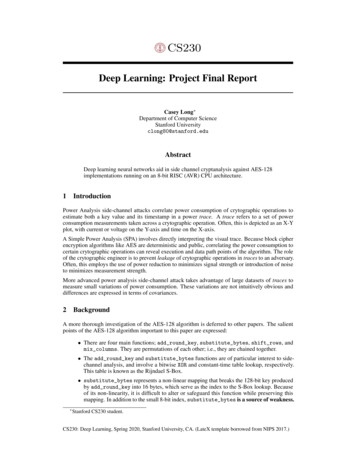
40M prior left arm DVT. Acute pain and swelling ofthe left upper arm, rule out DVT.Brachial arterycephalicbasilicbasiliccephalic1:5 dilution (20cc contrast 80cc NS) @ 3cc/sec. Tourniquet around bicepsregion, released 15 sec before initiation of scan.
CTV: Imaging Techniques Direct Venography (first pass):– Dilute contrast (1:5 or 1:6)– Fill veins of interest (50cc or more)– Slow infusion, 1-2cc/sec– Start acquisition towards end of infusion Indirect Venography (recirculation)– 100-150cc contrast needed for adequatevenous opacification– Empiric imaging delay 60 seconds: upper extremity and pelvic veins 3 to 3.5 min: lower extremity veins– Smart prep off vein of interestBaldt MM, et al. Radiology 1996;200:423-428
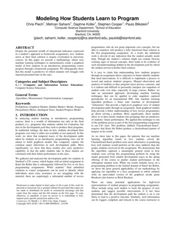
65M with metastatic lung ca and recent PEs. An IVC filter was placed but didnot fully deploy. A second IVC filter was placed above the first one.120cc contrast, diagnostic delay 70sec
CTV: Imaging Techniques Direct Venography (first pass):– Dilute contrast medium (1:5 or 1:6)– Fill veins of interest (50cc or more)– Slow infusion, 1-2cc/sec– Start acquisition towards end ofinfusionBaldt MM, et al. Radiology 1996;200:423-428
CTV: Imaging Techniques Indirect Venography (recirculation)– 150cc contrast needed for adequatevenous opacification (2 mL/kg)– Empiric imaging delay 60 sec: thoracic 70-80 sec: upper extremity 11- sec: pelvis 150 – 180 sec: lower extremity veins– ? Smart prep off vein of interest– Want veins 80HU to be diagnostic
INDIRECT CT VENOGRAPHY Large bolus of contrast followed by a delay toimage the recirculation phase– 150 mL (2 mL/kg BW) Empiric Delay (depends on venous territory) 60 seconds: thoracic 70-80 seconds: upper extremity 110 seconds: abdomen & pelvis 180 seconds: lower extremity NO Bolus Trigger Not an exact science, no target HU
Combo Direct / Indirect CTV R/O LUE venousmalformation; L handand arm swelling 120 mL @ 5 mL/sfollowed by 100 mL1:10 dilution at 2.5mL/s via L hand IV Caudocranialacquisition
Combo Direct / Indirect CTVProtocol and dataset courtesy of Scott Alexander, MD
Combo Direct / Indirect CTV
Combo Direct / Indirect CTV
CTA for TOS:Combo Direct / Indirect CTA Ipsilateral IV, arm over head w/ palm taped up Bolus: 120 mL full-strength @ 4ml/s Chase: 100 mL dilute (10%) contrast @2.5 ml/s Can inject contralateral arm at same time (dilute) 65 sec empiric delay, scan caudo-cranial Arm down, immediate re-scan cranio-caudal Volumetric Review
MRA for TOS: Blood Pool MRA Anatomic imaging: Oblique sag and cor T1/T2 Relaxed and Challenged imaging:§ Gadofosveset (blood pool agent)§ Breath-hold FSPGR, ECG-gated, high resolution (1.8mm ST, 448 x 448 matrix) CORONAL acquisition§ Challenged: Arm Abducted§ Relaxed: Arm Down
Arm UPArm DOWN
Venography: Common ClinicalIndicationsUpper Extremity /Chest– SVC syndrome(malignancy, post-XRT)– Catheter-relatedcomplications (clot,stenosis)– DVT– Thoracic Outlet syndrome– Dialysis accessLower Extremity– DVT ( /- PE study)– May-Thurner syndrome– Pre-transplant evaluationGeneral- Venous stent evaluation- Vascular Malformations –treatment planning
SVC ObstructionNOT A COMPREHENSIVE SYSTEM! Stanford, et al.: Venography series with4 main collateral pathwaysI. Partial SVC occlusion w/ patent Azygous v.II. Near complete obstruction SVC w/ antegradeflow azygous à RAIII. Near complete obstruction SVC w/retrograde flow azygousIV. Complete obstruction SVC one or moremajor tributaries (e.g. azygous v.)Stanford W, et al AJR 1987:148. 259-62.
SVC OcclusionMore common Mass / AdenopathyCatheter / Device (pacer / ICD leads)Fibrosing MediastinitisCatheter MassCatheter pleural effusionThrombusCatheter lymph nodesLess common
SVC Syndrome from Tumor
Classification of all collateral pathways one seriesFrom: Cihangiroglu: J Comput Assist Tomogr, Volume 25(1).January/February 2001.1-8
Most common venous collaterals listed in order of frequency (n 21).From: Cihangiroglu: J Comput Assist Tomogr, Volume 25(1).January/February 2001.1-8
A.1 superior vena cava2 inferior vena cava3 azygos vein4 hemiazygos vein5 accessory hemiazygos vein6 ascending lumbar vein7 lateral thoracic vein8 superficial epigastric vein9 internal mammary vein10 inferior epigastric vein11 pericardiophrenic vein12 right superior (highest) intercostal vein13 left superior (highest) intercostal vein14 intercostal vein15 inferior phrenic vein16 suprarenal veinFrom: Kim: J Comput Assist Tomogr, Volume28(1).January/February 2004.24-33B1 superior vena cava2 brachiocephalic (innominate) vein3 subclavian vein4 internal jugular vein5 external jugular vein6 jugular venous arch7 superior thyroidal vein8 middle thyroidal vein9 inferior thyroidal vein10 facial vein11 anterior jugular vein12 vertebral venous plexus13 vertebral vein, and14 deep cervical vein
Left Superior Intercostal VeinPericardiophrenic Vein
Inferior Phrenic v. (to IVC)
Intercostal veinsLat thoracic v.Azygousv.
Thoraco-acromioClavicular vv.Internal Mammary VeinsAreolar Venous Plexus
Systemic – portal collateralsCapsular / Liver surface vv.Paravertebral vv.
Venous collaterals organized by plexussystems – Easier, more complete to reportCihangiroglu: J Comput Assist Tomogr, Volume 25(1).January/February 2001.1-8
The poster child for revised venousplexus nomenclature
Chest / Upper extremity cases
Thoracic Outlet Syndrome (TOS) Symptoma(c compression/entrapmentof neurovascular structures by boneand/or so7 (ssue as they pass throughthe cervicoaxillary canal 90% Neurogenic (PT, postural Tx, NSAIDs) 10% Vascular Venous Arterial
Components of Cervico-AxillaryCanal Interscalene Triangle:#1 site of compression Costoclavicular Space:#1 site for vascularTOS Retro-pectoralis minorspace: #1 site formassesLinda D D et al. Radiographics 2010;30:1373-1400
CTA for TOS:Combo Direct / Indirect CTA Ipsilateral IV, arm over head w/ palm taped up Bolus: 120 mL full-strength @ 4ml/s Chase: 100 mL dilute (10%) contrast @2.5 ml/s Can inject contralateral arm at same time (dilute) 65 sec empiric delay, scan caudo-cranial Arm down, immediate re-scan cranio-caudal Volumetric Review
Bilateral Direct / IndirectCTA
Venous TOS:Effort Thrombosis Paget-Schroetter syndrome (PSS) AKA axillo-subclavian venousthrombosis Overhead athletes PE in up to 1/3!! * Post-thrombotic syndrome (later)* Perlowski AA. Vasc Med (2010) vol. 15 (6) pp. 469-79
Effort Thrombosis:36 YO weightlifter
Post-Op 1st rib resection
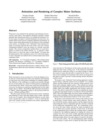
Arterial TOS Overhead athletes SX: Coolness, weakness, diffuse armpain (ischemic neuritis) Cause: Repetitive compression injury– Anatomic predisposition (tight CCS)– Post-traumatic, bony callus– Scalene hypertrophy
Arterial and Venous TOS:16 YO Volleyball AthleteRESTSTRESS
SVC and central veins
LUNG CAwith SVC syndrome
RSCV OCCLUSION – 47 F Dialysis Pt
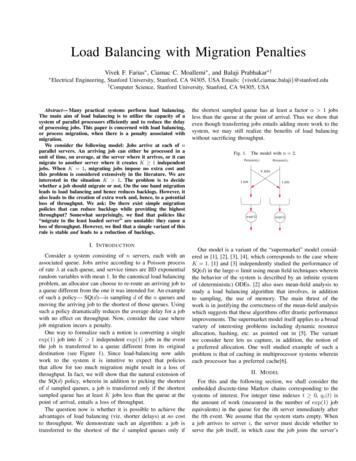
35M hx thigh sarcoma.Facial swelling & chestwall varicosities when hebent over to tie his shoes.Documented centralvenous obstruction.Treatment planning:Assess vascular access,particularly axillary &subclavian veins B/L.Simultaneous bilateral arminjection:1: 6 dilution (30cc contrast 170 cc NS, each arm) @2cc/sec.Courtesy of Anne Chin, MD
LT IJV90cc contrast, 60 sec diagnostic delay.Imaging range: angle of mandible to lesser trochanters.
SVC Occlusion from Aneurysm
RSCV Occlusion – PreviousCatheters
L innominate Occlusion - C-Arm CTRIMVSVCLT IJ injection 1:2 dilution (12cc contrast 12cc NS @ 2cc/sec) acquired on flatpanel detector Dyna-CT.Courtesy of Anne Chin, MD
60F ESRD, 3 overlapping stents placed for venousstenosis from previous catheters.Courtesy of Anne Chin, MDIV cannula in left arm. 100cc contrast 20ccNS flush, diagnostic delay 60sec.
LIV encasement – Adenopathy
In-stent LIV / SVC thrombus
LIV Occlusion – Dialysis Patient withLUE AVF
EJ arch, lat thoracic,and pharyngealcollaterals
62F central venous catheter forchemotherapy.100cc contrast,diagnostic delay 60sec
62F central venous catheter forchemotherapy.100cc contrast,diagnostic delay 60sec
MISC UE Cases
RUE Hemangiomatosis
UE AVMs
Pelvis / LE Cases
MAY-THURNER :SUPERFICIAL VENOUSVARICOSITIES
41 YO F, May - ThurnerLack of Augmentation – suspect upstream obstruction
LCIVExternal iliac vv.
S/P Mechnical Lysis, TPA, and PTA
Indirect Dx by arterial CTA
F/U stenting for May Thurner28F May-Thurner syndrome,CIV/EIV stent placement3 years agoCourtesy of Anne Chin, MD 120 cc contrast Monitoring delay 40sec Smart prep at infrarenal IVC
Vascular Mapping
ExtremityHemangiomatosisVenous MappingProtocol:CTA Runoff; 40 sec interscan delay;Caudocranial scan 16x0.75mmMajor drainage routes:LEFT 12th IC VEINLeft Gonadal V.Greater Saphenous V.
IVC Aneurysm
IVC Aneurysm Rare Saccular fusiform Cause unknown, may be related to anomalousconnections in embryologic venous systems– Acquired (trauma, AV fistulae)– May be associated with other congenital CVanomalies Sx: Thrombosis (7/16), pain, rupture, legswelling– Massive penile bleeding (1/16)– PE if thrombus
Conclusions CTV is a robust, non-invasive technique to visualize venousanatomy, and can be combined with arterial phase CTA Direct CTV: better opacification, less CM needed, but only theinjected and downstream veins will be visualized Indirect CTV: all venous anatomy is delineated, empiric delayor smart-prep at ROI, opacification occasionally unpredictable Combo CTV : Perhaps the best choice for excellent andconsistent venous opacification Provides accurate 3D visualization of venous anatomy fortreatment planning
Thanks to:Dominik Fleischmann, MDFrandics Chan, MD PhD
Key ReferencesLoud PA, et al. Radiology 2001; 219:498-502. (Sens / Spec of CTV goodcompared to Doppler)Begemann PG, et al. J Comput Assist Tomogr 2003; 27:399-409.(Sensitivity 100%; Specificity 97% compared with ultrasonography)Baldt MM, et al. Radiology 1996; 200:423-428. (Sensitivity 100%;Specificiity 96% compared with conventional venography)Sampson, FC, et al. Eur Radiol 2007; 17:175-181. (Pooled sensitivity 91.5%;Pooled specificity 94.8% compared with conventional venography)Kluge, A. et al. AJR 2006; 186:1686 – 1696 (Combo MRA/MRV for PE/DVT)Kim, HC, et al. J Comput Assist Tomogr 2004; 28:24-33 (Collateral Pathways)Cihangiroglu M, et al. J Comput Assis Tomogr 2001; 25: 1-8 (collaterals inSVC Obstruction)Lawler LP, et al. Radiographics 2002; 22:S45-S60 (normal and accessorychest venous pathways)
Key ReferencesKluge, A. et al. AJR 2006; 186:1686 – 1696 (Combo MRA/MRV for PE/DVT)Kim, HC, et al. J Comput Assist Tomogr 2004; 28:24-33 (Collateral Pathways)Cihangiroglu M, et al. J Comput Assis Tomogr 2001; 25: 1-8 (collaterals inSVC Obstruction)Lawler LP, et al. Radiographics 2002; 22:S45-S60 (normal and accessorychest venous pathways)Demos TC, et al. AJR 2004; 182:1139-1150 (Venous anomalies of chest)
CT venography (CTV) is a technique targeted to assess venous anatomy, determine venous . Exam may be lengthy Pt. cooperation? Spatial resolution (vs other choices) . Volumetric Review MRA for TOS: Blood P