Transcription
Margolskee et al. Diagnostic Pathology (2016) 11:27DOI 10.1186/s13000-016-0475-5RESEARCHOpen AccessHepatocellular adenoma classification: acomparative evaluation ofimmunohistochemistry and targetedmutational analysisElizabeth Margolskee1†, Fei Bao2†, Anne Koehne de Gonzalez1, Roger K. Moreira3, Stephen Lagana1,Anthony N. Sireci1, Antonia R. Sepulveda1, Helen Remotti1, Jay H. Lefkowitch1 and Marcela Salomao1*AbstractBackground: Four subtypes of hepatocellular adenomas (HCA) are recognized: hepatocyte-nuclear-factor-1αmutated (H-HCA), β-catenin-mutated type with upregulation of glutamine synthetase (b-HCA), inflammatory type(IHCA) with serum-amyloid-A overexpression, and unclassified type. Subtyping may be useful since b-HCA appear tohave higher risk of malignant transformation. We sought to apply subtype analysis and assess histological atypia,correlating these with next-generation sequencing analysis.Methods: Twenty-six HCA were stained with serum amyloid A (SAA), liver fatty acid-binding protein (LFABP),glutamine synthetase (GS), and β-catenin IHC, followed by analysis with a targeted multiplex sequencing panel.Results: By IHC, 4 HCA (15.4 %) were classified as b-HCA, 11 (42.3 %) as IHCA, 9 (34.6 %) as H-HCA, and two (7.7 %)unclassifiable. Eight HCA (30.8 %) showed atypia (3 b-HCA, 4 IHCA and 1 H-HCA). Targeted sequencing confirmedHNF1A mutations in all H-HCA, confirming reliability of LFABP IHC in identifying these lesions. CTNNB1 mutationswere detected in 1 of 4 (25 %) of GS/β-catenin-positive cases, suggesting that positive GS stain does not alwayscorrelate with CTNNB1 mutations.Conclusions: Immunohistochemistry does not consistently identify b-HCA. Mutational analysis improves thediagnostic accuracy of β-catenin-mutated HCA and is an important tool to assess risk of malignancy in HCA.Keywords: Hepatocellular adenoma, Atypical hepatocellular adenoma, Liver, HCA, b-HCABackgroundHepatocellular adenomas (HCA) are rare benign livertumors with the capacity to undergo malignant transformation. Epidemiologic studies report the prevalenceof HCA is approximately 3–4 cases per 100,000 peoplein Europe and North America, [1] and lower in Asiancountries [2]. The highest prevalence is described in females taking oral contraceptives, but other risk factors,such as anabolic steroid use, obesity and metabolic syndrome are described [2–5]. Inherited syndromes such as* Correspondence: marcelafa@hotmail.comElizabeth Margolskee and Fei Bao are co-first authors.†Equal contributors1Department of Pathology and Cell Biology, Columbia University MedicalCenter, 630 W 168th Street, VC14-238, New York, NY 10032, USAFull list of author information is available at the end of the articleglycogenosis type 1, maturity-onset diabetes of theyoung type 3 (MODY3), and familial adenomatouspolyposis have also been linked to the development ofHCA [3, 5, 6]. HCA can be solitary or multiple, with“liver adenomatosis” defined as presence of multipleHCA ( 10) of any size involving all liver segments [7].The majority of HCA are benign and some regress without surgical intervention. However, serious complications can occur. Notably, hemorrhage is described in upto one fourth of patients, mainly associated with large( 5 cm) lesions. Furthermore, 9 % of HCA may transform into hepatocellular carcinoma (HCC) with risk factors including male sex, androgen use, large tumors( 5 cm) and β-catenin-mutated HCA [5]. In these complicated cases, early surgical removal may improve 2016 Margolskee et al. Open Access This article is distributed under the terms of the Creative Commons Attribution 4.0International License (http://creativecommons.org/licenses/by/4.0/), which permits unrestricted use, distribution, andreproduction in any medium, provided you give appropriate credit to the original author(s) and the source, provide a link tothe Creative Commons license, and indicate if changes were made. The Creative Commons Public Domain Dedication o/1.0/) applies to the data made available in this article, unless otherwise stated.
Margolskee et al. Diagnostic Pathology (2016) 11:27patient outcomes [8, 9]. Therefore, there is interest indeveloping diagnostics to preemptively identify high-riskcases and guide clinical management. Imaging studiesmay identify a proportion of HCA with relatively highsensitivities and specificities, but some cannot be accurately differentiated from HCC [10, 11]. In these cases,histological evaluation becomes essential for diagnosis.In the last decade, four subtypes of HCA have beenidentified based on genotype-phenotype analyses [12].Inactivating mutations in the HNF1A (TCF1) gene causing loss of hepatocyte nuclear factor 1α (HNF-1α) expression define H-HCA, which are characterizedhistologically by marked steatosis and bland hepatocytecytology [12, 13]. A second subtype, with activatingmutations of β-catenin (b-HCA), is associated with anincreased risk of malignant transformation into HCC[9, 12, 14–16]. A third subtype (IHCA) is marked by inflammatory infiltrates or telangiectatic features by histology with increased serum amyloid A (SAA) and Creactive protein (CRP) expression by IHC. IHCA has beenlinked with activating mutations in several genes includingIL6ST, FRK, STAT3, JAK1, and GNAS [17, 18]. Whole exome sequencing has also demonstrated that there is overlap between b-HCA and IHCA in some adenomas thatharbor mutations in both the β-catenin and IL6ST genes[18]. Furthermore, in addition to previously reported mutations in exon 3, a smaller proportion of b-HCA carrymutations in exons 7 and 8 of CTNNB1; however no increased risk of malignant transformation was noted inthese patients [18]. Finally, a small proportion of HCAdoes not appear to fall into any of the above categoriesand were considered unclassifiable by current testing approaches (UHCA).Based on this classification, subtyping of HCA by immunohistochemistry has been used with some success [19].Briefly, loss of liver fatty-acid binding protein (LFABP)staining results from HNF-1A mutation in H-HCA, and increased SAA immunoreactivity serves as a marker forIHCA. Upregulation of a downstream CTNNB1 targetgene, glutamine synthase (GS), is seen in b-HCA, as well asnuclear β-catenin staining. It has been postulated that theimmunophenotypic subtypes closely parallel specific histologic features and molecular alterations, but limitationshave been observed by numerous studies and detailed studies correlating morphology, immunohistochemical profileand mutation analysis are lacking [3, 12, 20, 21]. The aimof our study was to apply the HCA classification systembased on histologic features and immunohistochemicalprofiles and correlate the findings with molecular analysis.MethodsCase selection and histopathological evaluationHCA cases diagnosed between January 1994 and December2012 were retrieved from our pathology departmentPage 2 of 10archives. For resection specimens, representative sectionsof tumor and non-tumorous liver were reviewed for histological features. The presence of multiple adenomas (2 ormore tumors) had been previously assessed by gross organreview and representative sections of each adenoma wereexamined by microscopy. For biopsies, only tumor tissuewas available for review and radiology reports werereviewed to determine the presence of multiple adenomas. Retrospective chart reviews were performed tocollect additional demographic data including age, gender, related medical history and clinical follow-up. Thestudy was approved by the Institutional Review Boardat Columbia University Medical Center.Hematoxylin and eosin (H&E) stained slides, reticulinand Masson’s trichrome stains as well as immunohistochemical studies (IHC) were used to evaluate generalmorphologic and immunophenotypic features. All caseswere reviewed by 3 pathologists (MAS, EM and FB).Tumor characteristics evaluated on routine H&E stainedslides included: steatosis (mild 0–33 %; moderate 33–66 %; marked 66 % of the lesion), inflammation, sinusoidal dilatation (telangiectasia), ductular proliferation, nuclear atypia (nuclear pleomorphism, increasednuclear:cytoplasmic ratio) and architectural atypia(gland-like or acinar growth). Atypia was defined as thepresence of any of the following: (1) nuclear atypia, (2)any degree of architectural atypia, and/or (3) focal lossof reticulin staining.ImmunohistochemistryImmunohistochemistry for LFABP (ABCAM, Cambridge,UK, 1:100 dilution), SAA (ABCAM, Cambridge, UK,1:100 dilution), β-catenin (BD Bioscience, San Jose, CA,1:50 dilution) and GS (Millipore, Billerica, MA, 1:2000)was performed in all cases using standard laboratory techniques in the Ventana Benchmark Ultra platform (Tucson,AZ, USA). GS IHC was scored as 0 (negative, or weakperivascular staining in 10 % of the tumor), 1 (perivascular staining or pseudo-maplike pattern of 10 % of thetumor), and 2 (diffuse strong staining), as previously described [19]. Pseudo-maplike GS pattern has been previously described as interconnected clusters of hepatocytesbeyond perivascular lesions, connected by inconspicuousbands of positive hepatocytes [22]. β-catenin IHC wasgraded as 0 (membranous staining) or 1 (nuclear stainingin any percentage of tumor cells). LFABP and SAA stainswere scored from 0 to 2 (Score of 0 negative or 10 %staining, 1 10–50 % staining, and 2 50 % positivestaining). In most cases, we used adjacent non-tumoralliver as internal negative controls, including negative SAAstaining, membranous β-catenin pattern, and normal centrilobular GS positivity. CD34 immunohistochemicalstains (DAKO, Carpinteria, CA, 1:200) were performed onatypical cases to evaluate for the presence of sinusoidal
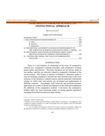
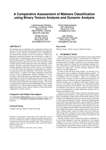
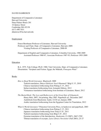
Margolskee et al. Diagnostic Pathology (2016) 11:27capillarization, as previously described [23]. In selectcases, glypican-3 (Cell Marque, Rocklin, CA, 1:100) immunohistochemistry was also performed.Molecular analysisMultiplex targeted DNA next generation sequencing wasperformed in 18 of 26 cases. DNA was extracted from frozen and/or formalin-fixed paraffin-embedded tumor tissueusing QIAamp DNA Mini Kit (QIAGEN, Germany) according to manufacturer’s specifications. DNA wasquantitated by fluorometry with the Invitrogen Qubitfluorometer and Quant-iT dsDNA BR Assay Kit (LifeSciences, Carlsbad, CA), as recommended by themanufacturer. The samples were sequenced using theTruSeq Amplicon Cancer Panel (MiSeq system, Illumina, CA), which covers 225 amplicons within 48cancer-related genes, including 2 amplicons corresponding to exons 3 and 4 of HNF1A gene, and one ampliconrepresenting exon 3 of CTNNB1 gene.Whole exome sequencing on an expanded panel of467 tumor-specific genes, including HNF1A, CTNNB1(all exons), IL6ST, STAT3, GNAS, and JAK1, was performed in a subset of cases. This test requires higheramount of DNA and could only be performed in 11cases with sufficient DNA (cases 2–4, 6–10, 13, 14 and26). Target capture and enrichment were performed withthe SureSelect Hybrid Capture system (Agilent Technologies, Santa Clara, CA) using custom probes. cDNA Libraries were then quantified using qPCR, diluted to 2nM andpooled for analysis on Illumina HiSeq 2500 using IlluminaTruSeq v3 chemistry (Illumina, San Diego, CA).Data deconvolution was performed using CASAVASoftware (Illumina, CA). Files meeting QC metrics wereused for mapping and variant calling using NextGENeSoftware (Softgenetics, State College, PA). Reads werealigned to the hg19 reference genome. Variant calls withallele prevalence 1 % in the 1000 Genome Project, 3variant reads, ambiguous alignments, quality score 10,and allele frequency 10 % were excluded. Variants werecross referenced with COSMIC, PROVEAN, and SIFTprediction tools [24].Statistical analysisFor categorical variables, Fisher’s exact test was used.One-way Kruskal-Wallis test was used for nonparametricdata, including immunohistochemical scoring. Continuousvariables were compared using a two-tailed student t-testor one-way ANOVA, as appropriate. P 0.05 was regardedas statistically significant.ResultsClinical characteristicsA total of 26 HCA cases (22 resections and 4 biopsies)were included in this study (male 5, female 21, meanPage 3 of 10age 36.2 16 years). Ten patients had multiple adenomas (38.5 %) with an average number of 6 lesions perpatient (range: 2–14). The average size of HCA was8.2 4.5 cm (range: 1-18 cm). Detailed clinical historyand follow-up data was available in 22 cases. One ormore risk factors for the development of HCA at thetime of resection (e.g. use of oral contraceptives, anabolic steroid use, obesity, prior pregnancy) was identified in the majority of cases (16/23, 70 %). Among thefemale patients, use of oral contraceptives (OCP) wasidentified in 12 of 21 (57.1 %) cases. One of 5 male patients (20 %) reported anabolic steroid use. We did notidentify any case of glycogen storage disease, vasculardiseases or mature-onset diabetes of the young type 3(MODY3). None of the 22 patients with follow-up datadeveloped hepatocellular carcinoma (mean overall survival of 4.2 years). One patient died of colon cancer.The patient’s demographics and follow-up data aresummarized in Table 1.Histopathological and immunohistochemical analysisFirst, we attempted to classify each adenoma based onIHC pattern, as previously described [19]. Briefly,LFABP-negative cases were classified as H-HCA (Fig. 1).HCA with strong and diffuse GS staining (score 2 )and/or β-catenin nuclear staining, regardless of the SAAstaining status, were categorized as b-HCA (Fig. 2). Theremaining HCA with SAA positivity (scores 1 to 2 )were classified as IHCA (Fig. 3).By applying the above criteria, we identified 4 b-HCA(15.4 %), 11 IHCA (42.3 %), and 9 H-HCA (34.6 %).Two cases (7.7 %) were negative or weakly positive forGS, negative for SAA, and positive for LFABP, and weretherefore unclassifiable (Table 2).By immunohistochemistry, we found significant overlap between the b-HCA and IHCA subtypes. Focal SAAstaining (1 , 50 % of cells) was present in 2 out of 4 bHCA (cases 1 and 2). Conversely, 5 IHCA with 1 or 2 SAA staining showed weak, heterogeneous GS expression (score 1 ) (cases 5–7, 10 and 13). We chose not toclassify these as mixed β-catenin/inflammatory HCAbased solely on immunohistochemistry because none ofthem demonstrated strong positivity for both GS andSAA. Cases with loss of LFABP did not show any staining for GS, except for one H-HCA case with strong (2 )GS staining (Table 2, case 16). In addition, strong (2 )SAA staining was present in 5 of 9 H-HCA (LFABPnegative) cases and might have resulted from proximityto areas of tumor necrosis or hemorrhage within thetumor, findings identified in at least 3 cases. Nuclear βcatenin staining was present in 3 of 5 (60 %) GS positive(2 ) cases, but was limited to rare to few cells corresponding to less than 5 % of total tumor area and predominantly located in perivascular areas (Fig. 2c).
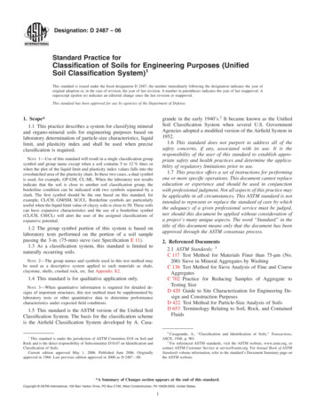
Margolskee et al. Diagnostic Pathology (2016) 11:27Page 4 of 10Table 1 Clinical information of hepatocellular adenoma casesCase Sex Age(years)MultipleadenomasAvg. Size Survival(cm)(days)Risk 246Prior pregnancy15F45Y9939OCP16M25N8.5730Androgen use17F39Y101095OCP, priorpregnancy18F34Y8n/aOCP, priorpregnancy19F42N5.5n/aOCP20F58Y42190OCP, obesity,prior pregnancy21F41Y4.52190OCP22F75N5.5deceased 6.31095OCPAbbreviations: F female, M male, N absent, Y Present, n/a data not available,OCP oral contraceptive use, aObesity is defined as BMI 30During the interpretation phase of this study, we encountered two technical difficulties. First, we found thatLFABP IHC can sometimes be faint in the nonneoplastic liver resulting in an inconspicuous gradientbetween native liver and tumor. In such cases, LFABPstaining was repeated in multiple tumor sections inorder to obtain acceptable results. Second, the few caseswith nuclear β-catenin positivity were difficult to evaluate since nuclear positivity could only be appreciated inless than 5 % of the tumor cells.Molecular analysisResults of targeted DNA sequencing analysis were correlated with the classification obtained by immunohistochemical analysis (Table 2). HNF1A mutations wereidentified in all analyzed H-HCA (5/5, 100 %) and in noother HCA subtype (p value 0.001). The codon alterationswere Gly207Asp, Asn237Ser, Asn237Lys, Leu281Gln andTyr286Cys, 3 of them previously reported in H-HCA [18].CTNNB1 gene alterations were identified in 2 cases:one b-HCA harboring both a CTNNB1 gene deletion(c.114 119del; p.38 40del) and a TP53 gene mutation(Ile251Phe) (case 1); and a second case originally classified as IHCA based on IHC results (ie. strong diffuseSAA positivity and focal GS staining), found to have amissense CTNNB1 mutation (Ser45Pro). This case alsocarried an IL6ST mutation (c.566 577del; p.189 193del)and was thus reclassified as a mixed β-catenin/inflammatory HCA (bIHCA) (case 7, Fig. 4b). CTNNB1 alterations in exons 7 and 8 were not identified in the 11tumors analyzed by whole-exome sequencing. Six tumors classified as IHCA due to 2 SAA staining had nomutations in the IHCA-related pathways.A total of 4 cases were reclassified following molecularanalysis. One case was the bIHCA described above. Theother 3 tumors, initially classified as b-HCA (2 GS staining), were reclassified because molecular analysis demonstrated no abnormalities in the CTNNB1 gene. Two ofthem carried IL6ST gene alterations (IL6ST c. C657A;Pro216His and IL6ST c.554 568del; p.185 190del) andFig. 1 Histological features of HNF-1a inactivated hepatocellular adenoma (H-HCA). a, H-HCA with steatosis and b, loss of LFABP expression (H&Eand IHC X 20). c, higher magnification showing marked steatosis (H&E x100)
Margolskee et al. Diagnostic Pathology (2016) 11:27Page 5 of 10Fig. 2 Histological features of beta-catenin activated hepatocellular adenoma (b-HCA). a, Lower power micrograph showing border of b-HCA(middle and right) with normal liver (left) (H&E x20). b, Diffuse GS expression in b-HCA (right) in contrast to the focal perivenular staining in thenormal liver tissue (left) (IHC X 20). c, diffuse and strong GS staining (right panel) vs. focal β-catenin nuclear staining (left panel) in b-HCA (IHC X 200)were reclassified as IHCA. The third case had no alterations in CTNNB1, HNF1A, IL6ST, STA3, GNAS or JAK1genes and final classification could not be confirmed bymolecular studies (Fig. 4a).Following classification by molecular testing (Table 2,last column), we analyzed the histological features ofeach subtype (Figs. 1, 2 and 3). All cases classified as HHCA by IHC showed steatosis of variable degree. IHCA(n 9) showed features of telangiectasia (n 9, 100 %),focal inflammation (n 6, 66.7 %) and ductular reaction(n 4, 44.4 %). Mild to moderate steatosis was occasionally seen IHCA (n 2, 22.2 %). Telangiectasia, a featurecommonly associated with IHCA, was seen in the bIHCA (case 7). No cases classified as b-HCA, IHCA orUHCA showed marked steatosis.In rare occasions, limited DNA concentration or DNAdegradation may limit detection of mutations and deletions in tumor samples. To circumvent this issue, DNAwas obtained from both frozen and formalin-fixed tumorsamples and sequenced using two different platforms.This approach was feasible in 12 of our 18 analyzedsamples and yielded reproducible sequencing results.By our sequencing methods, LFABP IHC captured allH-HCA cases and was 100 % specific, whereascombination of GS and β-catenin IHC predicted βcatenin mutations with a sensitivity of 50 %, specificityof 75 %, and PPV of 20 %. Correlation between histological features, IHC results and molecular analysisshowed that LFABP loss and tumor steatosis were significantly associated with H-HCA (p 0.001 and 0.036,respectively). Not surprisingly, telangiectasia was preferentially present in IHCA (p 0.0002) (Table 3).Evaluation of atypia in HCAEight cases (30.8 %) displayed one of more atypical features (Table 2, example in Fig. 5). By molecular testing,these cases were classified as b/b-IHCA (2 of 2, 100 %),IHCA (4 of 9, 44.4 %), H-HCA (1 of 5, 20 %), andUHCA (1 of 2, 50 %). Nuclear atypia was present in 7cases (87.5 %). Gland-like or acinar growth was seen in 4of 8 cases (50 %). Reticulin staining showed focal( 1 mm) loss of reticulin framework in 6 cases (75 %).By CD34 immunostaining, focal or patchy sinusoidalcapillarization was seen in 6 cases (75 %), but diffuseCD34 staining classically described in malignant hepatocellular lesions was not identified. Glypican-3 immunohistochemistry was negative in all atypical cases.Atypical HCA were more frequently present in maleFig. 3 Histological features of inflammatory hepatocellular adenoma (IHCA). a, IHCA with telangiectasia and focal inflammation (H&E X 20). b, Diffuseserum amyloid-A expression within the tumor (IHC X 20). c, Higher magnification micrograph highlighting inflammation (lower right) and telangiectasia(upper left) (H&E X 100)
Margolskee et al. Diagnostic Pathology (2016) 11:27Page 6 of 10Table 2 Histological, immunohistochemical and mutational analysis of hepatocellular adenomasMorphological featuresImmunohistochemistryCaseMorphologyAtypical featuresSAALFABPB-catGSMutational 10NuA, Lret1202CTNNB1 c.114 119del; p.38 40del1TP53 c.T751A; Ile251Phe1,2b-HCAb-HCA2TNuA, AG, Lret1212IL6ST c.C657A; Pro216His1,2,33T002b-HCAIHCA12IL6ST c.554 568del; p.185 190del1,3b-HCAIHCA40NuA, AG, Lret0212nm**b-HCAfavor b-HCA5a0AG, Lret1201n/aIHCAn/a6T, INuA, AG, Lret1201nm**IHCAIHCA7T, I, DRNuA2201CTNNB1 c.T133C; Ser45Pro1,3IL6ST c.566 577del; p.189 193del1,3IHCAbIHCA8T, I02200nm**IHCAIHCA9T, I, DR02200nm**IHCAIHCA10T, I, DR02201nm**IHCAIHCA11T, DRNuA, Lret22n/a0nmIHCAIHCA12T02200n/aIHCAn/aa13T, I, S (20 %)02201nm**IHCAIHCA14T, I, DR, S (60 %)022n/a0nm**IHCAIHCA15aT, I, DR02200n/aIHCAn/a16S (30 %)NuA2002HNF1A c.620G A; Gly207Asp1,3H-HCAH-HCA117S (20 %)02001HNF1A c.857A G;Tyr286CysH-HCAH-HCA18S (40 %)00000HNF1A c.842 T A;Leu281Gln1H-HCAH-HCA19S (70 %)00000n/aH-HCAn/a20S (80 %)00000HNF1A c.710A G; Asn237Ser1,3H-HCAH-HCA21S (70 %)02000HNF1A c.710A T; Asn237Lys1,3H-HCAH-HCA22S (10 %)020n/a0n/aH-HCAn/a23I, S (30 %)02000n/aH-HCAn/a24T, I, S (50 %)00000n/aH-HCAn/aa25S (10 : B-HCA β-catenin activated hepatic adenoma, IHCA inflammatory HCA, bIHCA HCA with mixed IHCA and b-HCA features, H-HCA HNF1A-inactivatedHCA, UHCA unclassifiable HCA, SAA serum amyloid A, LFABP liver fatty acid-binding protein, b-cat β-catenin, GS glutamine synthetase, 0 absent, T telangiectasia, Iinflammation, DR ductular reaction, S Steatosis (% fat), NuA nuclear atypia, AG acinar growth, Lret focal loss of reticulin stain, n/a not available, nm no mutation/deletion/insertion identified in HNF1A and CTNNB1 exon 3, nm** no mutation/deletion/insertion identified in HNF1A, IL6ST, STAT3, GNAS, JAK1 and CTNNB(expanded gene analysis)aOnly biopsy specimen available; bClassification based on IHC alone; cClassification following molecular evaluation (italic font is used to mark cases that werere-classified after molecular analysis); 1Deleterious alteration by PROVEAN prediction; 2 variant previously described in HCC; 3 variant previously described in HCApatients, but this result did not reach statistical significance (3 of 5 males [60 %] vs. 5 of 21 females [23.8 %],p 0.28). Similarly, patient age was not significantly different in patients with atypical HCA (mean age 28 vs.39.9 years, p 0.07).DiscussionHepatocellular adenomas have a small risk of malignant transformation with estimated rates of less than5 % [3, 25–27]. Recent advances in the morphologicand molecular classification of HCA identified b-HCAas the subtype most commonly associated to malignanttransformation. This finding warrants the use of robustand reliable approaches to identify such tumors [12, 18].In particular, the status of CTNNB1 mutations in HCAmay offer crucial information regarding management ofsome of these patients.The current HCA classification scheme is based onthe tumor’s morphologic and immunohistochemical characteristics and was originally validated with CTNNB1 andHNF1A gene sequencing [28]. More recently, highthroughput sequencing of a large HCA cohort led to thediscovery of novel genetic alterations, [17, 18] includingnew hot-spots in CTNNB1 gene in the b-HCA subtype,and defects in the IL-6/JAK/STAT3 pathway in IHCA.Our study is one of the first to use a comprehensive
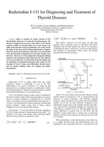
Margolskee et al. Diagnostic Pathology (2016) 11:27Page 7 of 10Fig. 4 Patterns of glutamine synthetase staining in HCA. a, Diffuse GS staining in CTNNB1 wild-type tumor (case 4) (upper right, x 10). b, GS staining ofbIHCA carrying both CTNNB1 and IL6ST alterations (case 7) shows perivascular and focal pseudomap-like pattern (lower aspect, x 10). Asinternal positive control, note the normal centrilobular pattern of GS staining of the adjacent non-neoplastic livertargeted next-generation sequencing panel to evaluate themutational status of HCA and correlate with immunohistochemistry results.As expected, our study demonstrated HNF1A genemutations in all analyzed H-HCA cases, confirming highaccuracy of this marker in the diagnosis of H-HCA.However, our results suggest limited reliability of GSand β-catenin immunohistochemistry in predicting βcatenin mutations. The utility of both markers has beenchallenged by other researchers [10, 21, 22, 29, 30].Lagana et al. showed strong GS positivity in over 50 %of HCA and Joseph et al. described patchy to diffuse GSstaining in 23 % of IHCA, but no correlation with molecular studies were available in these studies [22]. In thepresent study, seven of 12 GS-positive cases displayedfaint perivascular staining or heterogeneous positivity.Further molecular evaluation performed in 5 tumorsfailed to demonstrate CTNNB1 alterations in 4 cases,one of which was a biopsy specimen (case 13). Thesesomewhat controversial staining patterns and their correlation with β-catenin activation have not been fullycharacterized and may be a result of localized metabolicalterations within the tumor [20–22, 31].In addition, we identified strong GS positivity in fourβ-catenin wild-type tumors, including one H-HCA. Wespeculate whether these tumors may have activation ofan alternative pathway resulting in GS expression. Berryet al. described strong GS staining in a peliotic HCAand suggested that vascular flow alterations and hepaticparenchymal remodeling may explain increased GS expression [31, 32].In our study, GS staining failed to identify 1 of 2 confirmed CTNNB1-mutated HCA. Low GS expression inb-HCA has been previously reported in HCA carryingthe Ser45Pro mutation [33]. Interestingly, the same βcatenin mutation has been described in HCC and wasnot associated with nuclear β-catenin staining, suggesting that some CTNNB1 alterations may not disrupt GSand β-catenin expression patterns [34]. None of the 11cases evaluated by whole-exome sequencing carriedlarge CTNNB1 deletions or mutations in exons 7 or 8,the latter described in a minority of bHCA and previously shown to activate β-catenin in HCC cell lines [18].Caution is warranted in such cases as they show weakand patchy GS staining and lack nuclear staining of βcatenin [18].Table 3 Correlation between histological features and molecular classification of HCATelangiectasiaH-HCA (n 5)b-HCA/bIHCA (n 2)IHCA (n 9)UHCA (n 1)p value050 % (1)100 % (9)00.0002Inflammation20 % (1)50 % (1)67 % (6)100 % (1)NSDuctular Reaction050 % (1)44 % (4)0NSSteatosis (mean %)48 %09%00.04Atypical features20 % (1)100 % (2)44 % (4)0NSSAA positive (1 /2 )60 % (3)100 % (2)89 % (8)0NSGS positive (1 /2 )40 % (2)100 % (2)56 % (5)0NSLFABP loss100 % (5)0000.0001Abbreviations: SAA serum amyloid A, LFABP liver fatty acid-binding protein, GS glutamine synthetase, b-HCA beta-catenin activated hepatic adenoma, bIHCA HCAwith mixed IHCA and b-HCA features, IHCA inflammatory HCA, H-HCA HNF1A inactivated HCA, UHCA unclassifiable HCA, NS not statistically significant (p value 0.05)
Margolskee et al. Diagnostic Pathology (2016) 11:27Page 8 of 10Fig. 5 Histological atypia in HCAs. a, pseudoacinar formation (H&E X 200). b, nuclear atypia (H&E X 200). c, capillarization of the HCA on CD34immunohistochemical stain (X 100). d, focal loss of reticulin by reticulin silver stain (X 100)Following molecular analysis, four tumors were reclassified. One case phenotypically classified as IHCA wasreclassified as bIHCA because it carried both a βcatenin mutation and an IL6ST alteration. Three casesinitially categorized as b-HCA lacked CTNNB1 gene alterations. Two of them were re-subtyped as IHCA following identification of IL6ST gene alterations, all ofwhich previously described in IHCA [17, 35]. Theremaining case carried no mutations in HNF1A or anyof the IHCA-related genes. In this challenging case, thediagnosis of a β-catenin-activated HCA is favored, butthe lack of molecular evidence for CTNNB1 alterationssuggests alternative mechanisms of β-catenin activationand GS overexpression in the tumor [36]. This adenomawas originally resected from a 7-year-old patient with noknown medical conditions, who remains disease-free afterover 13 years of her surgery. HCA occurrence in preadolescence is extremely rare and little is known about the natural history and pathogenesis of such lesions. Six adenomasimmunophenotypically compatible with IHCA lacked identifiable mutations in IHCA-associated genes, a finding previously described by others [18, 27].A large percentage of HCA showed atypical features(30.8 %) but no correlation with a specific HCA type wasidentified. Other histological features, such as mild steatosis and telangiectas
TruSeq Amplicon Cancer Panel (MiSeq system, Illu-mina, CA), which covers 225 amplicons within 48 cancer-related genes, including 2 amplicons correspond- ing to exons 3 and 4 of HNF1A gene, and one amplicon representing exon 3 of CTNNB1 gene. Whole exome sequencing on an expanded panel of 467 tumor-specific genes, including HNF1A, CTNNB1 (all exons), IL6ST, STAT3, GNAS, and JAK1, was per-formed .