Transcription
Sign up to receive ATOTW weekly - email worldanaesthesia@mac.comRHEUMATOID ARTHRITIS AND ANAESTHESIA PART 2ANAESTHESIA TUTORIAL OF THE WEEK 26713TH AUGUST 2012Dr Rashmi MenonLeeds General InfirmaryCorrespondence to rashmimenon@doctors.org.ukQUESTIONS1.A patient with long standing rheumatoid arthritis (RA) presents with hoarseness of voice.What are the anaesthetic implications of this? Choose the single best answer.a. This history is not significantb. Hoarseness might worsen after endotracheal intubation-the patient should be warnedabout thisc. The patient may be difficult to intubate as the vocal cords may be swollend. The patient probably has cricoarytenoid involvement, which combined with airwayoedema post intubation can cause complete airway obstruction in the post operativeperiod.2.Regarding anaesthesia for patients with RA (true/false):a. Spinal and epidural anaesthesia should be used where possibleb. Regional nerve blocks are contraindicatedc. Awake fibreoptic intubation (AFOI) may be needed in cases of severe atlanto-axialsubluxationd. An LMA can be used for airway management to limit neck manipulatione. Cervical spine involement in RA is always accompanied by symptoms3.Regarding atlanto axial involvement in RA (true/false):a. The most common type of atlanto-axial subluxation (AAS) is posterior AASb. Subluxation exists when the distance between the atlas and the odontoid peg exceeds4 mm in patients older than 44 and 3 mm in younger patientsc. Flexion of the neck is more harmful than extensiond. AAS can be diagnosed only by a CT scane. Severe AAS may necessitate an awake tracheostomy for airway managementATOTW 267 Rheumatoid Arthritis and Anaesthesia – part 2 13/08/2012Page 1 of 8
Sign up to receive ATOTW weekly - email worldanaesthesia@mac.comINTRODUCTIONRheumatoid arthritis (RA) is a chronic systemic inflammatory disorder that affects 1-2% of thepopulation. The multisystem nature of the disease, issues with airway management and polypharmacymakes the anaesthetic management of these patients challenging. Anaesthetists may come acrosspatients with RA during orthopaedic surgery relating to their disease, joint injections, incidentalsurgery, pregnancy and delivery and ICU management.The aetiopathogenesis, clinical features, diagnosis and treatment of RA are discussed in “RheumatoidArthritis and Anaesthesia – part 1” (ATOTW 266). This article focuses on the anaestheticconsiderations in patients with RA.PRE-OPERATIVE ASSESSMENT AND PREPARATION A thorough history of the RA including duration of the disease, severity, drug treatments andsystemic complications should be taken.Rheumatoid patients are often in pain and suffer with deformities that restrict simplemovements; care should be taken during examination.The range of movement of all joints should be noted so as to optimise patient position andminimise injury during anaesthetic procedures (venous access, nerve blocks) and positioningfor surgery. Involvement of the joints of the wrists and fingers may limit function enough topreclude the use of a standard patient-controlled analgesia (PCA).These patients have very fragile skin that is easily damaged. Fragile veins makes peripheralvenous access unreliable and central venous access is often difficult due to neck deformity.Preoperative neurological deficit, if any, should be documented.Patients taking more than 10 mg prednisolone per day should be given appropriate perioperative steroid cover. Patients should continue their regular prednisolone and receivehydrocortisone intra-operatively to cover the stress response to surgery. Depending on thetype of procedure, hydrocortisone may need to be continued into the postoperative period asshown in Appendix 1.Patients are likely to have anaemia (normocytic normochromic anaemia due to chronic diseaseor microcytic anaemia due to gastric bleeding and drug toxicity of bone marrow) and theremay be increased requirement for red-cell transfusion.Patients may have cachexia, which is indicative of poorly controlled disease and an increasedlong-term cardiovascular risk. Cachexia and poor muscle bulk may also indicate myopathywhich may impair respiratory muscle function, and such patients may need to be consideredfor postoperative mechanical ventilation.Methotrexate can be continued in the peri-operative period without impaired wound healing ora substantially raised risk of peri-operative infection1. Furthermore, good disease control inthe peri-operative period is beneficial. There is lack of data regarding the use of otherimmunosuppressants. Peri-operative discontinuation of biological therapy for elective surgeryremains controversial. Recent studies have suggested that their use does not cause specificadverse effects and may improve recovery from postoperative anaemia.ATOTW 267 Rheumatoid Arthritis and Anaesthesia – part 2 13/08/2012Page 2 of 8
Sign up to receive ATOTW weekly - email worldanaesthesia@mac.comAirway assessment A detailed airway assessment including mouth opening, TMJ function, neck flexion andextension is essential.Temporomandibular joint involvement should be assessed pre-operatively using theMallampati score, mouth opening and mandibular protrusion.Any history of cervical surgery should be elicited, as cervical fixation devices may causeimpaired neck extension.There are no published evidence-based guidelines or general consensus on the need to obtaincervical spine X-rays before surgery in asymptomatic patients. However it may be prudent toobtain neck X-rays in long term patients with severe disease.If atlanto-axial subluxation (AAS) is suspected, plain X-ray films in flexion and extension ofthe neck are often diagnostic. Subluxation exists when the distance between the atlas and theodontoid peg exceeds 4 mm in patients older than 44 years and 3 mm in younger patients. Aflexion/extension CT or MRI should be considered if symptoms are associated with severepain, neurological signs are present or if there is significant abnormality noted on plain X-ray.Fibreoptic nasopharyngoscopy is indicated for patients with hoarseness because of thelikelihood of cricoarytenoid involvementManagement of the airwayThe issues in airway management are the increased prevalence of a difficult airway (RA patientsneeding cervical spine surgery have a high prevalence of grade 3 or 4 laryngoscopy) and the presenceof AAS, which may result in spinal cord injury during airway management. The extent of neck flexionand extension should be assessed and documented. Peri-operatively, care should be taken to avoid anymovement beyond this range.The risks are dependent on the type of AAS. The most common is anterior AAS (80%), which isworsened by C1/C2 flexion. Direct laryngoscopy is usually well tolerated. Subluxation is worsened bythe head moving anteriorly whilst the upper cervical spine remains static, e.g. putting a pillow behindthe head. A useful technique is to keep the upper cervical spine supported whilst the head is not movedanteriorly, e.g. using a doughnut head ring with a large enough hole to accommodate the occiput. Thissupports the cervical spine without anterior movement of the head, and is the best choice forrheumatoid patients.Posterior and vertical AAS both pose the risk of spinal cord compression during C1/C2 extension, themovement of which occurs during direct laryngoscopy, which should therefore be avoided. They aremuch less common than anterior AAS, and are mostly symptomatic.ATOTW 267 Rheumatoid Arthritis and Anaesthesia – part 2 13/08/2012Page 3 of 8Sam Shinde 18/7/12 3:02 PMDeleted: in flexion and extension
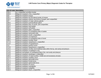
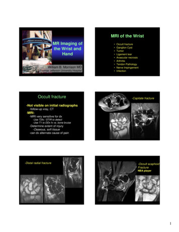
Sign up to receive ATOTW weekly - email worldanaesthesia@mac.comFigure 1. Cervical spine X-rays in a patient with severe rheumatoid arthritis on long termsteroids showing anterior atlanto-axial subluxation (with permission from Oxford UniversityPress). A- extension. B- flexion: Demonstrates severe anterior atlanto-axial subluxation. Notethe position of dens axis (arrow). There is a 13 mm dislocation between the dens and themain body of the axis. C3 shows degenerative changes.Cricoarytenoid arthritis is variable in frequency and often unrecognised. Patients have both joint andsoft tissue swelling causing stenosis. Symptoms may include hoarseness, stridor, a sense of pharyngealfullness when speaking and swallowing or dyspnoea. If cricoarytenoid involvement is suspected, avoidendotracheal inubation in favour of supraglottic airway devices. If intubation is essential, use thesmallest internal diameter tracheal tube possible. Problems occur post extubation, when the oedemacombined with an already narrow airway can cause complete obstruction. Consider the use of anairway exchange catheter at extubation, and observe patient in a high dependency area for some timepost extubation. In severe cases, a pre-operative tracheostomy may be required.Pre operative investigations Routine full blood count, renal function and electrolytes. Liver function tests in patients on long term disease modifying anti-rheumatic drugs(DMARDs). Low threshold for ordering respiratory investigations (chest radiographs, arterial blood gasesand lung function tests) due to the possibility of pulmonary involvement or respiratorymyopathy. An ECG should be performed to check for left ventricular hypertrophy and conductiondisturbances. An echocardiogram should be performed to look for evidence of diastolicdysfunction, left ventricular hypertrophy and valvular abnormalities. Left ventricular ejectionfraction may be normal and falsely reassuring in the presence of diastolic dysfunction.ATOTW 267 Rheumatoid Arthritis and Anaesthesia – part 2 13/08/2012Page 4 of 8
Sign up to receive ATOTW weekly - email worldanaesthesia@mac.comINTRA-OPERATIVE MANAGEMENTThere is no one recommended anaesthetic technique; choice of technique will depend on the patient’sgeneral condition, type of surgery, patient preference and skill of the anaesthetist. Regionalanaesthesia, general anaesthesia or a combination of the two may be employed.Regional anaesthesia, if possible, should always be considered, as it avoids airway manipulation,provides good postoperative pain relief and reduces polypharmacy. However, regional and neuraxialblocks may be technically difficult due to spinal arthritis and loss of anatomical landmarks fromcontractures or deformities. A higher than normal level should be anticipated during a subarachnoidblock1.If a general anaesthetic is indicated, there are several options for managing the airway depending onthe patient and the type and duration of surgery. Laryngeal mask airways (LMAs) and other supraglottic airway devices have the advantages ofrequiring minimal neck manipulation for insertion and causing lesser trauma and subsequentlaryngeal oedema compared with a tracheal tube. They should be used where possible. Theymay be difficult to insert in patients with fixed flexion deformities of the neck.If tracheal intubation is indicated, neck manipulation should be minimised, ideally withmanual in-line stabilisation, even in the absence of overt cervical spine instability. There areno case reports to date of spinal cord injury secondary to direct laryngoscopy, nor is there anyevidence of outcome difference with a particular technique. However, all due care must betaken during airway management. The intubating LMA could be used to aid intubation withminimal neck manipulation, although it is not a popular technique due to high risk of failureand associated trauma to the airway.Fibreoptic intubation is considered the appropriate and safer option in rheumatoid patientswith an anticipated difficult airway or known cervical spine instability. It generally carries lessrisk of major difficulties than tracheal intubation using direct laryngoscopy in this subgroup ofpatients. Awake fibreoptic intubation should be considered in patients with known cervicalspine subluxation, as it allows for assessment of neurological symptoms indicating spinalcompression. This technique is not useful under emergency conditions, or if excessive bloodor secretions are in the airway.A surgical tracheostomy under local anaesthesia may be indicated in emergency situations andin patients who have symptoms of upper airway obstruction.Intra-operatively, patients with RA present problems with positioning due to joint deformities andfragile skin due to steroid therapy. Pressure points should be meticulously padded, the neck adequatelysupported and skin handled with care. Excessive manipulation of stiff and fixed joints should beavoided.It is worth noting that the prolonged, relative spinal malposition during general anaesthesia, in a patientwith possible spinal stenosis, may cause cord injury. It has been suggested that the patients positionthemselves on the operating table before being anaesthetised. Attention to the maintenance of spinalcord perfusion may be important and hypotension should be avoided.The use of immunosuppressive treatments put patients with RA at an increased risk of infections. Strictaseptic technique should be adopted for intravascular access and regional blocks. Appropriateantibiotic prophylaxis should be given before starting surgery.ATOTW 267 Rheumatoid Arthritis and Anaesthesia – part 2 13/08/2012Page 5 of 8
Sign up to receive ATOTW weekly - email worldanaesthesia@mac.comPOSTOPERATIVE MANAGEMENTCareful observation of the airway and breathing are required in the immediate postoperative period.Previously asymptomatic cricoarytenoid arthritis may cause airway obstruction postoperatively as aresult of the additional oedema caused by tracheal intubation. This may occur several hourspostoperatively, necessitate reintubation or in some cases a tracheostomy.Pain should be adequately controlled to permit early mobilisation. Multimodal analgesia isrecommended. Some patients may have chronic pain issues and their pain may be difficult to manage.Opioid analgesia can be used in carefully monitored doses to avoid respiratory compromise. Patientswith joint deformities of the hand may not be able to use a standard PCA, but can be instructed in theuse of a mouth controlled device.Patients with RA tend to have a slower recovery and return to mobilisation, and also are considered tobe in a hypercoagulable state, hence appropriate thromboprophylaxis should be prescribed. Standardphysiotherapy and breathing exercises should be instituted as early as possible due to the increasedinfection risk. If signs of a postoperative infection develop, DMARDs should be suspended temporarilyand appropriate antimicrobials started.SUMMARYAnaesthesia for the patient with severe rheumatoid arthritis is challenging. It requires a goodknowledge of the multisystem effects of the disease, the drugs used to treat it, and meticulous attentionto detail, to safely anaesthetise these patients.ATOTW 267 Rheumatoid Arthritis and Anaesthesia – part 2 13/08/2012Page 6 of 8
Sign up to receive ATOTW weekly - email worldanaesthesia@mac.comANSWERSAnswer 1DHoarse voice in a patient with longstanding RA, should lead to suspicion of cricoarytenoid arthritis,which is variable in frequency and often unrecognised. Other symptoms include stridor, a sense ofpharyngeal fullness when speaking and swallowing or dyspnoea. Pre operative nasoendoscopy may aidin diagnosis. If cricoarytenoid involvement is suspected, avoid endotracheal int inubation in favour ofsupraglottic airway devices. If intubation is essential, use the smallest internal diameter tracheal tubepossible. Problems occur post extubation, when the oedema combined with an already narrow airwaycan cause complete obstruction. Consider the use of an airway exchange catheter at extubation, andobserve patient in a high dependency area for some time post extubation. In severe cases, a preoperative tracheostomy may be required.Answer 2TFTTFRegional anaesthesia in the form of neuraxial blocks or peripheral nerve blocks should always beconsidered as it avoids airway manipulation, provides good postoperative pain relief and reducespolypharmacy. However, regional and neuraxial blocks may be technically difficult due to spinalarthritis and loss of anatomical landmarks from contractures or deformities. LMAs and othersupraglottic airway devices should be used where possible as they require minimal neck manipulationfor insertion and cause lesser trauma and subsequent laryngeal oedema compared with a tracheal tube.AFOI is the technique of choice in patients with an expected difficult airway or known cervical spineinstability needing intubation. Cervical spine involvement is not always symptomatic.Answer 3FTTFTThe most common type of AAS is anterior AAS, occurring in 80% cases. Anterior AAS is worsenedby flexion. Subluxation exists when the distance between the atlas and the odontoid peg exceeds 4 mmin patients older than 44 and 3 mm in younger patients; this can be diagnosed by plain radiography ofthe neck. Severe cervical spine instability may necessitate an AFOI or a tracheostomy under localanaesthesia for safe management of the airway.REFERENCES AND FURTHER READING1.2.3.4.5.Samanta R, Shoukrey K, Griffiths R. Rheumatoid arthritis and anaesthesia. Anaesthesia. 2011;66: 1146–59.Fombon FN, Thompson JP. Anaesthesia for the adult patient with rheumatoid arthritis.CEACCP. 2006; 6: 235–9.Keith Allman, Barry Baker, Iain Wilson. Anaesthesia for the patient with rheumatoid arthritis.Oxford Handbook of Anaesthesia 3rd edition. Oxford University Press (2011)Temprano K. Rheumatoid arthritis. 1715-overviewCalder I. Airway management in cervical spine disease. In: Calder I, Pearce A, eds. CoreTopics in Airway Management, 2nd edn. Cambridge: Cambridge University Press, 2010:244–54.ATOTW 267 Rheumatoid Arthritis and Anaesthesia – part 2 13/08/2012Page 7 of 8
Sign up to receive ATOTW weekly - email worldanaesthesia@mac.comAPPENDIX 1Perioperative steroid supplementationPatients currently taking regular corticosteroids 10mg / day of prednisolone or equivalent - assume normal HPA axis – no additional coverrequired 10mg / day of prednisolone or equivalento Minor surgery e.g. hernia – routine preoperative steroid or 25mg IV hydrocortisoneat inductiono Intermediate surgery e.g. hysterectomy – routine preoperative steroid plus 25mg IVhydrocortisone at induction, then 6 hourly for 24 hours.o Major surgery e.g. cardiac – routine preoperative steroids plus 25mg IVhydrocortisone at induction, then 6 hourly for 48-72 hoursResume normal oral therapy when gastrointestinal function has returned. Patients not currently on corticosteroid therapy 3months since stopping steroids – Treat as if on steroids 3 months since stopping steroids – No perioperative steroids necessaryPatients on high dose immunosuppression Should continue usual immunosuppressive dose parenterally until able to revert to normal oralintake. For e.g. if a patient takes 60mgs of prednisolone / 24 hours, he/she should receive240mgs of hydrocortisone / 24 hours until oral intake can be resumed.Equivalent drug doses: 10mgs prednisolone dexamethasone 1.5mgs hydrocortisone40mgs methylprednisolone 8mgs betamethasone 1.5mgs.References:Allman KG, Wilson IH (2011) The patient on steroids. Oxford Handbook of Anaesthesia. 3rd ed.London: Oxford University Press.ATOTW 267 Rheumatoid Arthritis and Anaesthesia – part 2 13/08/2012Page 8 of 8
ATOTW 267 Rheumatoid Arthritis and Anaesthesia - part 2 13/08/2012 Page 1 of 8 RHEUMATOID ARTHRITIS AND ANAESTHESIA - PART 2 ANAESTHESIA TUTORIAL OF THE WEEK 267 13TH AUGUST 2012 Dr Rashmi Menon Leeds General Infirmary Correspondence to rashmimenon@doctors.org.uk QUESTIONS 1.