Transcription
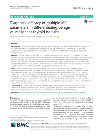
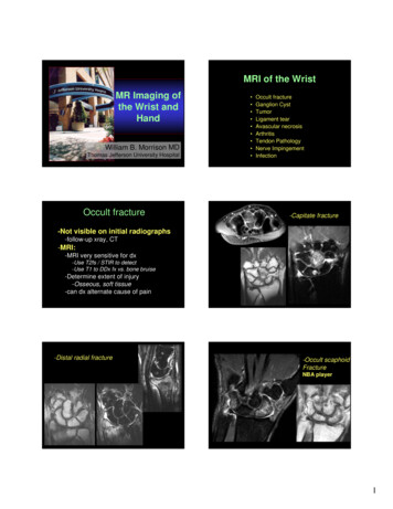
MRI of the WristMR Imaging ofthe Wrist andHandWilliam B. Morrison MDThomas Jefferson University HospitalOccult fracture Occult fractureGanglion CystTumorLigament tearAvascular necrosisArthritisTendon PathologyNerve ImpingementInfection-Capitate fracture-Not visible on initial radiographs-follow-up xray, CT-MRI:-MRI very sensitive for dx-Use T2fs / STIR to detect-Use T1 to DDx fx vs. bone bruise-Determine extent of injury-Osseous, soft tissue-can dx alternate cause of pain-Distal radial fracture-Occult scaphoidFractureNBA player1
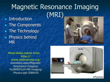
Ganglion CystGanglia: Common Locations- Common at wrist, esp. dorsal- May simulate mass, or maybe occult source of pain if smallor deep Dorsal––––Deep to tendonsAdjacent to lunate/capitate jointWeak area of capsuleExtends around dorsal intercarpal ligament Volar- Joint tendon sheath- MRI:-Lobulated-Fluid signal-Rim-enhancement-May indicate underlying ligament tear– Radial aspect off radioscaphoid joint– Adjacent to radial artery – may be confused forvessel / aneurysm Other areas– Into carpal tunnel– Off tendon sheathsVolar RadioscaphoidGanglionGanglion Cyst from JointExtending Around TendonsFluid signalDorsalintercarpalligamentRim enhancementExtensor TendonGanglionThe “Angry Ganglion”Tumor- MRI may help DDx:-Malignant / benign lesion vs. ‘pseudomass’- Most soft tissue ‘masses’ are benignlesions with characteristic MRI features-Lipomas-Ganglion cysts-Hemangiomas / vascular malformations-Giant cell tumor of tendon sheath- Osseous lesions-Radiographs important for DDx-MRI: solid vs. cystic (esp w contrast)2
-Giant cell tumor of tendon sheath (GCTTS)-LipomaLocation: tendon sheathSignal: low T1, T2Fat signalNo internal complexity-Glomus tumorLocation: distal digitSignal: ‘light bulb’ on T2, Gd-Nerve lesionFibrolipomatoushamartomaLocation: neuralSignal: high T1, fascicular patternMalignant lesion-Synovial sarcomaSolid, complex mass“Pseudomass”-Accessory muscleCharacteristic locationse.g., palmaris longusSignal: same as muscle3
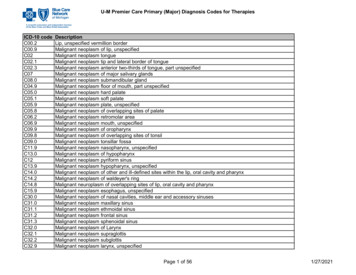
Ligament tear-Aneurysmalbone cystFluid-fluidlevels-Intrinsic ligaments-Scapholunate-Lunatotriquetral-Triangular fibrocartilage complex-central (radial aspect)-peripheral (ulnar side)-MR arthrography-Increases accuracy for dx of tearTriangular Fibrocartilage“Complex” (TFCC)Anatomy TFCCANATOMY TFC Dorsal andvolar RU lig UT ligament Meniscalhomologue ECU sheathTriangular fibrocartilageDorsal and volar radioulnar ligamentsUlnar-triquetral ligamentMeniscal homologueECU sheathTFCC - perforationCentral: toAttachesAttachestobone centrallycartilageUnliketheofRadiusTFCPeripheral: twoattachments-Look at slicewith styloidCentral TFCC TearPerforations may not be clinically significant4
-Peripheral TFCC tearECU Subluxation / PeripheralTFCC TearUlno-lunate AbutmentPeripheral TFCC Tear / LT TearECU Tenosynovitis / PeripheralTFCC TearIndirect Arthrogram – tear of central TFCwith ulnar-lunate abutment-Positive ulnarvariance-Cystic changein lunate-TFCC tear5
Scapholunate andLunatotriquetral LigamentsScapholunate Ligament TearDORSAL AND VOLAR BANDSThese bands are moremechanically importantthan central membraneDirect MR arthrogram –scapholunate tearSL or LT tearcan cause carpalmalalignmentDorsal tilt of lunate(DISI deformity)Scapholunate tearPalmarflexion of scaphoidScapholunate advanced collapse(SLAC wrist)Radiographic progressionOf SLACLateEarly-radioscaphoidjoint narrowingDISI deformityIntermediateProximal migration of capitateCarpal osteoarthritis6
SLAC wristSLAC secondary torheumatoid arthritisInflammatory arthropathiescan cause intrinsic ligamenttearsExtensive synovitisMarrow edemaLunatotriquetralligament tearLunatotriquetral Ligament TearLunate may tiltin palmar directionalong with scaphoid(VISI)Avascular necrosis-Keinbock’s diseaseReplacementof fat signal c/w AVN-Lunate (negative ulnar variance)-Scaphoid (fracture)Progression: density, fracture, collapse, OA7
“SNAC” Wrist-Scaphoid fracture with AVNof the proximal poleScaphoid Nonunion AdvancedCollapse-Scaphoid nonunion-Humpback deformity-Acts like an SL lig tear-Radiocarpal OA and AVN lunateArthritis-Scapholunate Advanced Collapse (SLAC)-Osteoarthritis-Subchondral cysts cartilage loss, spurs-Distribution depends on etiology-Trauma, instability, predisposing factors-Inflammatory arthropathies-Classic: rheumatoid arthritis-Carpus, MCPs-Diffuse involvement-Synovitis, erosions-Type 2 lunate with secondary OARheumatoid ArthritisMarked synovial proliferationLunate articulates with hamate8
Rheumatoid ArthritisRheumatoid ArthritisMRI can monitor activity, response to TxTenosynovitis in multiplesheaths suggests anInflammatory arthropathyErosionsTendon Pathology-Tenosynovitis-Tendon tear-Pulley lesionsDeQuervain’sTenosynovitis1st EXTENSORCOMPARTMENTExtensor TendonsCOMPARTMENT 4Extensor digitorumExtensor indicisCOMPARTMENT 3Extensor pollicis longusCOMPARTMENT 5Extensor digiti minimiCOMPARTMENT 2Extensor carpi radialis brevisExtensor carpi radialis longusCOMPARTMENT 6Extensor carpi ulnarisCOMPARTMENT 1Extensor Pollicis BrevisAbductor Pollicis LongusInflammation at distal forearm atcrossing point of first and secondextensor compartmentsIntersectionsyndrome9
Complete Tear – ExtensorTendonPartial Tear – FlexorCarpi RadialisLongitudinalTear-Chronicextensor tendontearPulleyInjuriesA series of pulleyssurround the flexortendons keepingthem apposed toboneTear due to chronicoveruse, esp inrock climbersA2 and A4 pulleys aremost commonly injured10
Flexor tendonpulley injuryStenosing TenosynovitisPulley Lesionpre / post stressThumb: UlnarCollateralLigament InjuryLine coronals up with sesamoidsSmall FOVTendon ‘catches’ insheath esp at pulleysDorsal Hood InjuryUlnar collateralligament tearNerve ImpingementDisrupted ‘sagittal band’allows ulnar subluxationof extensor tendon atMCP with flexionMedian nerveCommon in boxers-Guyon’s canal-Carpal tunnel syndromeUlnar nerve11
Carpal tunnel syndromeCarpal Tunnel– Pisiform / hamatemedially– Carpal bonesdorsal– Flexor retinaculumvolar– Median nerve deepto retinaculum– Flexor tendons– Flexor carpi radialis:outside the carpaltunnel-Flexortenosynovitis-Separation oftendons bysynovial tissueCTS: Flexor RetinaculumBowing-Mass effect frommuscle in carpal tunnelCTS: Proximal Enlargement andFasciculation-Volar ganglion cystin carpal tunnelFasciculation:Looks likedots insideproximalÎdistal12
PROXIMALGuyon’s CanalGUYON’SCANALDISTAL-Ganglion cyst withUlnar nerve impingementInfection-Septic arthritisGuyon’s canalJoint effusionSynovial thickening / enhancementSubchondral edema-OsteomyelitisMarrow edema / enhancement-Septic arthritisand osteomyelitisSeptic arthritis and osteomyelitis13
Routine MRI wrist:-Tendon pathologyMRI ProtocolTHANK YOU-Carpal tunnel syndrome-Ganglion cyst-Acute trauma-Osteoarthritis-AVNMRI wrist with IV contrast:-Mass-Infection-Inflammatory arthropathyMR arthrogram:-Ligament tear14
-Type 2 lunate with secondary OA Lunate articulates with hamate Rheumatoid Arthritis Marked synovial proliferation. 9 Rheumatoid Arthritis MRI can monitor activity, response to Tx Erosions Rheumatoid Arthritis Tenosynovitis in multiple shea