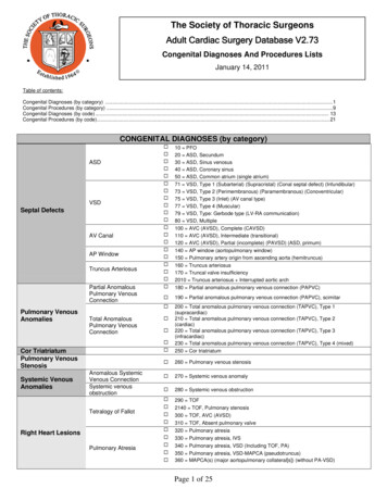
Transcription
Lyen et al. Journal of Congenital Cardiology (2017) 1:7DOI 10.1186/s40949-017-0008-4Journal ofCongenital CardiologyREVIEWOpen AccessAnomalous pulmonary venous drainage: apictorial essay with a CT focusStephen Lyen1*, Shalini Wijesuriya1, Eleanor Ngan-Soo1, Helen Mathias2, Michael Yeong1, Mark Hamilton1and Nathan Manghat1Abstract: The imaging of pulmonary venous anatomy has traditionally been performed with echocardiography andcatheter pulmonary angiography. Magnetic resonance imaging (MRI) and notably multi-detector row computedtomography (CT) have refined the usual imaging techniques.Anomalous pulmonary venous drainage (APVD) is the drainage of one or more pulmonary veins outside the leftatrium. Its detection is critical due to the strong association with congenital heart disease as well as other cardiacand respiratory anomalies, which have significant implications for patient management. The pervasive application ofCT combined with the relatively non-specific clinical presentation of APVD has resulted in the increased incidentaldetection of these anomalies. Knowledge is hence vital as the imaging specialist is now usually the first person tomake such a diagnosis. Furthermore, pulmonary veins are an important site for arrhythmogenic foci and radiofrequencyablation of such sites is used in the treatment of refractory atrial fibrillation. Hence an imaging road map of these veins iscrucial before any management can take place.This pictorial review will illustrate embryology, normal and variant pulmonary vein anatomy, and varied patterns of APVD.Finally, we discuss the implications in the treatment of atrial fibrillation.Keywords: Pulmonary veins, Drainage, Tomography X-ray computed, Magnetic resonance imagingBackgroundThere is a broad spectrum of pulmonary venous connections that deviate from the usual anatomical configurationand ranges from normal variant drainage to anomalouspulmonary-systemic communications, the latter arisingfrom a failure of separation of the pulmonary and systemiccirculations in-utero. Patients can be asymptomatic orpresent with non-specific cardiorespiratory symptoms.Diagnosis can be difficult, missed, or only made at lateclinical presentation in adulthood.Advances in non-invasive, cross-sectional imaging haverefined radiological evaluation. Electrocardiogram (ECG)gated cardiac CT allows low dose, detailed anatomical assessment with excellent spatial and temporal resolution.Cardiac magnetic resonance imaging enables anatomicaland physiological assessment, ventricular function andvolumetrics and three-dimensional multiphase angiographic techniques. In addition, it enables quantificationof shunt ratios (Qp:Qs). These techniques, including* Correspondence: smlyen@doctors.org.uk1Radiology Department, University Hospitals Bristol NHS Foundation Trust,Bristol Royal Infirmary, Upper Maudlin St, Bristol BS2 8HW, UKFull list of author information is available at the end of the articleechocardiography and chest radiography, must be regardedas complementary. The advantages and disadvantages ofeach technique in evaluating pulmonary venous connections are described in Table 1.Main textEmbryology of the pulmonary veinsUnderstanding of the embryology is important in appreciating the wide patterns of APVD. Lung development commences at approximately 26 days of gestation with therespiratory diverticulum, arising from the primitive foregut [1]. The associated venous plexus (splanchnic plexus)communicates with the systemic cardinal and umbilicovitelline veins, thus forming the initial route of pulmonaryvenous drainage. At 27 to 29 days, an out-pouching develops from the postero-superior wall of the primitive leftatrium [2]. This out-pouching subsequently begins tocommunicate with the pulmonary venous component ofthe splanchnic plexus. Simultaneously the systemic veinsinvolutes, thus separating the two venous systems. In normal embryos there is complete dissociation of these venoussystems which results in four separate pulmonary veins,which drain into the left atrium. Failure of this separation The Author(s). 2017 Open Access This article is distributed under the terms of the Creative Commons Attribution 4.0International License (http://creativecommons.org/licenses/by/4.0/), which permits unrestricted use, distribution, andreproduction in any medium, provided you give appropriate credit to the original author(s) and the source, provide a link tothe Creative Commons license, and indicate if changes were made. The Creative Commons Public Domain Dedication o/1.0/) applies to the data made available in this article, unless otherwise stated.
Lyen et al. Journal of Congenital Cardiology (2017) 1:7Page 2 of 13Table 1 Radiological assessment of the pulmonary veinsAdvantagesDisadvantagesEchocardiography Non-invasive No ionising radiation Accessible Portable Real-time imaging Haemodynamic data Limited evaluation of pulmonary veins and systemicconnections. Imperfect spatial resolution Operator dependentPulmonaryangiography Haemodynamic data Pulmonary pressures Chamber oxygensaturations Invasive Limited information on associated congenital cardiothoracicanomalies. Relationship to surrounding structures poorly defined. Radiation dose considerations Contrast allergyECG-gated MDCT Non-invasiveComprehensive thoracic anatomical assessmentHigh spatial resolutionRapid image acquisitionCan be used in patients with metallic implantsSimultaneous coronary artery and cardiac morphologicalevaluation Widely available No patient sedation. Cardiac MRI High temporal resolution No ionizing radiation Accurate ventricular functional analysis Accurate anatomical assessment Haemodynamic data 3D multiplanar reconstruction Quantification of shunt ratios. Non-contrast angiography is possible Available in specialised centres Longer duration of image acquisition and analysis Images suboptimal with cardiac dysrhythmia or inability of patientto breath hold Inferior spatial resolution to CT Patient claustrophobia Gadolinium contrast reactions. More prone to artefact.process leads to the various types of anomalous venousdrainage (Fig. 1). Failure of incorporation of the normalpulmonary veins into the left atrium may also occur (cortriatriatum), which will be discussed later.Normal pulmonary venous anatomyThe usual arrangement consists of four separate pulmonary veins: right and left superior and inferior veins, whichIonizing radiation exposurePotential adverse iodinated contrast reactionInferior temporal resolution to Echo and MRIPatient heart-rate control requireddrain individually into the left atrium (Fig. 2). The rightsuperior pulmonary vein (RSPV) drains the right upperand middle lobes of the lung with the right lower lobedrained by the right inferior pulmonary vein (RIPV). Theleft superior pulmonary vein (LSPV) drains the lingulaand left upper lobe, whilst the left lower lobe is drained bythe left inferior pulmonary vein (LIPV). This anatomicalarrangement is found in 60–70% of the population [3].Fig. 1 Embryology of anomalous pulmonary venous drainage. This diagram illustrates right sided pulmonary veins failing to establish a normalconnection with the left atrium and instead draining into the inferior vena cava. UV Umbilicovitelline vein; CCV Common cardinal vein;LB Lung buds; LA Left atrium; RA Right atrium; LV Left ventricle; RV Right ventricle; SVC Superior vena cava; IVC Inferior vena cava;ARPV Anomalous right pulmonary vein
Lyen et al. Journal of Congenital Cardiology (2017) 1:7Page 3 of 13Fig. 2 Normal pulmonary venous anatomy and normal variants. 3D CT reconstructions of the left atrium and pulmonary veins, posterior views.a Normal pulmonary venous anatomy. (1) Left superior pulmonary vein, (2) Left lower pulmonary vein, (3) Right superior pulmonary vein, (4)Right inferior pulmonary vein. b Normal variant right middle pulmonary vein (arrowhead). c Single conjoined left pulmonary vein (asterisk).d Anatomical variant with conjoined ostium (black arrow) of the left inferior pulmonary vein (2) and right inferior pulmonary vein (4)Normal variant pulmonary venous drainageA wide variation of normal variant pulmonary venousdrainage exists and is a common incidental finding. Normal variants typically consist of conjoined or accessoryveins [4]. A conjoined (or common) vein is present whenthe superior and inferior veins on the same side unite toform a single confluence before entering the left atrium,resulting in a single atriopulmonary venous junction(or ostium). They are more common on the left (Fig. 2).Rarely this can occur via a common superior or inferiorconjoined vein and can look similar to repaired totalanomalous pulmonary venous drainage (TAPVD) on imaging (Fig. 2).Accessory (or supernumerary) veins (Fig. 2) are separate from the superior or inferior pulmonary veins withan independent drainage (and atriopulmonary venousjunction) into the left atrium. In contrast to a conjoinedvein, these supernumerary variants usually have a narrower ostium than normal [5]. Accessory pulmonaryveins may be seen in 30% of patients [6]. In a studyexamining left atrial anatomy in patients with and without atrial fibrillation (AF) who had CT angiography,there was no significant difference in number of pulmonary veins, although the left atria and pulmonaryostial dimensions were larger in AF patients [7].Accessory veins are more common on the right, of whichthe most frequent variant is an accessory vein draining themiddle lobe, seen in up to 26% of patients [8].Anomalous pulmonary venous drainagePartial anomalous pulmonary venous drainage (PAPVD)describes the connection of at least one pulmonary vein,but not all, to the systemic venous system or right atrium(RA). Drainage of all pulmonary veins outside the leftatrium is termed total anomalous pulmonary venousdrainage (TAPVD).A. Partial anomalous pulmonary venous drainage(PAPVD)The prevalence of PAPVD has been reported to be between0.4–0.7% [9]. It is more common on the right and results inthe formation of a left-to-right shunt.Right-sided anomalous drainage may occur into any ofthe central veins. The most common form of PAPVD isthe anomalous drainage of the RSPV into the superiorvena cava (SVC) [10]. This frequently occurs at the junction of the SVC and RA (Fig. 3) [11]. Occasionally,drainage occurs at a higher level at the junction between
Lyen et al. Journal of Congenital Cardiology (2017) 1:7Page 4 of 13Fig. 3 Different variations of anomalous right superior pulmonary vein. (a) Axial image at the level of the main pulmonary artery. Right superiorpulmonary vein (RSPV) draining into the superior vena cava (SVC) only (arrow). No sinus venosus defect was demonstrated. (b) Axial oblique CT. RSPVwith a connection to the superior vena cava/right atrium (SVC/RA) confluence (arrow), but then continuing to drain into the left atrium (arrowhead).No direct sinus venosus defect. The right ventricle is dilated. (c) Axial oblique CT. RSPV draining into the SVC/RA confluence (white arrow) with a superiorsinus venosus defect (black arrowhead) and right ventricular dilatation. (d) Axial oblique CT image. Anomalous RSPV is seen draining into the SVC (whitearrow). Superior sinus venosus defect is demonstrated (black arrow) as well as an atrial secondum defect (black arrowhead)the SVC and brachiocephalic vein above the level of theazygos vein, or into the azygos vein itself (Fig. 4). Otherright-sided pulmonary-systemic connections includedrainage into the coronary sinus, inferior vena cava(IVC) or drainage of all right pulmonary veins into theRA. Anomalous pulmonary venous of the right superiorpulmonary veins is commonly associated with superiorsinus venosus atrial septal defects, an atrial septal defectbeing the most common associated congenital heartdefect (Figs. 3, 4 and 5) [12]. The identification of theseabnormalities has a significant bearing on surgical management as anomalous pulmonary veins could be reroutedto the left atrium via an atrial septal defect [13].Up to 18.2% of PAPVD cases affect the left-sidedpulmonary veins [12]. The commonest pattern on theleft is the LSPV connected to the left brachiocephalicvein (Figs. 6 and 7) [10]. In this condition, a verticallyorientated vessel courses lateral to the aortic arch priorto draining into the left brachiocephalic vein. Thisanomalous vein can sometimes be misdiagnosed as aleft-sided SVC on CT (Fig. 7) [10, 14]. The two anomalies can be differentiated at the level of the left hilum.Under normal conditions a single vessel is seen anteriorto the left main bronchus, the LSPV. In PAPVD this vessel is absent, whereas in persistent left SVC two vesselslie anterior to the left main bronchus, the anomalous leftsuperior pulmonary vein and the persistent left SVC.The course of the lobar intraparenchymal veins may alsobe appreciated as they drain into the anomalous vessel. Afurther feature of left upper lobe PAPVD is a normal or enlarged left brachiocephalic vein, whereas in a persistent leftSVC the left brachiocephalic vein may be absent or small.Other described left-sided connections include drainage into the hemiazygos vein or the coronary sinus [15].Careful scrutiny of the pulmonary veins is importantand the identification of less than four pulmonary veinsconnected to the left atrium should alert the reader to adiagnosis of PAPVD. However, ancillary features may bepresent which may also allude to this diagnosis. In thepresence of a left-to-right pulmonary shunt, enlargementof the right heart may occur due to increased volumeloading (Figs. 3, 4 and 5), dependent on the presence ofan intact atrial septum. In addition, the vessel receivingthe anomalous vein may be enlarged and there may beasymmetry of the pulmonary veins. The magnitude ofthe left-to-right shunt is dependent of the number
Lyen et al. Journal of Congenital Cardiology (2017) 1:7Page 5 of 13Fig. 4 Anomalous drainage into the azygos vein. Images a and b are from an adolescent patient with anomalous pulmonary venous drainageinto the azygos vein. Images c and d are from a paediatric patient with anomalous pulmonary venous drainage identified on echocardiography.a Sagittal maximum intensity projection (MIP) CT image showing the right superior pulmonary vein (white arrow) draining into a dilated azygosvein (labelled AZ). b Three-dimensional (3D) CT reconstruction of patient in (a), posterior view. The black arrow indicates the right superior pulmonaryvein draining into the azygos vein. c 3D CT reconstruction. Most of the right sided pulmonary veins are seen draining into a dilated azygos vein (labelledAZ). d Axial CT image of the patient described in (c). Only a very small right middle pulmonary vein is seen draining normally into the left atrium (whitearrow). Note the dilated right atrium and right ventricle consistent with a significant left-to-right shuntPAPVD. In a series looking at single PAPVD, the shuntis modest (Qp:Qs 1.3–1.6) associated with mild dilatation of the right ventricle [16]. On top of volume loadingto the right ventricle, APVD is one of the treatablecauses of pulmonary hypertension in adults [17].Congenital cardiac and thoracic anomalies, for example thoracic hypoplasia and cardiac dextro/levoversion may also occur, the presence of which shouldprompt examination of the pulmonary venous anatomy (Figs. 8, 9 and 10).Fig. 5 Anomalous right superior and middle pulmonary veins. (a) Coronal oblique maximum intensity projection CT image. The right superior pulmonaryvein is seen draining into the superior vena cava (arrow). Just inferior to this the right superior and inferior middle pulmonary veins are also seen drainingseparately into the superior vena cava/right atrium confluence (arrowheads). b Axial oblique MIP CT 5-chamber view, demonstrating a superior sinus venosusdefect (black arrow), anomalous middle pulmonary vein (white arrowhead) and marked right ventricular dilatation secondary to the left-to-right shunt
Lyen et al. Journal of Congenital Cardiology (2017) 1:7Page 6 of 13Fig. 6 Anomalous left superior pulmonary vein. a Axial oblique maximum intensity projection (MIP) CT image. Anomalous left superior pulmonary vein(LSPV) drains into the left brachiocephalic vein (LBCV) (white arrow). AO – Aorta. b Three-dimensional CT reconstruction demonstrating the anomalousLSPV draining into the LBCV (white arrow)Cross-sectional imaging with CT and MRI is widelyused for the diagnosis of APVD in the adult population.CT has superior spatial resolution and thus is useful inpresurgical planning in complex lesions. However, MRI isoften sufficient for diagnosis and has the advantages ofproviding functional assessment and shunt calculation.In addition, MRI does not require ionising radiationwhich is of particular importance in young patients.Scimitar (hypogenetic lung/venolobar) syndromeScimitar syndrome consists of right-sided anomalouspulmonary venous drainage, hypoplasia of the lung andFig. 7 Anomalous left superior pulmonary vein (LSPV) may mimic a left sided superior vena cava (SVC) on axial CT images. Patient 1. Bilateral SVCs withanomalous left SVC draining into the left atrium (a and b). a Bilateral SVCs. Right SVC (arrowhead) and small left sided SVC (white arrow). b Coronal MIP CTin the same patient showing the anomalous left SVC draining into the left atrium (white arrow). Normal right-sided SVC (thin white arrow). Patient 2. Partialanomalous pulmonary venous drainage (c and d). c Anomalous LSPV (white arrow), which on single axial slice is similar in appearance to the left-sided SVCin (a). d Coronal MIP CT showing the anomalous LSPV (white arrow) draining into the left brachiocephalic vein (LBCV). Normal right SVC (thin white arrow)
Lyen et al. Journal of Congenital Cardiology (2017) 1:7Page 7 of 13Fig. 8 Anomalous right-sided pulmonary venous drainage with a superior sinus venosus defect. a Axial oblique CT MPR images show an anomalousright superior pulmonary vein (RSPV) draining into the superior vena cava/right atrium (SVC/RA) junction (white arrow) with an associated superiorsinus venosus defect (SSVD, arrowhead) connecting the left atrium (LA) and right atrium (RA). b Post surgical repair the right superior pulmonary veinis now directed into the left atrium with closure of the SSVD. c Sagittal oblique CT images showing the SVC overriding a SSVD between the two atria.d Post surgery the SSVD has been repaired, the SVC (black arrow) drains directly into the right atriumpulmonary artery, cardiac dextroposition (Fig. 9) andsystemic arterial supply to the right lower lobe. The latter may occur either from the descending thoracic orupper abdominal aorta [18]. The anomalous right-sideddrainage occurs most commonly into the subdiaphragmatic IVC (Fig. 9) [19]. Drainage may also occur intothe porto-hepatic veins, azygos vein, coronary sinus,right atrium or the suprahepatic portion of the IVC.Scimitar syndrome can also be associated with pulmonary arteriovenous malformations, abnormalities ofthe diaphragm and genitourinary tract. Whilst usuallyright-sided, left-sided cases have been described in theliterature [20].The scimitar is a curved traditional sword held byPersian and Turkish warriors, from which the syndromereceives its name. The imaging findings on chest x-ray areclassic. The anomalous vein is seen as a curved, tubularstructure noted lateral to the right heart border, coursinginferiorly (Scimitar sign) (Fig. 9).A pseudoscimitar syndrome occurs when the aboveconstellation of findings is associated with an anomalousvein that takes a tortuous course and drains into the leftatrium, rather than the IVC, producing a false positiveScimitar sign on chest radiography. This has been coinedthe pseudoscimitar vein, also known as a meanderingright pulmonary vein [10, 21].PAPVD: Chest radiographyPatients with PAPVD often have non-specific cardiac orrespiratory symptoms and are initially investigated withchest radiography, which can be completely normal.However, PAPVD should be suspected with the following findings: An usual course of a central venous catheter [22]. Focal mediastinal widening to the left of the aorticknob (Fig. 11).This may be caused by an anomalous left superior pulmonary vein. Scimitar vein and other associated findings such asright lung hypoplasia and cardiac dextroposition. Unusual patterns of pulmonary oedema.Left upper lobe PAPVD will spare vascular congestionof the left upper lobe in left heart failure, as the left upper
Lyen et al. Journal of Congenital Cardiology (2017) 1:7Page 8 of 13Fig. 9 Scimitar Syndrome. a Coronal oblique maximum intensity projection (MIP) CT images. A large right sided pulmonary vein is seensupradiaphragmatic inferior vena cava (IVC) in a patient with Scimitar Syndrome. b Axial CT image of same patient as in (a). The white arrowindicates the anomalous right pulmonary vein. The asymmetry of the pulmonary vasculature provides a clue to identifying the abnormality. Theright hemidiaphragm is elevated consistent with hypoplasia of the right lower lobe, a common association with anomalous pulmonary venousdrainage. c Chest radiograph of a paediatric patient, demonstrating a tubular opacity in the right paracardiac region consistent with a scimitarvein (arrow). d Coronal MIP CT image showing a large right pulmonary vein draining into the infradiaphragmatic IVCFig. 10 Anomalous right inferior pulmonary vein drain. Adult patient presenting with breathlessness. a Three-dimensional (3D) CT reconstruction.A large right inferior pulmonary vein is seen draining most of the right upper lobe into the supradiaphragmatic inferior vena cava (arrow). Mostof the lower lobe is seen draining normally into the left atrium (arrowhead). b Sagittal MIP CT image shows a hypoplastic right lower lobe (arrow– oblique fissure). The large curvilinear right pulmonary vein is seen draining the right upper lobe
Lyen et al. Journal of Congenital Cardiology (2017) 1:7Page 9 of 13Fig. 11 Chest radiograph findings in anomalous pulmonary venous drainage- Focal mediastinal widening to the left of the aortic knob. a Patient1. Postero-anterior (PA) chest radiograph (CXR). Vascular shadow adjacent to the aortic knuckle corresponds to anomalous left superior pulmonaryvein (LSPV) in (b). b Coronal maximum intensity projections (MIP) CT image demonstrating anomalous LSPV (black arrow) draining intothe brachiocephalic vein (LBCV). This is the most common left sided partial anomalous pulmonary venous drainage pattern. c Patient 2.Posteroanterior chest radiography. The LSPV can be seen as a prominent vascular shadow lateral to the aortic knuckle and left subclavianartery (black arrow). d Coronal MIP MR angiography demonstrating LSPV (arrow) draining into the left BCV (LBCV)lobe pulmonary vein does not drain into the leftatrium. Similarly, in right heart failure the sameanomaly causes pulmonary oedema isolated to the leftupper lobe [23]. Effects of longstanding left-to-right shunts (Fig. 12).These include enlargement of the cardiac silhouette(in particular right ventricular dilatation), pulmonaryplethora, right hilar vascular prominence and dilatationof the central pulmonary arteries.The above findings are summarised in Table 2.Cor TriatriatumThis rare condition refers to failure of incorporationof normal pulmonary veins into the left atrium [9]and accounts for up to 0.1% of congenital heart disease [24]. There is separation of the left atrium intotwo chambers, separated by a thin, fenestrated membrane. The posterior chamber drains the pulmonaryveins and the anterior chamber (true left atrium)drains into the left ventricle via the mitral valve,overall resulting in an obstruction to the drainage ofthe pulmonary veins. Patients can present with failureto thrive and congestive heart failure.B. Total anomalous pulmonary venous drainage (TAPVD)This refers to drainage of all four of the pulmonary veinsto another cardiovascular structure other than the leftatrium [25] and accounts for about 2% of cardiac malformations. Four types exist, depending on the level ofdrainage:Type 1 – supracardiac: This accounts for up to 55% ofcases and is the most common. It typically involves convergence of all four pulmonary veins behind the heart toform a common vein (Fig. 13). This then drains into theleft brachiocephalic vein and henceforth into the SVC.This can cause the classic ‘snowman’ or ‘figure-of eightappearance’ on chest radiography.Type 2 - abnormal communication at the cardiac level:drainage occurs into the RA or coronary sinus.Type 3 – drainage below the level of the heart or diaphragm, for example into the IVC, portal or hepaticveins.
Lyen et al. Journal of Congenital Cardiology (2017) 1:7Page 10 of 13Fig. 12 Effects of a longstanding left-to-right shunt in patients with PAPVD. a Right superior pulmonary vein (RSPV) draining into thesuperior vena cava (SVC). Right sided pulmonary plethora is seen. b Right atrial dilatation, cardiomegaly and features of pulmonaryoedema. Subsequent computed tomographic pulmonary angiography (CTPA) demonstrated a RSPV draining into the SVC with dilatedright heart chambers. c Patient with anomalous right superior and middle pulmonary veins draining into the superior vena cava/rightatrium confluence. A chest radiograph demonstrates cardiomegaly and central pulmonary artery dilatation secondary to long-termleft-to-right shuntType 4 - drainage at more than one level (Fig. 13).In patients with TAPVD, a right-to-left shunt is crucialfor early survival and this manifests usually in the formof a patent foramen ovale or an atrial septal defect.Patients present early in the neonatal period with symptoms of congestive cardiac failure and cyanosis. Promptsurgical repair is essential, thus it is rarely encounteredon CT or MRI. In the vast majority of cases, echocardiography is the mainstay of diagnosis and anatomicaldescription of TAPVD. Cross-sectional imaging is usually reserved for when the diagnosis is unclear, orincomplete definition of the pulmonary veins at echocardiography [26].TAPVD is commonly associated with other congenitalcardiac lesions. A large international multicentre studyconducted by Seale et al. (n 422) showed 14% of patients had associated cardiac lesions, and that if definitive repair was not performed at the time of surgery thiswas an independent risk factor for death [27]. Inaddition, pre- and postoperative pulmonary vein obstruction or stenosis were also independent risk factorsfor death in multivariate analysis. This highlights theTable 2 Features of PAPVD on chest radiography May appear normal Unusual course of a central venous catheter Focal widening of the mediastinum to the left of the aortic knob Unusual patterns of pulmonary oedema The Scimitar veinimportance of accurate pre- and postoperative anatomical evaluation in this cohort of patients.Relationship between pulmonary veins and atrialfibrillationAtrial fibrillation (AF) is the most common cardiacarrhythmia, affecting 5% of the population of theUnited Kingdom over the age of 65 years, increasingto 10% above the age of 75 years [28]. Patients withAF are at a greater risk of stroke, increasing from1.5% from ages 50–59 years to 23.5% in those aged80–89 years [29]. Mortality rates are also higher inthese patients [30].One of the mechanisms believed to trigger the onset of AF is the production of ectopic atrial beats[4]. The pulmonary veins have been found to be animportant site of such arrhythmogenic activity, withup to 96% of foci triggering paroxysmal episodes ofAF arising from them [31]. This may be explained bythe observation of sleeves of myocardial tissue fromthe left atrium extending into the pulmonary veins,with distances varying from 2 to 17 mm [32]. Ectopicfoci most commonly arise from the left vssuperiorpulmonary vein. This may be explained by two observations: myocardial sleeves are longer in thesuperior pulmonary veins and thicker at the atriopulmonary venous junction [33]. Radiofrequency ablation (RFA) has been successfully used to disconnectthe electrical connection between the pulmonaryveins and the left atrium, thereby treating AF in affected patients [34]. Effects of a longstanding shunt - dilatation of the right heart chambersand central pulmonary arteries.The role of CT in the mapping of pulmonary veinanatomy - what the electrophysiologist needs to know Cardiac and thoracic associations eg: pulmonary hypoplasia andcardiac dextroposition in Scimitar syndrome.Given the variability of pulmonary venous anatomy,detailed pre-procedural evaluation of the left atrium
Lyen et al. Journal of Congenital Cardiology (2017) 1:7Page 11 of 13Fig. 13 Total anomalous pulmonary venous drainage (TAPVD) repair. a Three-dimensional (3D) CT reconstruction. An adult patient presentingwith breathlessness had a computed tomographic pulmonary angiogram (CTPA). A single left pulmonary vein (LPV) crosses the midline to form asingle confluence with the right pulmonary veins (RPV) and joins the left atrium (LA) via a baffle (white arrow). A residual anomalous left superiorpulmonary vein (white arrowhead) is seen draining into the left brachiocephalic vein (LBCV); presumed previous type 4 TAPVD. Incidentally thepulmonary vessels in the right lower lobe were tortuous and dilated with a suspected arteriovenous malformation. b Three dimensional (3D) CTreconstruction. A paediatric patient with a Type 2 TAPVD repair. The pulmonary veins (white arrowheads) can be seen forming a single venousconfluence posterior to the LA. This has been reconnected to the LA via a small baffle (white arrow). c Sagittal CT image. Type 2 TAPVD. A singlevenous confluence of the pulmonary veins is present (asterisk) posterior to the left atrium (LA) with a focal narrowing where this hasbeen surgically connected to the LA (black arrow). d Axial CT image. This again shows the posterior venous confluence of the p
Anomalous pulmonary venous drainage (APVD) is the drainage of one or more pulmonary veins outside the left atrium. Its detection is critical due to the strong association with congenital heart disease as well as other cardiac . sel is absent, whereas in persistent left SVC two vessels lie anterior to the left main bronchus, the anomalous left .